. 2007 Jan 8:7:1.
doi: 10.1186/1471-2261-7-1.

The effects of second-hand smoke on biological processes important in atherogenesis

Affiliations

The effects of second-hand smoke on biological processes important in atherogenesis

Hongwei Yuan et al. BMC Cardiovasc Disord. .

Abstract

Background: Atherosclerosis is the leading cause of death in western societies and cigarette smoke is among the factors that strongly contribute to the development of this disease. The early events in atherogenesis are stimulated on the one hand by cytokines that chemoattract leukocytes and on the other hand by decrease in circulating molecules that protect endothelial cells (ECs) from injury. Here we focus our studies on the effects of "second-hand" smoke on atherogenesis.

Methods: To perform these studies, a smoking system that closely simulates exposure of humans to second-hand smoke was developed and a mouse model system transgenic for human apoB100 was used. These mice have moderate lipid levels that closely mimic human conditions that lead to atherosclerotic plaque formation.

Results: "Second-hand" cigarette smoke decreases plasma high density lipoprotein levels in the blood and also decreases the ratios between high density lipoprotein and low density lipoprotein, high density lipoprotein and triglyceride, and high density lipoprotein and total cholesterol. This change in lipid profiles causes not only more lipid accumulation in the aorta but also lipid deposition in many of the smaller vessels of the heart and in hepatocytes. In addition, mice exposed to smoke have increased levels of Monocyte Chemoattractant Protein-1 in circulation and in the heart/aorta tissue, have increased macrophages in the arterial walls, and have decreased levels of adiponectin, an EC-protective protein. Also, cytokine arrays revealed that mice exposed to smoke do not undergo the switch from the pro-inflammatory cytokine profile (that develops when the mice are initially exposed to second-hand smoke) to the adaptive response. Furthermore, triglyceride levels increase significantly in the liver of smoke-exposed mice.

Conclusion: Long-term exposure to "second-hand" smoke creates a state of permanent inflammation and an imbalance in the lipid profile that leads to lipid accumulation in the liver and in the blood vessels of the heart and aorta. The former potentially can lead to non-alcoholic fatty liver disease and the latter to heart attacks.


Figures
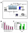
Figure 1
Figure 1
Schematic representation of the Smoking System, smoking dose calibration, and effects of cigarette smoke on blood carboxyhemoglobin (CoHb) levels. (A) Schematic representation of the smoking system. Smoke was generated by using a puffer box built by the University of Kentucky [33] and chambers built by Teague Enterprises. Each smoke type can be mixed with fresh air before it was puffed through a tube into the exposure chambers. In this study only the SSW chamber was used. The air and smoke in the chamber were exhausted using a blower fan (air moving fan). A vacuum pump/gas meter was used for dose calibration only. (B) The smoking dose was calibrated to 25 mg TPM/m3 by adjusting the intake and exhaust valves. Smoke samples taken from the SSW chamber were passed through a filter unit; the particulate material deposited onto the filter was measured by weighing the filter before and after smoke filtration. The vacuum pump gas meter recorded the smoke sample volume passed through the filter. Total particulate matter (TPM) was calculated by dividing the total particle weight trapped on the filter by the volume passed through the filter. w.c. = water column. (C) Mice were exposed to SSW smoke for 6 hrs intermittently and blood was drawn to test for carboxyhemoglobin. Blood was treated with ammonium hydroxide, absorbance measured at 420 nm and 413 nm and the ratio plotted. C = control.
Figure 2
Figure 2
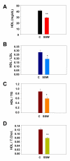
Effect of cigarette smoke on the HDL and its ratio to other plasma lipids. Mice were exposed to SSW 6 hrs/day intermittently, 5 days a week. Blood samples were collected from the tail vein and, after centrifugation, the plasma was used to analyze for levels of HDL and other lipids by enzymatic reaction using colorimetric methods with commercially available kits (Wako Chemicals). After incubating with coloring reagent that only reacts with a specific cholesterol or type of lipid, the absorbance was measured and the concentration of each was calculated by plotting the measured values against a standard curve. (A) The levels of HDL in the blood of mice exposed to SSW are significantly lower than in the non-exposed mice. The ratios of HDL to LDL (B), HDL to TG (C), and HDL to TC (D) are all significantly lower in the SSW exposed mice.
Figure 3
Figure 3
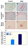
Lipid accumulation in arterial blood vessels. Oil red O was used to detect the presence of lipids in tissue sections of the heart and aorta. (A) & (B) Presence of higher lipid deposition in the root of the aorta in the smoke-exposed mice. (C) & (D) Similar to A&B but in the proximal part of ascending aorta. (E) & (F) Small blood vessels in the heart muscle of SSW- exposed mice show much more lipid accumulation than in control mice. Thin black lines outline the outside of the vessel walls. (G) The lesion area in both control and SSW-exposed mice was measured as described in the Materials and Methods. The results are expressed as percentage of total area of aorta wall in cross section. Data are mean ± SEM for each group. Statistical analyses were done according to unpaired t test. *P < 0.05 vs. control. The mice were exposed to SSW for 1 year. The images are representative of 2 mice per group.
Figure 4
Figure 4
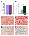
Lipid accumulation in liver tissue. About equal amounts of snap-frozen liver from the sacrificed mice of both control and SSW mice were used for lipid extraction with chloroform: methanol (2:1). The extracted lipids were dried and redissolved in 1% Triton X-100. Commercial kits (WAKO Chemicals USA, Inc., Richmond, VA) were used for determination of (A) total cholesterol and (B) triglycerides. (C-F) Oil Red O staining of frozen liver sections reveals the lipid contents in the liver of control mice (C) and SSW mice (D). The liver of control mice (E) and SSW mice (F) were also stained with Diff-Quick to show their histological structure. The mice were exposed to SSW for 1 year. Representative images of one mouse out of three.
Figure 5
Figure 5
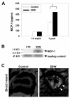
MCP-1 in the plasma and heart/aortic tissues in human apoB100 transgenic mice on high fat diet and exposed to SSW: (A) Levels of MCP-1 in the plasma of smoking mice at two different time points during smoking. 30 μl of cytokine standard or plasma sample were added into the appropriate wells of a Mouse Cytokine array (Quansys Biosciences, Utah, USA) containing an antibody to this chemokine. The images were captured by a CCD camera and analyzed using the Quansys image analysis software. The concentration of MCP-1 was calculated and plotted using a standard curve. (B) Immunoblot analysis for MCP-1 in the heart/aortic tissue (the upper half of the heart together with ascending aorta). The protein extractions were quantified using a DC protein assay kit (Bio-Rad, Hercules, CA) and equal amounts of protein were loaded in each lane of SDS-Polyacrylamide Gel. A 45 KD protein was used to evaluate equal loadings because this protein is always constant, independent of the treatment when the gels are stained with Commasie blue. (C): Immunolabeling for MCP-1 in an arterial vessel of the heart tissue shows that this chemokine is produced by the endothelial cells (arrowheads) and by some of the cells in the adventitia (arrow). The mice were exposed to SSW periodically for 1 year. Representative images of one mouse out of three.
Figure 6
Figure 6
Macrophages and neutrophils in apoB100 transgenic mice on high fat diet: (A) & (B) Tissue sections of arterial blood vessels in the heart were immunolabeled with the antibody to F4/80 which is specific to macrophages, and (C) & (D) with an antibody to myeloperoxidase to detect neutrophils. Macrophages were observed in the walls of arterial vessels in mice exposed to SSW smoke but not in the control mice. However, neutrophils were not found in the arterial vessel walls of either control mice or mice exposed to SSW. The mice were exposed to SSW for 1 year as described in the Materials and Methods. Representative images of one mouse out of three.
Figure 7
Figure 7
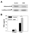
Plasma levels of adiponectin and TNFα. (A) Immunoblot analysis shows the levels of adiponectin in the plasma. Blood sample from both control and SSW smoking mice were applied to 7.5% polyacrylamide electrophoresis gel and transferred to the nitrocellulose membrane. Adiponectin was probed with a goat anti-mouse adiponectin antibody and with a secondary conjugated to HRP for subsequent use with the enhanced chemiluminescence system (ECL, Amersham Pharmacia). Less adiponectin was detected in the plasma of mice exposed to SSW. Samples representative of 2 animals per group. (B) Cytokine array results showing TNFα levels in the plasma. At early stages of smoking, there was no significant difference between the TNFα levels in the control and SSW smoked mice, whereas at late stages of SSW smoking, the TNFα level in plasma was significantly increased. This increment was correlated with the decrease of adiponectin and increment of MCP-1 in the plasma. Samples were done in triplicate.
Figure 8
Figure 8
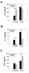
Plasma levels of specific cytokines related to the immune response in atherosclerosis. Cytokine array results show the changes of the cytokines IL-12 (A), IL-4 (B), and IFNγ (C) in the plasma of hApoB100 transgenic mice that were exposed to SSW smoke. The cytokines in the plasma were captured by antibodies coated on the bottom of 96-well plates (Quansys Biosciences, Utah, USA). After washing, probing with streptavidin-HRP, and reacting with substrate, the images were captured with CCD camera (Roper Micromax 1300B). The strength of signal was translated into concentration of cytokine by plotting against a standard curve.
Figure 9
Figure 9
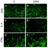
SSW smoke exposure favors a Th1 immune response in human apoB100 Transgenic mice. Tissue sections of the spleen were labeled with rat anti-mouse CD4+, IL-4, and IFNγ antibodies, and detected by FITC-labeled goat anti-rat IgG. (A-F) Immunolabeling showing the peripheral lymphocytes in spleen expressing CD4+ (A, B), IL-4 (C, D), and IFNγ (E, F) in hApoB100 transgenic mice that were exposed to normal air (A, C, E) or SSW smoke (B, D, F).

Similar articles

Cited by

References

    1. Glass CK, Witztum JL. Atherosclerosis. the road ahead. Cell. 2001;104:503–516. doi: 10.1016/S0092-8674(01)00238-0. - DOI - PubMed
    1. Ross R. The pathogenesis of atherosclerosis: a perspective for the 1990s. Nature. 1993;362:801–809. doi: 10.1038/362801a0. - DOI - PubMed
    1. Bruce I, McNally J, Bell A. Enhanced monocyte generation of reactive oxygen species in primary systemic vasculitis. J Rheumatol. 1997;24:2364–2370. - PubMed
    1. Terkeltaub R, Boisvert WA, Curtiss LK. Chemokines and atherosclerosis. Curr Opin Lipidol. 1998;9:397–405. doi: 10.1097/00041433-199810000-00003. - DOI - PubMed
    1. Cybulsky MI, Gimbrone MA., Jr. Endothelial expression of a mononuclear leukocyte adhesion molecule during atherogenesis. Science. 1991;251:788–791. doi: 10.1126/science.1990440. - DOI - PubMed

Publication types

MeSH terms