. 2007 Apr 1;580(Pt 1):255-74.
doi: 10.1113/jphysiol.2006.120832. Epub 2007 Jan 4.

C-type natriuretic peptide activates a non-selective cation current in acutely isolated rat cardiac fibroblasts via natriuretic peptide C receptor-mediated signalling

Affiliations

C-type natriuretic peptide activates a non-selective cation current in acutely isolated rat cardiac fibroblasts via natriuretic peptide C receptor-mediated signalling

R A Rose et al. J Physiol. .

Abstract

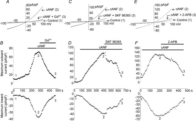
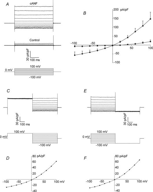
In the heart, fibroblasts play an essential role in the deposition of the extracellular matrix and they also secrete a number of hormonal factors. Although natriuretic peptides, including C-type natriuretic peptide (CNP) and brain natriuretic peptide, have antifibrotic effects on cardiac fibroblasts, the effects of CNP on fibroblast electrophysiology have not been examined. In this study, acutely isolated ventricular fibroblasts from the adult rat were used to measure the effects of CNP (2 x 10(-8) M) under whole-cell voltage-clamp conditions. CNP, as well as the natriuretic peptide C receptor (NPR-C) agonist cANF (2 x 10(-8) M), significantly increased an outwardly rectifying non-selective cation current (NSCC). This current has a reversal potential near 0 mV. Activation of this NSCC by cANF was abolished by pre-treating fibroblasts with pertussis toxin, indicating the involvement of G(i) proteins. The cANF-activated NSCC was inhibited by the compounds Gd(3+), SKF 96365 and 2-aminoethoxydiphenyl borate. Quantitative RT-PCR analysis of mRNA from rat ventricular fibroblasts revealed the expression of several transient receptor potential (TRP) channel transcripts. Additional electrophysiological analysis showed that U73122, a phospholipase C antagonist, inhibited the cANF-activated NSCC. Furthermore, the effects of CNP and cANF were mimicked by the diacylglycerol analogue 1-oleoyl-2-acetyl-sn-glycerol (OAG), independently of protein kinase C activity. These are defining characteristics of specific TRPC channels. More detailed molecular analysis confirmed the expression of full-length TRPC2, TRPC3 and TRPC5 transcripts. These data indicate that CNP, acting via the NPR-C receptor, activates a NSCC that is at least partially carried by TRPC channels in cardiac fibroblasts.


Figures

Figure 1
Figure 1. Properties of a weakly outwardly rectifying ionic current identified using voltage ramps in acutely isolated rat cardiac fibroblasts
The voltage-clamp protocol consisted of a 1 s ramp from –100 to +100 mV from a holding potential of 0 mV. Under these conditions, and in the absence of any external pharmacological compounds, a small weakly outwardly rectifying current was identified. A and B, representative recordings and time-course data (at –100 and +100 mV) of the effects of replacing external Cl with CH3SO3. C, summary data (mean ± s.e.m.; n = 6 fibroblasts) for control conditions (black bars) and external Cl replacement (shaded bars) at –100 and +100 mV. There was no significant effect of external Cl replacement on this current indicating that it is not a Cl conductance. D and E, representative recordings and time-course data of the effects of replacing external Na+ with NMDG+. F, summary data (mean ± s.e.m.; n = 10 fibroblasts) for control conditions and external Na+ replacement. Replacement of external Na+ with NMDG+ significantly decreased inward current at –100 mV without altering outward current at +100 mV. As shown in the time-course data, this effect was completely reversible following a return to Na+-containing superfusate. G and H, representative recordings and the time course of the effects of divalent cation removal. I, summary data (mean ± s.e.m.; n = 9 fibroblasts) for the effects of removing external Ca2+ and Mg2+. Note that divalent cation removal significantly increased both inward and outward currents and linearized the current–voltage (I–V) curve. As shown in the time-course data, this effect was completely reversible.
Figure 2
Figure 2. CNP (2 × 10⁻⁸m) and the NPR-C agonist, cANF (2 × 10⁻⁸m), increase a non-selective cation current in acutely isolated rat cardiac fibroblasts
Currents were elicited by 1 s voltage ramps from –100 to +100 mV from a holding potential of 0 mV. A and B, representative recordings and time-course data for the effects of C-type natriuretic peptide (CNP) on the non-selective cation current (NSCC). The representative traces for control conditions (1) and CNP treatment (2) correspond to the points indicated in the time-course figure. Note that CNP significantly increased the weakly outwardly rectifying NSCC. C and D, representative recordings and time-course data for the effects of cANF, a selective agonist for the natriuretic peptide C receptor (NPR-C) that activates Gi proteins, on the NSCC. Note that cANF mimics the effect of CNP on isolated rat cardiac fibroblasts, suggesting the effect of CNP is via the NPR-C receptor. E and F, representative recordings and time-course data for the effects of carbachol (CCh, 1 × 10⁻⁵m) on the NSCC. CCh, which activates Gi proteins via M2 muscarinic receptors, elicits a similar effect to CNP and cANF. Summary data (mean ± s.e.m.) for the effects of CNP, cANF and CCh (n values shown in parentheses) on maximum inward and outward current are shown in G. In these histograms, black bars are control conditions, and shaded bars represent drug treatment. Each compound significantly (*) increased inward and outward current at –100 and +100 mV, respectively (P < 0.05) compared with control conditions. There was no significant difference between the effects of CNP, cANF or CCh (P > 0.05) on the NSCC in rat ventricular fibroblasts.
Figure 4
Figure 4. RT-PCR analysis of the molecular identity of transient receptor potential (TRP) channel transcripts in acutely isolated fibroblasts obtained from adult rat ventricles
Total RNA was isolated from adult rat fibroblasts (see Methods), and RT-PCR was used to assay the mRNA transcripts of ion channels from the TRPC (A), TRPV (B) and TRPM (C) subfamilies. PCR products were generated from rat gene specific primers. A 100 bp molecular mass marker was used to estimate the size of the amplicon. PCR products were sequenced to confirm their identity. Summary data are means ± s.e.m.
Figure 10
Figure 10. RT-PCR detection of mRNA transcripts covering the full coding regions of TRPC2, TRPC3 and TRPC5 in rat cardiac fibroblasts
PCR products were generated through the use of gene-specific primers for TRPC2 (lanes 1–4 from the left), TRPC3 (lanes 5–7), and TRPC5 (lanes 8–11). A molecular mass marker was used to estimate the size of the amplicon. RT-PCR was also performed in the presence of glyceraldehyde-3-phosphate dehydrogenase (GAPDH), an internal standard (lane 12). Similar results were obtained from four independent cardiac fibroblast cDNA preparations. Refer to Table 1 for primer details.
Figure 3
Figure 3. Effect of PTX on the non-selective cation current in acutely isolated rat ventricular fibroblasts
Fibroblasts were pre-treated with pertussis toxin (PTX; 0.5 μg ml−1; 37°C) for a minimum of 3 h to inactivate Gi/o proteins. A and B, representative recordings and time-course data for the effects of cANF (2 × 10⁻⁸m) on current amplitude following PTX pre-treatment. Fibroblasts were superfused with cANF for a minimum of 10 min, during which cANF failed to elicit the typical increase in NSCC. C, summary data (mean ± s.e.m.; n values indicated in parentheses) for the effects of cANF, as well as CCh (1 × 10⁻⁵m), on maximum inward and outward current at –100 and +100 mV following PTX pre-treatment. Black bars denote control data, the grey bars illustrate cANF or CCh results. There was no significant effect of cANF or CCh on inward and outward current (P > 0.05) indicating the involvement of Gi/o proteins in the activation of the NSCC in rat ventricular fibroblasts.
Figure 5
Figure 5. Effects of Gd3+, SKF 96365 and 2-aminoethoxydiphenyl borate (2-APB) on the cANF-activated non-selective cation current in rat cardiac fibroblasts
Currents were elicited with a 1 s voltage ramp from –100 to +100 mV (holding potential was 0 mV). A, representative current changes in control conditions (1), following the application of cANF (2 × 10⁻⁸m) (2), and during superfusion with cANF and Gd3+ (1 × 10⁻⁵m) (3). B, time course of the effects of cANF and Gd3+ on inward and outward currents in the cardiac fibroblast. The representative traces correspond to the time points indicated in the time-course figure. C, representative current recordings in control conditions (1), following the application of cANF (2 × 10⁻⁸m) (2), and during superfusion with cANF and SKF 96365 (5 × 10⁻⁵m) (3). D, time course of the effects of cANF and SKF 96365 on inward and outward currents in the cardiac fibroblast. E, representative current recordings in control conditions (1), following superfusion with cANF (2 × 10⁻⁸m) (2), and during the application of cANF and 2-APB (1 × 10⁻⁴m) (3). F, time course of the effects of cANF and 2-APB on the NSCC in the cardiac fibroblast. Note that each pharmacological compound potently inhibited the NSCC activated by cANF. Data are representative of measurements made on six fibroblasts for the Gd3+ experiment, seven fibroblasts for the SKF 96365 experiment, and five fibroblasts for the 2-APB experiment (refer to Fig. 9 for summary data).
Figure 9
Figure 9. Summary of effects of selected pharmacological compounds on the cANF-activated NSCC in acutely isolated rat ventricular fibroblasts
In these histograms, black bars denote control data, darkly shaded bars depict results following the application of cANF, and lightly shaded bars correspond to data following the application of cANF in combination with the indicated compound. Concentrations of all drugs and chemicals are given in the previous figures. *The value in the presence of cANF is significantly different from control. †The value in the presence of cANF and the indicated compound is significantly different than in cANF alone. Data are means ± s.e.m., n values are indicated in parentheses.
Figure 6
Figure 6. Properties of the cANF-activated non-selective cation current (NSCC) evaluated using rectangular voltage-clamp step protocols
A, families of currents recorded using 500 ms voltage-clamp steps from –100 to +100 mV (holding potential, 0 mV) in control conditions and following the application of cANF (2 × 10⁻⁸m). Note that the currents show the same weak outward rectification properties as seen in response to voltage ramps. B, summary data illustrating the I–V curves in control conditions (▪) and following cANF superfusion (•) obtained from voltage-step protocols as illustrated in A. cANF significantly increased inward and outward currents (mean ± s.e.m.; n = 5 fibroblasts). C, two-pulse protocol consisting of a prepulse to +100 mV followed by a series of voltage-clamp steps from –100 to +100 mV (in 20 mV increments) recorded in the same cell as shown in A. The current responses were recorded in the presence of cANF. Note that the inward and outward current densities measured during the test pulses are unchanged from those in A. D, the I–V curve, plotted from the voltage-clamp steps illustrated in C. Note that it has not significantly changed from that presented in B. E, two-pulse protocol consisting of a family of voltage-clamp steps from –100 to +100 mV followed by a step to –100 mV recorded in the same fibroblast. The sample data shown were recorded in the presence of cANF. F, representative I–V curve plotted from the family of voltage-clamp steps in E. Note the absence of tail currents in C and E.
Figure 7
Figure 7. Effect of the phospholipase C (PLC) antagonist U73122, and its inactive analogue U73343, on the cANF-activated non-selective cation current in rat cardiac fibroblasts
Currents were studied using 1 s voltage ramps from –100 to +100 mV (holding potential was 0 mV). A, representative current recordings in control conditions (1), following superfusion with cANF (2 × 10⁻⁸m) (2), and during the application of cANF and U73122 (5 × 10⁻⁶m) (3). B, time course of the effects of cANF and U73122. Representative traces correspond to the time points indicated in time-course figure. C, representative current recordings in control conditions (1), following superfusion with cANF (2 × 10⁻⁸m) (2), and during application of cANF in combination with U73343 (5 × 10⁻⁶m) (3). Note that U73343 is a non-functional analogue of U73122. It has no effect on PLC activity. D, time course of the effects of cANF and U73343. These data are representative of measurements made on five fibroblasts for the U73122 experiment, and six fibroblasts for the U73343 experiment (refer to Fig. 9 for summary data). U73122 significantly inhibited the cANF-activated non-selective cation current, while U73343 had no significant effect.
Figure 8
Figure 8. Effects of intracellular application of OAG on isolated rat cardiac fibroblasts
OAG (1-oleoyl-2-acetyl-sn-glycerol), which is an analogue of diacylglycerol, was included in the recording pipette at a concentration of 1 × 10⁻⁴m. It entered the cell by diffusion under conventional whole-cell recording conditions. Currents were studied using a voltage ramp from –100 to +100 mV from a holding potential of 0 mV. A, representative current recordings under control conditions (1), and after OAG has entered the fibroblast (2). B, time course of the effects of OAG on cardiac fibroblasts. Representative traces correspond to the time points indicated in the time-course figure. Because OAG was in the recording pipette, it entered the fibroblasts upon establishment of the whole-cell recording condition at time 0 s. C, representative current recordings under control conditions (1), and after OAG had entered the fibroblast (2) following pretreatment with staurosporine (1 × 10⁻⁶m). Cells were preincubated with staurosporine for 30 min in order to block protein kinase C (PKC) activity before being voltage clamped. D, time course of the effects of OAG following pretreatment of fibroblasts with staurosporine. E, summary data (mean ± s.e.m.) showing the effects of OAG (n = 5 fibroblasts), and OAG following staurosporine pretreatment (n = 7 fibroblasts), on maximum inward and outward currents in these acutely isolated ventricular fibroblasts. Black bars denote control data, shaded bars depict results following the application of OAG. *Significant difference between control and OAG application. Note that OAG mimics the ability of cANF to activate the NSCC in cardiac fibroblasts, and that this occurs independently of PKC activity.

Similar articles

Cited by

References

    1. Allessie M, Schotten U, Verheule S, Harks E. Gene therapy for repair of cardiac fibrosis: a long way to Tipperary. Circulation. 2005;111:391–393. - PubMed
    1. Anand-Srivastava MB, Sairam MR, Cantin M. Ring-deleted analogs of atrial natriuretic factor inhibit adenylate cyclase/cAMP system. Possible coupling of clearance atrial natriuretic factor receptors to adenylate cyclase/cAMP signal transduction system. J Biol Chem. 1990;265:8566–8572. - PubMed
    1. Anand-Srivastava MB, Sehl PD, Lowe DG. Cytoplasmic domain of natriuretic peptide receptor-C inhibits adenylyl cyclase. Involvement of a pertussis toxin-sensitive G protein. J Biol Chem. 1996;271:19324–19329. - PubMed
    1. Bandyopadhyay BC, Swaim WD, Liu X, Redman RS, Patterson RL, Ambudkar IS. Apical localization of a functional TRPC3/TRPC6-Ca2+ signaling complex in polarized epithelial cells. Role in apical Ca2+ influx. J Biol Chem. 2005;280:12908–12916. - PubMed
    1. Barr AJ, Ali H, Haribabu B, Snyderman R, Smrcka AV. Identification of a region at the N-terminus of phospholipase Cβ3 that interacts with G protein βγ subunits. Biochemistry. 2000;39:1800–1806. - PubMed

Publication types

MeSH terms

Substances

LinkOut - more resources