. 2007 Mar;27(5):1745-57.
doi: 10.1128/MCB.01974-06. Epub 2006 Dec 28.

Specific phosphorylation of p120-catenin regulatory domain differently modulates its binding to RhoA

Affiliations

Specific phosphorylation of p120-catenin regulatory domain differently modulates its binding to RhoA

Julio Castaño et al. Mol Cell Biol. 2007 Mar.

Abstract

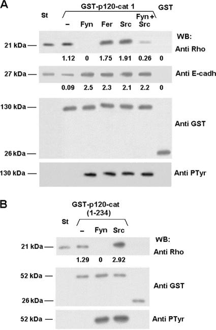
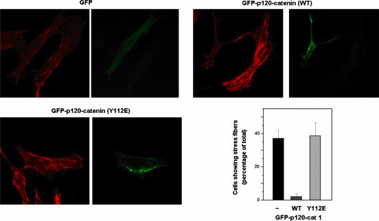
p120-catenin is an adherens junction-associated protein that controls E-cadherin function and stability. p120-catenin also binds intracellular proteins, such as the small GTPase RhoA. In this paper, we identify the p120-catenin N-terminal regulatory domain as the docking site for RhoA. Moreover, we demonstrate that the binding of RhoA to p120-catenin is tightly controlled by the Src family-dependent phosphorylation of p120-catenin on tyrosine residues. The phosphorylation induced by Src and Fyn tyrosine kinases on p120-catenin induces opposite effects on RhoA binding. Fyn, by phosphorylating a residue located in the regulatory domain of p120-catenin (Tyr112), inhibits the interaction of this protein with RhoA. By contrast, the phosphorylation of Tyr217 and Tyr228 by Src promotes a better affinity of p120-catenin towards RhoA. In agreement with these biochemical data, results obtained in cell lines support the important role of these phosphorylation sites in the regulation of RhoA activity by p120-catenin. Taken together, these observations uncover a new regulatory mechanism acting on p120-catenin that contributes to the fine-tuned regulation of the RhoA pathways during specific signaling events.


Figures

FIG. 1.
FIG. 1.
RhoA interacts with the N-terminal regulatory domain of p120-catenin. (A) Diagram of the different domains of p120-catenin. Alternative splicing in the N-terminal domain gives rise to isoforms 1, 2, 3, and 4, each initiating at distinct ATG start codons (1, 55, 102, and 340, respectively) (arrows). The regulatory domain contains nine tyrosines (Y96, Y112, Y217, Y228, Y257, Y280, Y291, Y296, and Y302) capable of being phosphorylated in vitro (15, 17). The length and composition of the protein fragments used in this work are shown. (B) GST fusion proteins (1.5 pmol) were incubated with 5 pmol of RhoA. Protein complexes were affinity purified with glutathione-Sepharose and analyzed by sodium dodecyl sulfate-polyacrylamide gel electrophoresis and Western blotting (WB) with anti-RhoA MAb. RhoA (1.2 pmol) was included as an internal reference standard (St). When indicated, the assay was performed in the presence of a 100-fold molecular excess of cytoEcad. Blots were reanalyzed with anti-GST to ensure that similar amounts of fusion proteins were present in all cases. (C) GST-p120-catenin fusion proteins or GST-β-catenin (7 pmol) was incubated with 270 μg of whole-cell extracts from RWP-1 cells. Protein complexes were affinity purified as described above and analyzed with anti-E-cadherin and anti-GST MAbs. In the Input lane, 5% of the total cell extract used for the assay was loaded. (D) Either GST-cytoEcad or GST (1.2 pmol) was incubated with 30 pmol of RhoA in the presence of 3 pmol of p120-catenin-1 when indicated. Protein complexes were affinity purified with glutathione-Sepharose and analyzed with specific MAbs. p120-catenin (0.6 pmol) was included as an internal reference standard. In panels B, C, and D, the numbers below the lanes correspond to the amounts of bound RhoA, E-cadherin, or p120-catenin, respectively, determined after densitometry, compared to the standard or to the amount bound to full-length GST-p120-catenin. The results presented in this figure correspond to an experiment representative of three performed.
FIG. 2.
FIG. 2.
Tyrosine phosphorylation of p120-catenin modulates its interaction with RhoA. Full-length GST-p120-catenin (A) or GST-p120-catenin(1-234) (B) was phosphorylated with Fyn, Fer, Src, or Fyn and Src tyrosine kinases, as described in Materials and Methods. Control and phosphorylated GST-p120-catenin proteins or GST as a control (1.5 pmol) were incubated with 5 pmol of RhoA (upper panel) or with 2 pmol cytoEcad (middle panel) in a final volume of 200 μl. Protein complexes were affinity purified with glutathione-Sepharose and analyzed by sodium dodecyl sulfate-polyacrylamide gel electrophoresis and Western blotting (WB) with specific MAbs. RhoA (1 pmol) (A and B) or cytoEcad (0.1 pmol) (A) were included as internal reference standards (St). The numbers below the lanes correspond to the amounts of RhoA or E-cadherin bound to GST-p120-catenin fusion proteins compared to the standard. The results presented in this figure correspond to an experiment representative of at least three performed.
FIG. 3.
FIG. 3.
Phosphorylation of Tyr112 inhibits the p120-catenin-RhoA interaction. (A) Diagram of the N-terminal regulatory domain of p120-catenin(1-347). Tyrosine residues phosphorylated by tyrosine kinases are indicated (Y96, Y112, Y217, Y228, Y257, Y280, Y291, Y296, and Y302). (B) Mapping of the residues involved in Src and Fyn phosphorylation of p120-catenin. Various amounts (2 pmol in the case of Fyn and 4 pmol in the case of Src) of different GST-p120-cat(1-234) point mutants were phosphorylated with Fyn (left panel) or Src (right panel). Phosphorylation was analyzed by Western blotting (WB) with an anti-PTyr MAb. The membrane was stripped and reanalyzed against GST to check that similar levels of the different GST-p120-cat(1-234) point mutants were used. (C) RhoA-binding assays were performed with 5 pmol of RhoA and 5 pmol of GST or GST-p120-cat(1-234) fusion proteins corresponding to the wild-type (WT) form, the Y96F Y228F double mutant, or the Y96F Y228F Y112F triple mutant. When indicated, the fusion proteins were phosphorylated with recombinant Fyn or Src tyrosine kinases. Protein complexes were affinity purified with glutathione-Sepharose and analyzed by Western blotting with anti-RhoA. RhoA (1 pmol [left panel] or 0.4 pmol [right panel]) was included as an internal reference standard (St). (D) GST-p120-catenin and Y112E, Y217E, and Y228E point mutants (1.5 pmol) were incubated with 5 pmol of RhoA in a final volume of 200 μl, as indicated above. RhoA (1 pmol [left panel] or 2.5 pmol [right panel]) was included as an internal reference standard. (E) Pull-down assays were performed by incubating 6 pmol of the different GST-p120-catenin point mutants with 300 μg of whole-cell extracts from RWP-1 cells; protein complexes were affinity purified with glutathione-Sepharose and analyzed by sodium dodecyl sulfate-polyacrylamide gel electrophoresis and Western blotting with anti-E-cadherin. Blots were reanalyzed with anti-GST antibodies to ensure equal loading of samples. In the Input lane, a sample containing 5% of the total cell extracts used for the assay was loaded. (F) RWP-1 cells were cotransfected with different GFP-p120-catenin fusion proteins and pEF-Bos-active Fyn or the empty plasmid. After 48 h, cell extracts were prepared, 300 μg of each cell extract was immunoprecipitated with anti-GFP, and the associated proteins were analyzed with specific MAbs against GFP, RhoA, and Fyn. The numbers below the lanes correspond to the amounts of RhoA or E-cadherin bound to GST-p120-catenin fusion proteins, compared to the standard or to the amount bound to wild-type GST-p120-catenin. The results presented in this figure correspond to an experiment representative of at least three performed.
FIG. 4.
FIG. 4.
p120-catenin interacts better with the RhoA-bound GDP form. (A) p120-catenin (1 pmol) was incubated with 2 pmol of GDP- or GTP-loaded GST-RhoA. Protein complexes were affinity purified with glutathione-Sepharose and analyzed by sodium dodecyl sulfate-polyacrylamide gel electrophoresis and Western blotting (WB) with an anti-p120-catenin MAb. p120-catenin (0.25 pmol) was included as an internal reference standard (St). (B) NIH 3T3 cells were transfected with two RhoA mutants: AU5-RhoAQ63L (which is always active) or AU5-RhoAT19N (inactive). After 48 h, cell extracts were prepared and incubated (300 μg) with GST-p120-catenin. Protein complexes were purified with glutathione-Sepharose and associated proteins analyzed with a monoclonal antibody specific to the AU5 tag. Blots were reanalyzed with anti-GST to ensure that similar levels of fusion protein forms were present in all samples. In the Input lane, a sample containing 5% of the total cell extracts used for the assay was loaded. The results presented in this figure correspond to an experiment representative of three (A) or two (B) performed.
FIG. 5.
FIG. 5.
Phosphorylation of p120-catenin on Tyr112 blocks its RhoGDI activity. [3H]GDP-bound RhoA (20 pmol) was incubated with equimolar amounts of the different GST-p120-catenin fusion proteins for 10 min. An excess of GTP (10 μM) was then added to allow the exchange of bound [3H]GDP-RhoA for GTP, and samples were incubated for an additional 10 min. [3H]GDP-bound RhoA was separated from free nucleotide by filtration, and the amount of [3H]GDP bound to RhoA was determined and compared to the radionucleotide bound at time zero. The figure shows the averages ± standard deviations of three experiments performed in duplicate.
FIG. 6.
FIG. 6.
Phosphorylation of p120-catenin on Tyr112 by Fyn prevents repression of RhoA activity by p120-catenin. SW480 cells were cotransfected with 7.5 μg of pcDNA3.1-Myc-His-RhoA wild type (WT) and (A) with 7.5 μg of pEF-Bos-active Fyn, pEF-Bos-active v-Src, or pEF-Bos alone, (B) with pEF-Bos-active Fyn and 7.5 μg of GFP, GFP-p120-catenin wild type, or GFP-p120-catenin Y112F, or (C) with the same amounts of GFP, GFP-p120-catenin wild type, or GFP-p120-catenin Y112E. After 48 h, cell extracts were prepared and added to a suspension of GST-rhotekin-glutathione-Sepharose. The amount of active RhoA was analyzed by Western blotting (WB) with a specific MAb against RhoA. Blots were reanalyzed with anti-Fyn and -Src (A), with anti-Fyn and anti-GFP (B), or with anti-GFP (C). In the Input lane, a sample of 5% of the total cell extract used for the assay was loaded. The right panels show the representation of active Rho with respect to the total amount of this protein. The averages ± standard deviations of the three different experiments performed in each case are presented.
FIG. 7.
FIG. 7.
Tyr112 controls formation of stress fibers. NIH 3T3 cells were transfected with 5 μg of GFP vector alone, the GFP-p120-catenin wild type (WT), or the GFP-p120-catenin Y112E mutant. The presence of stress fibers was determined after staining F-actin with phalloidin-rhodamine and visualizing cells by fluorescence microscopy. Note that transfection of GFP or the GFP-p120-catenin Y112E mutant did not substantially affect the presence of stress fibers with respect to control untransfected cells not positive for GFP, whereas the p120-catenin wild type almost totally prevented it. The averages (±standard deviations) of the results obtained in two experiments in which at least 40 cells were examined are also included.
FIG. 8.
FIG. 8.
Phosphorylation of p120-catenin on Tyr112 prevents its RhoA-dependent effects in vivo. NIH 3T3 cells were transfected with 1 μg of the different phrGFP vectors in order to express the indicated GFP fusion proteins. When appropriate, 5 μg of pCFP-E-cadherin (D) or pEF-Fyn (I and J) was also transfected. After 24 h, cells were plated on glass coverslips, fixed with 4% paraformaldehyde, and washed with phosphate-buffered saline. Coverslips were mounted on glass slides and the morphology of transfected cells visualized by confocal microscopy. Arrows indicate the membrane protrusions typically induced by the expression of wild-type (WT) p120-catenin. The most predominant morphology observed in each case is shown; other relevant phenotypes are also displayed in the case of cells transfected with GFP-p120-catenin (wild type). The percentages of cells showing the different phenotypes are included (K); this diagram corresponds to the result of one experiment representative of the three performed. Fifty transfected cells were observed under each condition. A cell was scored as having a branched phenotype (examples in panels B and E) if it presented three or more extensions longer than the cell body. Cells presenting one or two extensions at least twice the cell body were scored as having a partial branched phenotype (intermediate phenotype, examples in panels C and I).
FIG. 9.
FIG. 9.
Effect of K-Ras overexpression in IEC-18 cells on RhoA distribution and activity. (A) Activity of RhoA in IEC or IEC-K-ras cells was determined using chromatography with GST-rhotekin-glutathione-Sepharose as indicated in Materials and Methods. Bound proteins were analyzed with a RhoA-specific MAb. The lower panel shows the representation of active Rho with respect to the total amount of this protein. The averages ± standard deviations of the three different experiments performed in each case are presented. (B) Cytosolic and membrane-associated fractions from IEC and IEC-K-ras cells were prepared as detailed in Materials and Methods. Fractions were precipitated with an anti-p120-catenin MAb and analyzed by Western blotting (WB) with the indicated MAbs. In parallel, an aliquot of the four different inputs was also analyzed. (C) IEC and IEC-K-ras cells were cotransfected with inactive AU5-RhoA(T19N) and GFP-p120-catenin, either the wild type or the Y112F mutant, or with GFP as a control. Cytosolic and membrane fractions were prepared as described above, and the exogenous p120-catenin was immunoprecipitated (IP) with an anti-GFP antibody. The immunoprecipitates and inputs were analyzed by Western blotting with the indicated antibodies. The results presented in panels B and C correspond to an experiment representative of three performed.
FIG. 10.
FIG. 10.
Proposed model for the regulation of RhoA activity by p120-catenin. p120-catenin protein is composed by the regulatory and the armadillo domains (1). After binding of RhoA to the regulatory domain, the armadillo repeats are disposed in the close vicinity of the nucleotide binding site of RhoA, hindering the release of GDP and therefore contributing to the GDI effect of p120-catenin (3). In cells, most p120-catenin is located at the membrane due to its interaction with the juxtamembrane domain (JMD) of E-cadherin (2). This membrane-associated p120-catenin is unable to work as a GDI due to either a conformational change induced in the armadillo domain by E-cadherin or the recruitment of a RhoA GEF (not depicted) by this protein. Therefore, GDP bound to RhoA is exchanged by GTP, and the affinity of the GTPase by p120-catenin decreases and is rapidly released (4). This effect of E-cadherin or E-cadherin-associated factor is not present in the cytosol; therefore, only cytosolic p120-catenin can work as a GDI (3). Activation of Fyn tyrosine kinase leads to phosphorylation of tyrosines 112, 228, and others in the regulatory domain of the p120-catenin located in the membrane fraction, where Fyn is normally present (5), and not in the cytosol (7). Phosphorylation of Tyr112 prevents interaction of GDP-bound RhoA with p120-catenin (6), therefore precluding the activation of this protein and increasing the levels of this GTPase at the cytosol, where it can be bound and inhibited by unphosphorylated p120-catenin (8).

Similar articles

Cited by

References

    1. Anastasiadis, P. Z., S. Y. Moon, M. A. Thoreson, D. J. Mariner, H. C. Crawford, Y. Zheng, and A. B. Reynolds. 2000. Inhibition of RhoA by p120 catenin. Nat. Cell Biol. 2:637-644. - PubMed
    1. Brembeck, F. H., T. Schwarz-Romond, J. Bakkers, S. Wilhelm, M. Hammerschmidt, and W. Birchmeier. 2004. Essential role of BCL9-2 in the switch between beta-catenin's adhesive and transcriptional functions. Genes Dev. 18:2225-2230. - PMC - PubMed
    1. Bryant, D. M., and J. L. Stow. 2004. The ins and outs of E-cadherin trafficking. Trends Cell. Biol. 14:427-434. - PubMed
    1. Carnac, G., M. Primig, M. Kitzmann, P. Chafey, D. Tuil, N. Lamb, and A. Fernandez. 1998. RhoA GTPase and serum response factor control selectively the expression of MyoD without affecting Myf5 in mouse myoblasts. Mol. Biol. Cell 9:1891-1902. - PMC - PubMed
    1. Castaño, J., I. Raurell, J. Piedra, S. Miravet, M. Duñach, and A. García de Herreros. 2002. β-Catenin N-and C-tails modulate the coordinated binding of adherens junction proteins to β-catenin. J. Biol. Chem. 277:31541-31550. - PubMed

Publication types

MeSH terms