. 2007 Jan 1;176(1):113-24.
doi: 10.1083/jcb.200607021. Epub 2006 Dec 26.

Synaptotagmin-12, a synaptic vesicle phosphoprotein that modulates spontaneous neurotransmitter release

Affiliations

Synaptotagmin-12, a synaptic vesicle phosphoprotein that modulates spontaneous neurotransmitter release

Anton Maximov et al. J Cell Biol. .

Abstract

Central synapses exhibit spontaneous neurotransmitter release that is selectively regulated by cAMP-dependent protein kinase A (PKA). We now show that synaptic vesicles contain synaptotagmin-12, a synaptotagmin isoform that differs from classical synaptotagmins in that it does not bind Ca(2+). In synaptic vesicles, synaptotagmin-12 forms a complex with synaptotagmin-1 that prevents synaptotagmin-1 from interacting with SNARE complexes. We demonstrate that synaptotagmin-12 is phosphorylated by cAMP-dependent PKA on serine(97), and show that expression of synaptotagmin-12 in neurons increases spontaneous neurotransmitter release by approximately threefold, but has no effect on evoked release. Replacing serine(97) by alanine abolishes synaptotagmin-12 phosphorylation and blocks its effect on spontaneous release. Our data suggest that spontaneous synaptic-vesicle exocytosis is selectively modulated by a Ca(2+)-independent synaptotagmin isoform, synaptotagmin-12, which is controlled by cAMP-dependent phosphorylation.


Figures

Figure 1.
Figure 1.
Expression of native synaptotagmin-12 in various tissues and neuronal cultures. (A) Lysates of COS7 cells transfected with cDNAs encoding full-length synaptotagmins 1–13 (Syt 1–13) were analyzed with indicated polyclonal antibodies to various synaptotagmins and valosin-containing protein (VCP; used as a loading control). (B–F) Various tissues, cultured cell lines, and primary neuronal cultures were analyzed by immunoblotting with antibodies to synaptotagmin-12 (Syt 12), -1 (Syt 1), and VCP as a loading control. Equal amounts of total protein were loaded into each lane. (B) Synaptotagmin-12 is expressed in adult mouse brain and adrenal medulla. (C) Expression of synaptotagmin-12 could not be detected in cultured cell lines. (D) Analysis of indicated rat tissues isolated at different stages of development ranging from embryonic day 18 to postnatal day 40 (as shown on the right side of each image), shows that expression of synaptotagmin-12 is restricted to postnatal brain and displays progressive increase during development. (E and F) Relative expression levels of synaptotagmin-1 and -12 were determined by quantitative immunoblotting of total brain homogenates isolated from mouse at different stages of postnatal development and cortical neurons isolated from newborn mouse pups and cultured in vitro for 5–15 d. (E) Raw immunoblotting data. (F) Quantification of expression with I125-labeled secondary antibodies. The relative expression levels of each protein were plotted as the percentage of expression in adult brains (n ≥ 4). Similar results were obtained with cultured hippocampal neurons (not depicted). Data are shown as the mean ± the SEM.
Figure 2.
Figure 2.
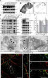
Expression and localization of synaptotagmin-12. (A) Total homogenates of cortex, cerebellum, hippocampus, olfactory bulb, brain stem, and spinal cord isolated from the brains of adult rats were analyzed with antibodies to synaptotagmin-12, -1, and VCP (loading control). 20 μg of total protein was loaded into each lane. Note that synaptotagmin-12 is expressed at different levels in various brain regions, with the highest expression level in cerebellum. (B and C) Sections of brains isolated from adult mice were labeled with antibody to synaptotagmin-12. (B) Labeling of brain sections indicates that synaptotagmin-12 is abundantly expressed in various brain regions. (C) Brain sections were labeled with antibody to synaptotagmin-12 mixed with ∼1 mg/ml recombinant GST-linker fusion protein that was used for immunization. Note that synaptotagmin-12 immunoreactivity is greatly reduced in the presence of the antigen. (D) Distribution of native synaptotagmin-12 in subcellular fractions of adult brain separated by centrifugation. Similar to synaptotagmin-1 (Syt 1) and synaptobrevin/VAMP2 (Syb 2), synaptotagmin-12 is enriched in synaptic vesicle fraction (crude SVs). Asterisks indicate proteolitically cleaved fragment of synaptotagmin-1 and various splice variants of synaptotagmin-7. (E) Expression of indicated proteins in brains of adult wild-type and synapsin double-knockout mice (DKO) was analyzed by quantitative immunoblotting with I125-labeled secondary antibodies. (F) The relative expression levels of indicated proteins in DKO brains were normalized to VCP and presented as the percentage of wild type. Note that expression of synaptotagmin-12 and other synaptic vesicle proteins is reduced by ∼40%. Data are shown as the mean ± the SEM. (G and H) Electron micrographs of adult mouse hippocampal sections labeled with synaptotagmin-12 antibody alone (G) or in the presence of ∼1 mg/ml of the antigen (synaptotagmin-12 linker region) used for immunization (H). The postsynaptic densities are marked by white arrowheads. Note that synaptotagmin-12 immunoreactivity is restricted to presynaptic sites (G; black arrowheads) and inhibited in the presence of the antigen (H). (I and J) Localization of recombinant synaptotagmin-12 in cultured hippocampal neurons. The neurons were infected at 5 d after plating (5 DIV) with lentivirus expressing full-length synaptotagmin-12 cDNA, and double labeled at 14 DIV with antibodies to synaptotagmin-12 and the dendritic marker MAP2 (I; Syt 12, green; MAP2, red) or Syt 12 and synaptic marker synaptophysin (J; Syt 12, green; synaptophysin, red). Inset (I′) illustrates enlarged fragment of dendrite marked by white box. Note that recombinant synaptotagmin-12 is concentrated in discrete clusters apposed to dendrites and colocalized with synaptophysin. Bars: (G) 100 nM; (I and J") 20 μM.
Figure 3.
Figure 3.
Synaptotagmin-12 is not glycosylated and lacks phospholipid binding. (A) Deglycosylation analysis of synaptotagmin-12. Proteins from rat brain homogenate were treated with sialidase (S), o-glycanase (O), PNGase (N), or different combinations of these enzymes to remove neuraminic acid and O- and N-linked sugars. The samples were then analyzed by immunoblotting with the antibodies to synaptotagmin-1 and -12. Treatment with deglycosylation enzymes results in a substantial change in mobility of synaptotagmin-1 in SDS-PAGE, but has no effect on mobility of synaptotagmin-12, indicating that synaptotagmin-12 is not glycosylated. (B) C2 domains of synaptotagmin-12 do not bind phospholipids in a Ca2+-dependent manner. Recombinant C2AB domains of synaptotagmins-1 and -12 were expressed as GST-fusion proteins and tested in phospholipid-binding assay at different concentrations of free Ca2+ (as indicated on the top of the gel). Unlike C2 domains of synaptotagmin-1 (Syt 1 C2AB; top), C2 domains of synaptotagmin-12 (Syt 12 C2AB) do not bind phospholipids when tested alone or in the presence of Syt 1 C2AB.
Figure 4.
Figure 4.
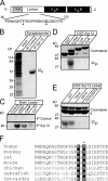
Synaptotagmin-12 is phosphorylated in a cAMP-dependent manner at serine97. (A) Schematic diagram of synaptotagmin-12 domain structure. A fragment of linker sequence containing two putative sites for PKA-dependent phosphorylation (T87 and S97, marked by asterisks) is illustrated below. (B) In vivo phosphorylation of native synaptotagmin-12. The proteins from rat brain synaptosomes preincubated with [32P]orthophosphate were extracted in detergent and immunoprecipitated with antibody to synaptotagmin-12 or preimmune serum. Incorporation of 32P was measured with x-ray film. Note that a single ∼50-kD band is observed in synaptotagmin-12, but not in control immunoprecipitation. (C) In vitro phosphorylation of native synaptotagmin-12. Proteins extracted from brain were immunoprecipitated with synaptotagmin-12 antibody or preimmune serum attached to Sepharose beads. The beads were incubated with brain cytosol mixed with 1 mM ATP and 10 μCi γ-[32P]ATP alone or with addition of 0.1 mM cAMP, 0.1 mM cGMP, or 1 mM Ca2+. Note that synaptotagmin-12 is strongly phosphorylated in a cAMP-dependent manner. (D and E) Identification of a functional PKA phosphorylation site in synaptotagmin-12 sequence. (D) Indicated fragments of synaptotagmin-12 were expressed as recombinant GST fusion proteins (top shows Coomassie staining of all fusion proteins; the asterisk corresponds to GST) and phosphorylated in vitro in the presence of catalytic subunit of PKA and γ-[32P]ATP. Note that only linker region of synaptotagmin-12 is phosphorylated. (E) Phosphorylation of wild-type synaptotagmin-12 linker GST fusion protein and linkers containing single alanine substitutions in residues T87, S97, or a combination of these mutations. Note that phosphorylation is completely abolished in S97A mutant. The asterisk corresponds to GST. (F) Multiple sequence alignment of synaptotagmin-12 homologues from different species shows the fragments of the linker sequences containing conserved PKA phosphorylation site (marked by black boxes).
Figure 5.
Figure 5.
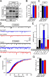
Synaptotagmin-12 regulates spontaneous transmitter release. (A) Analysis of expression and phosphorylation of native and recombinant synaptotagmin-12 in cultured cortical neurons. Noninfected neurons or neurons infected with wild-type or S97A synaptotagmin-12 lentiviruses were analyzed by immunoblotting with antibodies to synaptotagmin-12, -1, Rab3, and VCP as a loading control. Radioimmunoprecipitation analysis (bottom) shows that recombinant wild-type synaptotagmin-12 is phosphorylated in cultured neurons and phosphorylation is abolished in S97A mutant. (B and C) Expression levels of synaptotagmin-12 and -1 in noninfected neurons and neurons infected with wild-type and S97A synaptotagmin-12 lentiviruses were determined by quantitative immunoblotting with I125-labeled secondary antibodies, normalized to VCP, and plotted as the percentage of expression in P15 brains. (D–G) Overexpression of wild-type synaptotagmin-12 increases the rate of spontaneous release in inhibitory synapses. Spontaneous mIPSCs were monitored in the absence or presence of 50 μM forskolin from noninfected neurons (control) or neurons infected with the lentiviruses expressing wild-type synaptotagmin-12 or S97A mutant. mIPSCs were recorded in the presence of CNXQ and APV to block excitatory responses and Tetrodotoxin (TTX) to block spontaneous firing. (D) Typical mIPSCs recorded from control neurons (shown in black) or neurons infected with wild-type (shown in blue) or S97A (shown in red) synaptotagmin-12 lentiviruses. mIPSCs were recorded in control conditions (traces 1–3) or in the presence of forskolin (traces 4–6). Scale bars apply to all traces. (E) Mean frequencies of mIPSCs monitored in various conditions (indicated on the bottom of the graph) from three different cortical cultures. The total numbers of analyzed neurons are shown on each column. (F and G) Overexpression of synaptotagmin-12 does not affect the amplitudes of spontaneous mIPSCs. F shows a cumulative amplitude histogram constructed from 550 individual mIPSCs collected from control noninfected neurons and neurons infected with wild-type and S97A synaptotagmin-12 lentiviruses, and G shows the plot of average mIPSC amplitudes. For each group, n ≥ 12 neurons (data from three independent cultures). Data are shown as the mean ± the SEM.
Figure 6.
Figure 6.
Analysis of evoked IPSCs in neurons expressing synaptotagmin-12. Primary cortical neurons isolated from newborn pups were infected with lentivirus expressing wild-type synaptotagmin-12 and analyzed at 14–17. DIV Noninfected neurons or neurons infected with GFP-lentivirus were used as a control. Inhibitory synaptic responses were evoked by local extracellular stimulation and recorded in a whole-cell mode. AMPA and NMDA currents were suppressed by the addition of CNQX and APV to the extracellular bath solution. Recordings were performed in 0.5 or 2 mM extracellular Ca2+. The holding potential was –70 mV. The data are shown as the mean ± the SEM. (A–D) Amplitudes and the time course of individual IPSCs recorded form control neurons (shown in black) or neurons infected with synaptotagmin-12 lentivirus (shown in blue). IPSCs were triggered by single-action potentials applied at low frequency (0.1 Hz). (A and B) IPSCs recorded in 0.5 (A) or 2 mM (B) extracellular calcium from the neurons cultured for 14–16 DIV. The scale bars apply to all traces. (C) Mean amplitudes of IPSCs monitored in indicated concentrations of extracellular calcium from three different cortical cultures. The total numbers of analyzed neurons are shown on each column. (D) Cumulative histograms illustrate the time course of total charge transferred over 1.5 s after single-action potential stimulation. (E) Paired pulse IPSC ratios measured in 2 mM extracellular calcium in control neurons and neurons infected with synaptotagmin-12 lentivirus. IPSCs were triggered by two action potentials spaced by various interpulse periods, as indicated on the plot. Control, n = 10; Syt 12, n = 11. (F and G) Typical inhibitory responses recorded in 0.5 (F) or 2 mM (G) extracellular calcium from control (current traces on the top, shown in black) or synaptotagmin-12–infected neurons (current traces on the bottom, shown in blue) triggered by 100 action potentials applied at 10 Hz. The scale bars apply to all traces. The plots on the bottom show the average amplitudes of synchronous IPSCs during stimulus trains. (H) Average total charges transferred during trains of 100 action potentials applied at 10 Hz. The total numbers of analyzed neurons are shown on each column. Data are shown as the mean ± the SEM.
Figure 7.
Figure 7.
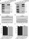
Synaptotagmin-12 forms a Ca2+-independent complex with synaptotagmin-1. Brain proteins were extracted in 1% of Triton X-100 and immunoprecipitated with polyclonal antibody to synaptotagmin-12 (A), monoclonal antibody to synaptotagmin-1 (B), corresponding preimmune serum or irrelevant control antibody in the presence of 1 mM Ca2+ or EGTA. Immunoprecipitates were extensively washed and analyzed by immunoblotting with various antibodies, as indicated on the right side of each image. Input lines were loaded with 1% of total protein extract used for immunoprecipitation. (A) Synaptotagmin-1, but not SNARE, proteins are coimmunoprecipitated with synaptotagmin-12. (B) Synaptotagmin-12 and syntaxin-1 are coimmunoprecipitated with synaptotagmin-1 in a Ca2+-independent manner. (C–F) Synaptotagmin-1 and -12 were coimmunoprecipitated form nontreated brain homogenates or homogenates incubated with PKA activator 8-Br-cAMP (1 mM), PKA inhibitor H-89 (5 μM), or PKC activator PDBu (1 μM). Immunoblots (C and D) and protein quantifications with I125-labeled secondary antibodies (E and F) show the relative amounts of synaptotagmin-1 and -12 in total protein extracts and immunoprecipitations under indicated conditions. Data are shown as the mean ± the SEM.
Figure 8.
Figure 8.
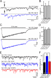
Synaptotagmin-12 regulates spontaneous transmitter release independently from synaptotagmin-1. Primary cortical neurons isolated from newborn pups of synaptotagmin-1–deficient mice were infected with lentiviruses expressing GFP (control), wild-type synaptotagmin-12, or S97A mutant and analyzed at 15–17 DIV. (A–D) Evoked IPSCs triggered in 2 mM of extracellular Ca2+ by single-action potentials or trains of 100 action potentials applied at 10 Hz. (A) Typical asynchronous IPSCs triggered by single-action potentials in control neurons (black) or neurons expressing wild-type synaptotagmin-12 (blue). Scale bars apply to both traces. (B) Average charge transferred over 1.5 s by asynchronous responses triggered by single action potentials. The total numbers of analyzed neurons are shown on each column. (C) Asynchronous responses triggered by high frequency trains of 100 action potentials applied at 10 Hz. Scale bars apply to both traces. (D) Average total synaptic charge transfer during trains or 100 potentials in control neurons or neurons expressing wild-type synaptotagmin-12. The total numbers of analyzed neurons are shown on each column. (E and F) Synaptotagmin-12 increases the rate or spontaneous release in synaptotagmin-1–deficient neurons. (E) Typical mIPSCs recorded from control neurons (black) or neurons infected with wild-type (blue) or S97A (red) synaptotagmin-12 lentiviruses. mIPSCs were recorded in 0.2 mM of extracellular Ca2+ in the presence of tetrodotoxin, APV, and CNQX. Scale bars apply to all traces. (F) Average frequencies of mIPSCs monitored from two different cortical cultures. The total numbers of analyzed neurons are shown on each column. Data are shown as the mean ± the SEM.

Similar articles

Cited by

References

    1. Adolfsen, B., S. Saraswati, M. Yoshihara, and J.T. Littleton. 2004. Synaptotagmins are trafficked to distinct subcellular domains including the postsynaptic compartment. J. Cell Biol. 166:249–260. - PMC - PubMed
    1. Angleson, J.K., and W.J. Betz. 2001. Intraterminal Ca(2+) and spontaneous transmitter release at the frog neuromuscular junction. J. Neurophysiol. 85:287–294. - PubMed
    1. Banerjee, A., J.A. Kowalchyk, B.R. DasGupta, and T.F. Martin. 1996. SNAP-25 is required for a late postdocking step in Ca2+-dependent exocytosis. J. Biol. Chem. 271:20227–20230. - PubMed
    1. Bennett, M.K., N. Calakos, and R.H. Scheller. 1992. Syntaxin: a synaptic protein implicated in docking of synaptic vesicles at presynaptic active zones. Science. 257:255–259. - PubMed
    1. Bowen, M.E., K. Weninger, J. Ernst, S. Chu, and A.T. Brunger. 2005. Single-molecule studies of synaptotagmin-1 and complexin binding to the SNARE complex. Biophys. J. 89:690–702. - PMC - PubMed

MeSH terms