

Dissociation of estrogen receptor expression and in vivo stem cell activity in the mammary gland
- PMID: 17190790
- PMCID: PMC2063618
- DOI: 10.1083/jcb.200604065
Dissociation of estrogen receptor expression and in vivo stem cell activity in the mammary gland
Abstract
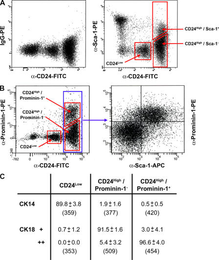
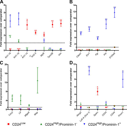
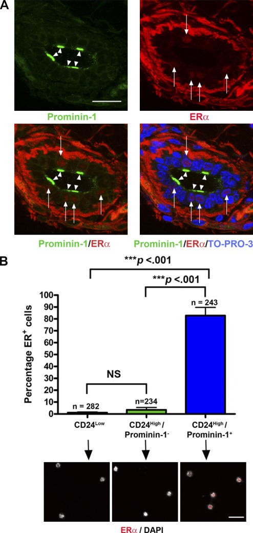
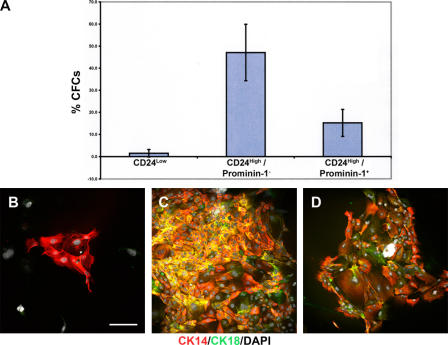
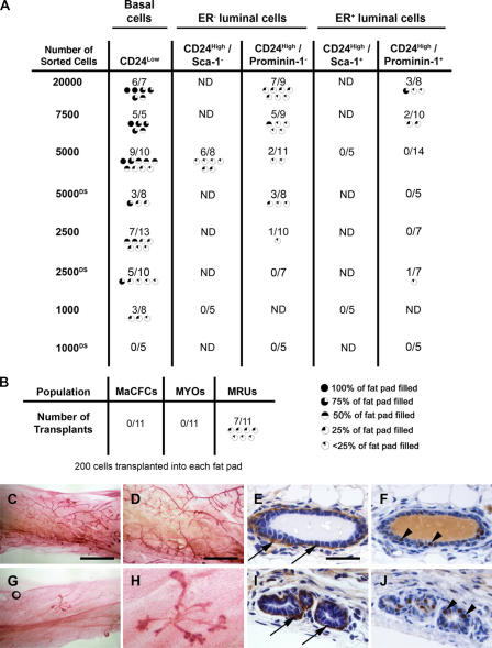
The role of estrogen in promoting mammary stem cell proliferation remains controversial. It is unclear if estrogen receptor (ER)-expressing cells have stem/progenitor activity themselves or if they act in a paracrine fashion to stimulate stem cell proliferation. We have used flow cytometry to prospectively isolate mouse mammary ER-expressing epithelial cells and shown, using analysis of gene expression patterns and cell type-specific markers, that they form a distinct luminal epithelial cell subpopulation that expresses not only the ER but also the progesterone and prolactin receptors. Furthermore, we have used an in vivo functional transplantation assay to directly demonstrate that the ER-expressing luminal epithelial subpopulation contains little in vivo stem cell activity. Rather, the mammary stem cell activity is found within the basal mammary epithelial cell population. Therefore, ER-expressing cells of the mammary epithelium are distinct from the mammary stem cell population, and the effects of estrogen on mammary stem cells are likely to be mediated indirectly. These results are important for our understanding of cellular responses to hormonal stimulation in the normal breast and in breast cancer.
Figures
Similar articles
-
SCA-1 Labels a Subset of Estrogen-Responsive Bipotential Repopulating Cells within the CD24+ CD49fhi Mammary Stem Cell-Enriched Compartment.Stem Cell Reports. 2017 Feb 14;8(2):417-431. doi: 10.1016/j.stemcr.2016.12.022. Epub 2017 Jan 26. Stem Cell Reports. 2017. PMID: 28132885 Free PMC article.
-
c-Kit is required for growth and survival of the cells of origin of Brca1-mutation-associated breast cancer.Oncogene. 2012 Feb 16;31(7):869-83. doi: 10.1038/onc.2011.289. Epub 2011 Jul 18. Oncogene. 2012. PMID: 21765473
-
Phenotypic and functional characterisation of the luminal cell hierarchy of the mammary gland.Breast Cancer Res. 2012 Oct 22;14(5):R134. doi: 10.1186/bcr3334. Breast Cancer Res. 2012. PMID: 23088371 Free PMC article.
-
Delineating the epithelial hierarchy in the mouse mammary gland.Cold Spring Harb Symp Quant Biol. 2008;73:469-78. doi: 10.1101/sqb.2008.73.020. Epub 2008 Nov 6. Cold Spring Harb Symp Quant Biol. 2008. PMID: 19022771 Review.
-
Form and function: how estrogen and progesterone regulate the mammary epithelial hierarchy.J Mammary Gland Biol Neoplasia. 2015 Jun;20(1-2):9-25. doi: 10.1007/s10911-015-9337-0. Epub 2015 Jul 19. J Mammary Gland Biol Neoplasia. 2015. PMID: 26188694 Free PMC article. Review.
Cited by
-
Stem cells and the developing mammary gland.J Mammary Gland Biol Neoplasia. 2013 Jun;18(2):209-19. doi: 10.1007/s10911-013-9284-6. Epub 2013 Apr 27. J Mammary Gland Biol Neoplasia. 2013. PMID: 23624881 Free PMC article. Review.
-
Hormone-sensing cells require Wip1 for paracrine stimulation in normal and premalignant mammary epithelium.Breast Cancer Res. 2013 Jan 31;15(1):R10. doi: 10.1186/bcr3381. Breast Cancer Res. 2013. PMID: 23369183 Free PMC article.
-
PIK3CA(H1047R) induces multipotency and multi-lineage mammary tumours.Nature. 2015 Sep 3;525(7567):114-8. doi: 10.1038/nature14669. Epub 2015 Aug 12. Nature. 2015. PMID: 26266975
-
Patient-derived luminal breast cancer xenografts retain hormone receptor heterogeneity and help define unique estrogen-dependent gene signatures.Breast Cancer Res Treat. 2012 Sep;135(2):415-32. doi: 10.1007/s10549-012-2164-8. Epub 2012 Jul 24. Breast Cancer Res Treat. 2012. PMID: 22821401 Free PMC article.
-
The Met oncogene and basal-like breast cancer: another culprit to watch out for?Breast Cancer Res. 2010;12(4):208. doi: 10.1186/bcr2617. Epub 2010 Aug 23. Breast Cancer Res. 2010. PMID: 20804567 Free PMC article. Review.
References
-
- Clarke, R.B., A. Howell, C.S. Potten, and E. Anderson. 1997. Dissociation between steroid receptor expression and cell proliferation in the human breast. Cancer Res. 57:4987–4991. - PubMed
-
- Clarke, R.B., K. Spence, E. Anderson, A. Howell, H. Okano, and C.S. Potten. 2005. A putative human breast stem cell population is enriched for steroid receptor-positive cells. Dev. Biol. 277:443–456. - PubMed
-
- DeOme, K.B., L.J. Faulkin Jr., H.A. Bern, and P.B. Blair. 1959. Development of mammary tumors from hyperplastic alveolar nodules transplanted into gland-free mammary fat pads of female C3H mice. Cancer Res. 19:515–520. - PubMed
Publication types
MeSH terms
Substances
Grants and funding
LinkOut - more resources
Full Text Sources
Other Literature Sources
Medical
Miscellaneous
