Spontaneous hepatocarcinogenesis in farnesoid X receptor-null mice
- PMID: 17183066
- PMCID: PMC1858639
- DOI: 10.1093/carcin/bgl249
Spontaneous hepatocarcinogenesis in farnesoid X receptor-null mice
Abstract
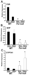
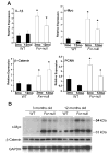
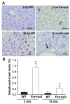
The farnesoid X receptor (FXR) controls the synthesis and transport of bile acids (BAs). Mice lacking expression of FXR, designated Fxr-null, have elevated levels of serum and hepatic BAs and an increase in BA pool size. Surprisingly, at 12 months of age, male and female Fxr-null mice had a high incidence of degenerative hepatic lesions, altered cell foci and liver tumors including hepatocellular adenoma, carcinoma and hepatocholangiocellular carcinoma, the latter of which is rarely observed in mice. At 3 months, Fxr-null mice had increased expression of the proinflammatory cytokine IL-1beta mRNA and elevated beta-catenin and its target gene c-myc. They also had increased cell proliferation as revealed by increased PCNA mRNA and BrdU incorporation. These studies reveal a potential role for FXR and BAs in hepatocarcinogenesis.
Figures


Similar articles
-
Prevention of spontaneous hepatocarcinogenesis in farnesoid X receptor-null mice by intestinal-specific farnesoid X receptor reactivation.Hepatology. 2015 Jan;61(1):161-70. doi: 10.1002/hep.27274. Epub 2014 Oct 30. Hepatology. 2015. PMID: 24954587
-
Spontaneous development of liver tumors in the absence of the bile acid receptor farnesoid X receptor.Cancer Res. 2007 Feb 1;67(3):863-7. doi: 10.1158/0008-5472.CAN-06-1078. Cancer Res. 2007. PMID: 17283114
-
Farnesoid X receptor antagonizes nuclear factor kappaB in hepatic inflammatory response.Hepatology. 2008 Nov;48(5):1632-43. doi: 10.1002/hep.22519. Hepatology. 2008. PMID: 18972444 Free PMC article.
-
FXR, a multipurpose nuclear receptor.Trends Biochem Sci. 2006 Oct;31(10):572-80. doi: 10.1016/j.tibs.2006.08.002. Epub 2006 Aug 14. Trends Biochem Sci. 2006. PMID: 16908160 Review.
-
The role of FXR in disorders of bile acid homeostasis.Physiology (Bethesda). 2008 Oct;23:286-95. doi: 10.1152/physiol.00020.2008. Physiology (Bethesda). 2008. PMID: 18927204 Review.
Cited by
-
Bile acid and receptors: biology and drug discovery for nonalcoholic fatty liver disease.Acta Pharmacol Sin. 2022 May;43(5):1103-1119. doi: 10.1038/s41401-022-00880-z. Epub 2022 Feb 25. Acta Pharmacol Sin. 2022. PMID: 35217817 Free PMC article. Review.
-
Role of bile acids and their receptors in gastrointestinal and hepatic pathophysiology.Nat Rev Gastroenterol Hepatol. 2022 Jul;19(7):432-450. doi: 10.1038/s41575-021-00566-7. Epub 2022 Feb 14. Nat Rev Gastroenterol Hepatol. 2022. PMID: 35165436 Review.
-
Suppression of Hepatic Bile Acid Synthesis by a non-tumorigenic FGF19 analogue Protects Mice from Fibrosis and Hepatocarcinogenesis.Sci Rep. 2018 Nov 21;8(1):17210. doi: 10.1038/s41598-018-35496-z. Sci Rep. 2018. PMID: 30464200 Free PMC article.
-
Baohuoside I inhibits FXR signaling pathway to interfere with bile acid homeostasis via targeting ER α degradation.Cell Biol Toxicol. 2023 Aug;39(4):1215-1235. doi: 10.1007/s10565-022-09737-x. Epub 2022 Jul 8. Cell Biol Toxicol. 2023. PMID: 35802278
-
Bile-ology: from bench to bedside.J Zhejiang Univ Sci B. 2019 May;20(5):414-427. doi: 10.1631/jzus.B1900158. J Zhejiang Univ Sci B. 2019. PMID: 31090267 Free PMC article. Review.
References
-
- Lee FY, Lee H, Hubbert ML, Edwards PA, Zhang Y. FXR, a multipurpose nuclear receptor. Trends Biochem Sci 2006 - PubMed
-
- Sinal CJ, Tohkin M, Miyata M, Ward JM, Lambert G, Gonzalez FJ. Targeted disruption of the nuclear receptor FXR/BAR impairs bile acid and lipid homeostasis. Cell. 2000;102:731–44. - PubMed
-
- Cariou B, van Harmelen K, Duran-Sandoval D, van Dijk TH, Grefhorst A, Abdelkarim M, Caron S, Torpier G, Fruchart JC, Gonzalez FJ, Kuipers F, Staels B. The farnesoid X receptor modulates adiposity and peripheral insulin sensitivity in mice. J Biol Chem. 2006;281:11039–49. - PubMed
-
- Ward JM, Goodman DG, Squire RA, Chu KC, Linhart MS. Neoplastic and nonneoplastic lesions in aging (C57BL/6N x C3H/HeN)F1 (B6C3F1) mice. J Natl Cancer Inst. 1979;63:849–54. - PubMed
Publication types
MeSH terms
Substances
Grants and funding
LinkOut - more resources
Full Text Sources
Other Literature Sources
Medical
Molecular Biology Databases
Miscellaneous

