Comparative Study
. 2006 Dec 20;26(51):13202-12.
doi: 10.1523/JNEUROSCI.4608-06.2006.

Connecdenn, a novel DENN domain-containing protein of neuronal clathrin-coated vesicles functioning in synaptic vesicle endocytosis

Affiliations
Comparative Study

Connecdenn, a novel DENN domain-containing protein of neuronal clathrin-coated vesicles functioning in synaptic vesicle endocytosis

Patrick D Allaire et al. J Neurosci. .

Abstract

Clathrin-coated vesicles (CCVs) are responsible for the endocytosis of multiple cargo, including synaptic vesicle membranes. We now describe a new CCV protein, termed connecdenn, that contains an N-terminal DENN (differentially expressed in neoplastic versus normal cells) domain, a poorly characterized protein module found in multiple proteins of unrelated function and a C-terminal peptide motif domain harboring three distinct motifs for binding the alpha-ear of the clathrin adaptor protein 2 (AP-2). Connecdenn coimmunoprecipitates and partially colocalizes with AP-2, and nuclear magnetic resonance and peptide competition studies reveal that all three alpha-ear-binding motifs contribute to AP-2 interactions. In addition, connecdenn contains multiple Src homology 3 (SH3) domain-binding motifs and coimmunoprecipitates with the synaptic SH3 domain proteins intersectin and endophilin A1. Interestingly, connecdenn is enriched on neuronal CCVs and is present in the presynaptic compartment of neurons. Moreover, connecdenn has a uniquely stable association with CCV membranes because it resists extraction with Tris and high-salt buffers, unlike most other CCV proteins, but it is not detected on purified synaptic vesicles. Together, these observations suggest that connecdenn functions on the endocytic limb of the synaptic vesicle cycle. Accordingly, disruption of connecdenn interactions with its binding partners through overexpression of the C-terminal peptide motif domain or knock down of connecdenn through lentiviral delivery of small hairpin RNA both lead to defects in synaptic vesicle endocytosis in cultured hippocampal neurons. Thus, we identified connecdenn as a component of the endocytic machinery functioning in synaptic vesicle endocytosis, providing the first evidence of a role for a DENN domain-containing protein in endocytosis.


Figures
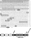
Figure 1.
Figure 1.
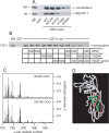
Identification of connecdenn. The amino acid sequence of mouse connecdenn (from gi31542027) and a domain/motif model are presented. The DENN domain is composed of uDENN, DENN, and dDENN modules indicated by solid, dotted, and dashed lines, respectively. Potential AP-2-binding motifs are indicated by ovals with dark shading, and proline-rich regions containing PXXP cores are indicated by boxes with light shading. The annotated mouse connecdenn has been entered into GenBank under the accession number DQ448594.
Figure 2.
Figure 2.
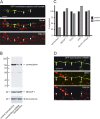
Interaction of connecdenn with AP-2. A, Soluble brain lysates were incubated with protein G beads alone or antibody AP.6 against the α-adaptin subunit of AP-2 and protein G beads. Proteins specifically bound to the beads were processed for Western blot with antibodies against the indicated proteins. B, Soluble brain lysates were incubated with GST or GST fused to the ear domains of α-, β2-, and γ-adaptin, precoupled to glutathione-Sepharose beads. Proteins specifically bound to the beads were processed for Western blot with antibody against connecdenn. C, Lysates from HEK-293 cells transfected with Flag-tagged connecdenn were incubated with GST or GST–α-ear precoupled to glutathione-Sepharose beads, and proteins specifically bound to the beads were processed for Western blot with antibody against the Flag-epitope tag. D, COS-7 cells transfected at low levels with full-length Flag-tagged connecdenn and processed with a polyclonal Flag antibody (red channel) and a monoclonal antibody against AP-2 (AP.6, green channel). The blend of the two images is shown in the left panel, and a blend and individual images of the region indicated by the box is shown in the right three panels. Scale bar: lower-magnification image, 10 μm; higher-magnification images, 4.3 μm.
Figure 3.
Figure 3.
Identification of the connecdenn-binding site on AP-2. A, Soluble brain lysates were incubated with GST or GST fused to wild-type (WT) α-ear or a variety of α-ear point mutants as indicated, precoupled to glutathione-Sepharose beads. Proteins specifically bound to the beads were processed for Western blot with antibodies against the indicated proteins. B, Soluble brain lysates were incubated with GST or GST–α-ear, precoupled to glutathione-Sepharose beads. Incubations were without or with a DPW peptide (CSDPWGSDPWG) from epsin 1, a FXDXF peptide (CSFFEDNFVPE) from amphiphysin1, or a WXXF-acidic peptide (CQAPQPSNWVQF-COO) from NECAP 1 at the molar ratios of peptide to fusion protein indicated. Proteins specifically bound to the beads were processed for Western blot with antibody against connecdenn. For all above experiments, an aliquot of starting material (SM) equal to 1/10 of that added to the beads was run in parallel. C, Magnitude of the amide chemical shift changes [{(Δ1H shift)2 + (Δ15N shift × 0.2)2}1/2 in parts per million (ppm)] of the α-ear during binding CQAPQPSNWVQF-COO (WVQF-COO, NECAP 1; top) and CVEQLRRQWETFE (WETFE-COO, connecdenn; bottom). The residue numbers correspond to mouse α-adaptin. D, HADDOCK modeled structure of WETFE/α-ear complex. The backbone trace of the α-ear is colored according to the size of the amide chemical shift changes during binding of the connecdenn peptide.
Figure 4.
Figure 4.
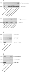
Connecdenn binds to SH3 domains from endocytic proteins. A, Soluble lysates from Flag–connecdenn-transfected HEK-293 cells were incubated with GST or GST fused to the SH3 domain of either amphiphysin I or II, PACSIN 1 or 2, endophilin A1, and the five individual SH3 domains of intersectin, precoupled to glutathione-Sepharose beads. Proteins specifically bound to the beads were processed for Western blot with antibodies against the indicated proteins. B, Soluble brain lysates were incubated with anti-intersectin serum or preimmune serum from the same rabbit along with protein A-Sepharose beads. Proteins specifically bound to the beads were processed for Western blot with antibodies against the indicated proteins. C, Soluble brain lysates were incubated with anti-connecdenn serum (3775) or preimmune serum from the same rabbit along with protein A-Sepharose beads. Proteins specifically bound to the beads were processed for Western blot with antibodies against the indicated proteins. For B and C, Western blots with polyclonal antibodies were revealed with protein A-HRP. For all experiments, an aliquot of starting material (SM) equal to 1/10 of that added to the beads was run in parallel.
Figure 5.
Figure 5.
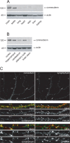
Connecdenn is brain enriched and is detected at the synapse of hippocampal neurons in culture. Equal protein extracts from various rat tissues including skeletal muscle (skel.) (A) and rat brain regions (B) were blotted with antibodies against connecdenn and actin. C, Rat hippocampal neurons at 21 DIV were processed for indirect immunofluorescence with a polyclonal antibody against connecdenn (3775) and monoclonal antibodies against either synaptophysin or PSD-95. For the bottom 12 panels, the blend of connecdenn (red) and synaptophysin or PSD-95 (green) or the individual images are shown. Scale bars: top two panels, 10 μm; middle six panels, 3.33 μm; bottom six panels, 2.33 μm. The percentage of connecdenn-positive punctate that colocalize with synaptophysin-positive punctae is 71% in C, above the average of 45.0% for all cells examined.
Figure 6.
Figure 6.
Connecdenn is enriched on CCVs. Equal protein aliquots of the successive fractions of a procedure leading to highly enriched CCVs were analyzed by Western blot with antibodies against CHC, α-adaptin, and connecdenn (A) and CHC, synaptotagmin, and synaptophysin (B). H, Homogenate; P, pellet; S, supernatant; SG, sucrose gradient.
Figure 7.
Figure 7.
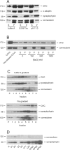
Connecdenn is stably associated with CCV membranes. A, CCVs isolated from rat brain were resuspended in buffer A, 0.5 m Tris buffer, or sodium carbonate buffer at pH 11.0. The samples were centrifuged at high g, and proteins in the supernatant (S) and pellet (P) fractions were analyzed by Western blot with antibodies against the indicated proteins. B, CCVs were stripped of their coats with two successive rounds of incubation with 0.5 m Tris. The resulting vesicles were split in five aliquots, pelleted at high g, and resuspended in Laemmlli sample buffer (Tris-stripped CCVs) or in 10 mm HEPES, pH 7.4, containing 0, 150, 500, or 1000 mm NaCl. Samples were again centrifuged, and the pellet (P) and supernatant (S) fractions were analyzed by Western blot with antibodies against the indicated proteins. C, CCVs in buffer A and CCVs extracted with Tris buffer were loaded on the top of linear 20–50% sucrose gradients prepared in buffer A and Tris buffer, respectively, and were centrifuged at 145,000 × g for 1.5 h. The gradients were fractionated from the bottom, and equal volume aliquots of each fraction along with the pellet (P) were analyzed by Western blot with the indicated antibodies. D, Equal protein aliquots of the successive fractions of a procedure leading to highly enriched SVs were analyzed by Western blot with the indicated antibodies. H, Homogenate; P, pellet; S, supernatant; LP, lysed pellet; LS, lysed supernatant; CPG, controlled pore glass. CPG3 contains purified SVs.
Figure 8.
Figure 8.
Disruption of connecdenn function reduces CME of SVs in neurons. A, An axon labeled with GFP-connecdenn peptide motif domain is seen to run along a dendrite from a nonexpressing cell. KCl-induced endocytosis of FM4-64 is indicated in red. Note the punctate staining pattern, indicative of FM dye uptake into nonlabeled axons. Note the reduced (arrowheads) or lack of (square heads) uptake in varicosities of the axon expressing GFP–connecdenn peptide motif domain. Scale bars, 5 μm. B, Hippocampal neurons at 14 DIV were transduced with FIV expressing GFP alone (control transduction) or GFP and an shRNA specific for connecdenn. At 21 DIV, protein extracts were made, and equal protein aliquots were analyzed by Western blot with antibodies against NECAP 1 and CLCa/b as well as actin and Na+/K+-ATPase (data not shown). C, Blots were scanned, and the relative amounts of the indicated proteins in control versus shRNA transduced neurons was plotted. D, A representative image of an axon from a neuron transduced with an FIV virus driving expression of GFP and an shRNA specific for connecdenn. The GFP-labeled axon is seen to cross an axon from a nonexpressing cell. KCl-induced endocytosis of FM4-64 is indicated in red. Note the punctate staining pattern, indicative of FM dye uptake into nonlabeled axons. Note the reduced (arrowheads) or lack of (square heads) uptake in varicosities of the axon expressing GFP. Scale bars, 5 μm.

Similar articles

Cited by

References

    1. Barik S. Site-directed mutagenesis by double polymerase chain reaction. Mol Biotechnol. 1995;3:1–7. - PubMed
    1. Blondeau F, Ritter B, Allaire PD, Wasiak S, Girard M, Hussain NK, Angers A, Legendre-Guillemin V, Roy L, Boismenu D, Kearney RE, Bell AW, Bergeron JJ, McPherson PS. Tandem MS analysis of brain clathrin-coated vesicles reveals their critical involvement in synaptic vesicle recycling. Proc Natl Acad Sci USA. 2004;101:3833–3838. - PMC - PubMed
    1. Brett TJ, Traub LM. Molecular structures of coat and coat-associated proteins: function follows form. Curr Opin Cell Biol. 2006;18:395–406. - PubMed
    1. Burman JL, Wasiak S, Ritter B, de Heuvel E, McPherson PS. Aftiphilin is a component of the clathrin machinery in neurons. FEBS Lett. 2005;579:2177–2184. - PubMed
    1. Conner SD, Schmid SL. Identification of an adaptor-associated kinase, AAK1, as a regulator of clathrin-mediated endocytosis. J Cell Biol. 2002;156:921–929. - PMC - PubMed

Publication types

MeSH terms

Substances

Associated data