. 2007 Mar;27(5):1545-57.
doi: 10.1128/MCB.00773-06. Epub 2006 Dec 18.

Requirement of Nhp6 proteins for transcription of a subset of tRNA genes and heterochromatin barrier function in Saccharomyces cerevisiae

Affiliations

Requirement of Nhp6 proteins for transcription of a subset of tRNA genes and heterochromatin barrier function in Saccharomyces cerevisiae

Priscilla Braglia et al. Mol Cell Biol. 2007 Mar.

Abstract

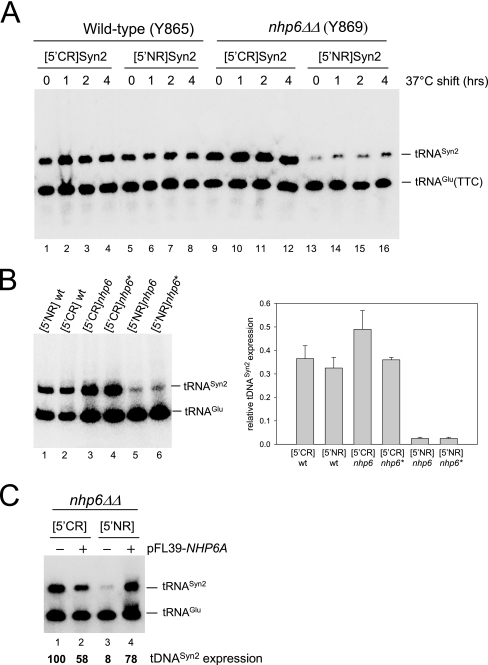
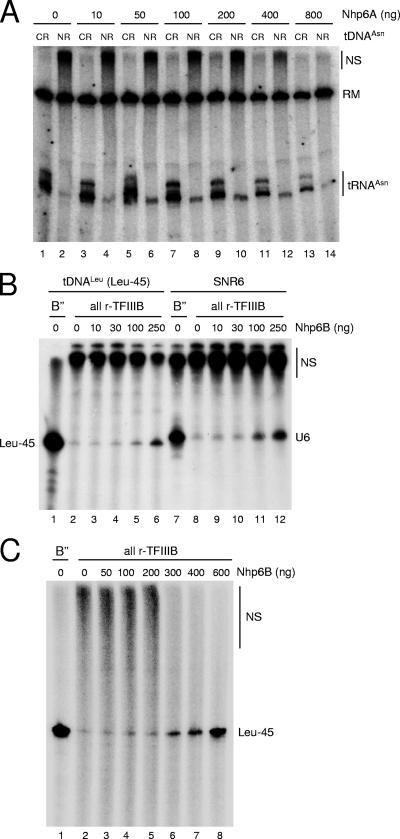
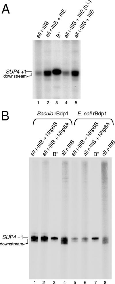
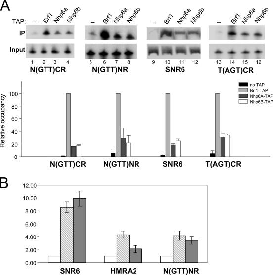
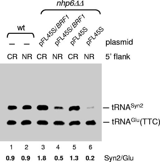
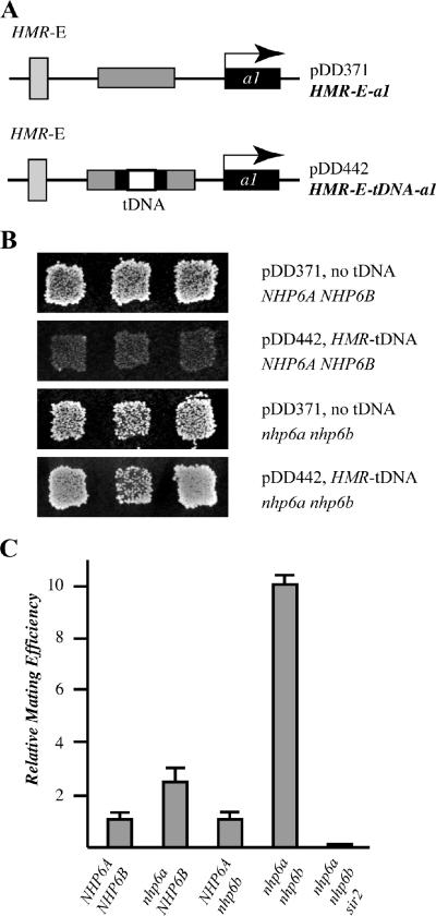
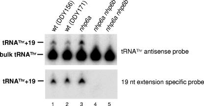
A key event in tRNA gene (tDNA) transcription by RNA polymerase (Pol) III is the TFIIIC-dependent assembly of TFIIIB upstream of the transcription start site. Different tDNA upstream sequences bind TFIIIB with different affinities, thereby modulating tDNA transcription. We found that in the absence of Nhp6 proteins, the influence of the 5'-flanking region on tRNA gene transcription is dramatically enhanced in Saccharomyces cerevisiae. Expression of a tDNA bearing a suboptimal TFIIIB binding site, but not of a tDNA preceded by a strong TFIIIB binding region, was strongly dependent on Nhp6 in vivo. Upstream sequence-dependent stimulation of tRNA gene transcription by Nhp6 could be reproduced in vitro, and Nhp6 proteins were found associated with tRNA genes in yeast cells. We also show that both transcription and silencing barrier activity of a tDNA(Thr) at the HMR locus are compromised in the absence of Nhp6. Our data suggest that Nhp6 proteins are important components of Pol III chromatin templates that contribute both to the robustness of tRNA gene expression and to positional effects of Pol III transcription complexes.


Figures

FIG. 1.
FIG. 1.
Nhp6 stimulates tRNA gene transcription in vivo in a context-dependent manner. (A) WT (lanes 1 to 8) and nhp6ΔΔ strains (lanes 9 to 16) were transformed with pFL46S containing either [5′CR]Syn2 (lanes 1 to 4 and 9 to 12) or [5′NR]Syn2 (lanes 5 to 8 and 13 to 16) tDNA, grown on selective medium at 30°C, and then shifted to 37°C for the indicated periods of time. Total RNA was isolated and subjected to Northern blot analysis with a probe complementary to both the tDNASyn2 transcript and the endogenous tRNAGlu(TTC). The migration positions of the two RNA species are indicated on the right. (B) Total RNA was extracted from NHP6A NHP6B strains carrying an integrated copy of either the [5′NR]Syn2 (lane 1) or the [5′CR]Syn2 (lane 2) reporter tDNA (strains DDY3529 and DDY3524, respectively; see Table 1) and from two different nhp6ΔΔ strains carrying either an integrated copy of [5′CR]Syn2 (lanes 3 and 4; strains DDY3532 and DDY3534, respectively) or an integrated copy of [5′NR]Syn2 (lanes 5 and 6; strains DDY3535 and DDY3536, respectively. The bar graph on the right reports the results of phosphorimager quantification of the gel image shown and of an image derived from an identical Northern blotting experiment conducted in parallel. tDNASyn2 transcript levels are expressed as ratios of each tRNASyn2 signal to the tRNAGlu(TTC) signal in the same lane. (C) nhp6ΔΔ strain Y869 containing plasmid-borne [5′CR]Syn2 (lanes 1 and 2) or [5′NR]Syn2 (lanes 3 and 4) was transformed with either the empty pFL39 vector (lanes 1 and 3) or pFL39 carrying the NHP6A gene (lanes 2 and 4). Total RNA was extracted and subjected to Northern blot analysis as for panel A. The migration positions of the tDNASyn2 transcript and the endogenous tRNAGlu(TTC) are indicated on the right. The values reported below each lane derive from phosphorimager quantification of the gel image after normalization with the tRNAGlu(TTC) signal as an internal standard and are relative to the value measured in lane 1, which was arbitrarily set to 100.
FIG. 2.
FIG. 2.
Stimulation of tRNA gene transcription in vitro by recombinant Nhp6. (A) In vitro transcription of either N(GTT)CR (odd-numbered lanes) or N(GTT)NR (even-numbered lanes) was carried out in a reconstituted system containing recombinant Bdp1 protein from baculovirus-infected insect cells and in the presence of the indicated amounts of recombinant Nhp6A protein. The migration position of tRNAAsn transcripts is indicated on the right, together with the position of a radiolabeled DNA fragment used as a recovery marker (RM). The migration position of large-size nonspecific transcription products is also indicated on the right (NS). (B) In vitro transcription of either a shortened variant (Leu-45) of the L(CAA)CL tDNA (lanes 1 to 6) or the SNR6 template (lanes 7 to 12) was carried out in a reconstituted system containing either the crude B" fraction (lanes 1 and 7) or recombinant Bdp1 protein purified from overexpressing E. coli cells (lanes 2 to 6 and 8 to 12) and supplemented with the indicated amount of recombinant Nhp6B protein. The migration position of the shortened tRNALeu transcript is indicated on the left (Leu-45). The migration position of the SNR6 transcript is indicated on the right (U6), as is the position of large-size nonspecific transcription products (NS). (C) In vitro transcription of the Leu-45 template was carried out in a reconstituted system containing either the crude B" fraction (lane 1) or recombinant Bdp1 protein purified from overexpressing E. coli cells (lanes 2 to 8) and supplemented with the indicated amounts of recombinant Nhp6B protein. The migration position of the shortened tRNALeu transcript is indicated on the left (Leu-45), as is the position of large-size nonspecific transcription products (NS).
FIG. 3.
FIG. 3.
TFIIIE stimulates SUP4 transcription without influencing TSS selection. (A) In vitro transcription of the SUP4 tRNATyr template was carried out in a reconstituted system containing either TFIIIB reconstituted with the crude B" fraction (lane 3) or all-recombinant TFIIIB containing E. coli-expressed Bdp1 protein (lanes 1, 2, 4, and 5). The reaction mixtures in lanes 2 and 5 were supplemented with the TFIIIE fraction; the reaction mixture in lane 4 was supplemented with the heat-inactivated (h.i.) TFIIIE fraction. The migration positions of SUP4 transcripts resulting from initiation at +1 and from initiation events at downstream sites are indicated on the left. (B) In vitro transcription of the SUP4 tRNATyr template was carried out in a reconstituted system containing the crude B" fraction (lanes 3 and 7), baculovirus-expressed rBdp1 (lanes 1, 2, and 4), or E. coli-expressed rBdp1 (lanes 5, 6, and 8). The reaction mixtures in lanes 1 and 5 were supplemented with rNhp6B protein (100 ng); the reaction mixtures in lanes 2 and 6 were supplemented with rNhp6A protein. Only one-fifth of the total reaction products were loaded in lanes 3 and 7 to improve the resolution of the SUP4 tRNA signal and thus more easily compare transcript sizes in the different lanes.
FIG. 4.
FIG. 4.
Nhp6 association with SNR6 and tRNA genes in vivo. (A) ChIP analysis was performed with an untagged reference strain (lanes 1, 5, 9, and 13) and with BRF1-TAP-, NHP6A-TAP-, and NHP6B-TAP-tagged strains, as indicated above the lanes. The extent of association of each of the three tagged proteins with the N(GTT)CR, N(GTT)NR, SNR6, and T(AGT)CR loci was assessed by PCR in the presence of radiolabeled dATP. The phosphorimager quantification of gel images is reported below as a bar graph of data derived from three independent experiments (error bars indicate standard deviations). PCR signals from immunoprecipitated (IP) DNA were normalized to the PCR signals obtained in the input DNA reaction mixture. The calculated values were then expressed relative to the values obtained with BRF1-TAP, which were arbitrarily set to 100. (B) The extent of association of Nhp6A and Nhp6B with the SNR6, HMRA2, and N(GTT)NR loci was quantitatively evaluated by real-time PCR. Signals obtained with immunoprecipitated DNA were normalized to the input, and the calculated values are expressed relative to the background signal obtained with the untagged strain, which was arbitrarily set to 1. Two independent PCRs were conducted in triplicate, and error bars indicate standard deviations of average values obtained in each experiment. NHP6A-TAP, light gray bars; NHP6B-TAP, dark gray bars; untagged reference strain, open bars.
FIG. 5.
FIG. 5.
BRF1 overexpression partially rescues the tDNA transcriptional defect in the nhp6ΔΔ strain. WT (lanes 1 and 2) and nhp6ΔΔ (lanes 3 to 6) strains were transformed with pFL46S containing either [5′CR]Syn2 (lanes 1, 3, and 5) or [5′NR]Syn2 (lanes 2, 4, and 6) tDNA together with the empty pFL45S vector (lanes 5 and 6), pFL45S carrying the BRF1 gene (lanes 3 and 4), or no additional plasmid (lanes 1 and 2). Total RNA was isolated and subjected to Northern blot analysis with a probe complementary to both the tDNASyn2 transcript and the endogenous tRNAGlu(TTC). The migration positions of the two RNA species are indicated on the right.
FIG. 6.
FIG. 6.
Nhp6 is required for the heterochromatin barrier function of the HMR-tDNA. (A) Plasmid pDD371 contains the HMR locus of S. cerevisiae lacking the I silencer. Plasmid pDD442 contains the HMR-tDNA cloned into the a2 gene between the E silencer and a1, which efficiently blocks silencing from repressing the transcription of a1. (B) Strains DDY171 and DDY591 (nhp6ΔΔ) were transformed with plasmid pDD371 or pDD442, and mating assays were performed as previously described (22). (C) Quantitative analysis of the effects of nhp6 mutations on mating efficiency. The strains used in this assay contain the HMR-E-tDNA constructs integrated back into chromosome III and are DDY689 (NHP6A NHP6B), DDY714 (nhp6a NHP6B), DDY669 (NHP6A nhp6b), DDY671 (nhp6a nhp6b), and DDY705 (nhp6a nhp6b sir2).
FIG. 7.
FIG. 7.
Nhp6 is required for transcription of HMR-tDNAThr. WT and nhp6 mutant strains (each with HMR and the HMR-tDNA deleted) were transformed with plasmid pDD570, which carries a marked HMR-tDNA boundary (containing an additional 19 bp between the end of the coding sequence and the terminator) cloned into the HMR locus between the E silencer and the a1 gene. Total RNA was isolated and subjected to Northern blot analysis with either a bulk tRNAThr antisense oligonucleotide (upper panel) or an oligonucleotide specific for the 19-base extension marking the HMR-tDNA (lower panel) as the probe.

Similar articles

Cited by

References

    1. Andrau, J. C., and M. Werner. 2001. B"-associated factor(s) involved in RNA polymerase III preinitiation complex formation and start-site selection. Eur. J. Biochem. 268:5167-5175. - PubMed
    1. Bachman, N., M. E. Gelbart, T. Tsukiyama, and J. D. Boeke. 2005. TFIIIB subunit Bdp1p is required for periodic integration of the Ty1 retrotransposon and targeting of Isw2p to S. cerevisiae tDNAs. Genes Dev. 19:955-964. - PMC - PubMed
    1. Baker, R. E., S. Camier, A. Sentenac, and B. D. Hall. 1987. Gene size differentially affects the binding of yeast transcription factor τ to two intragenic regions. Proc. Natl. Acad. Sci. USA 84:8768-8772. - PMC - PubMed
    1. Bianchi, M. E., and A. Agresti. 2005. HMG proteins: dynamic players in gene regulation and differentiation. Curr. Opin. Genet. Dev. 15:496-506. - PubMed
    1. Biswas, D., A. N. Imbalzano, P. Eriksson, Y. Yu, and D. J. Stillman. 2004. Role for Nhp6, Gcn5, and the Swi/Snf complex in stimulating formation of the TATA-binding protein-TFIIA-DNA complex. Mol. Cell. Biol. 24:8312-8321. - PMC - PubMed

Publication types

MeSH terms