. 2007 Jan;147(1):164-75.
doi: 10.1111/j.1365-2249.2006.03249.x.

Txk, a member of the non-receptor tyrosine kinase of the Tec family, forms a complex with poly(ADP-ribose) polymerase 1 and elongation factor 1alpha and regulates interferon-gamma gene transcription in Th1 cells

Affiliations

Txk, a member of the non-receptor tyrosine kinase of the Tec family, forms a complex with poly(ADP-ribose) polymerase 1 and elongation factor 1alpha and regulates interferon-gamma gene transcription in Th1 cells

T Maruyama et al. Clin Exp Immunol. 2007 Jan.

Abstract

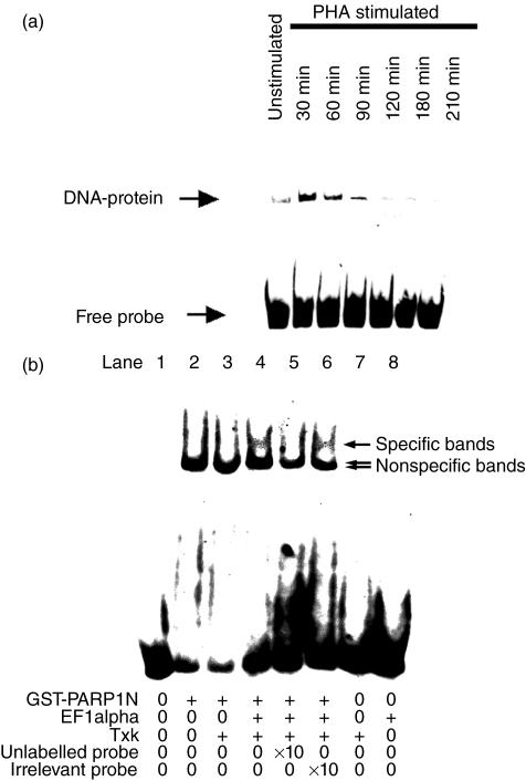
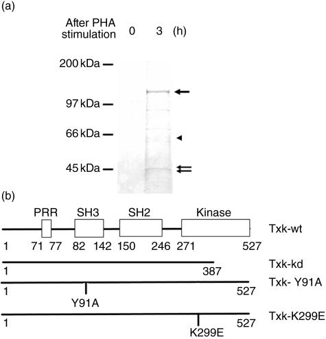
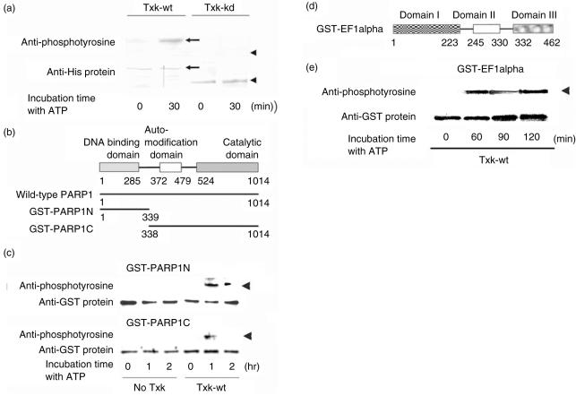
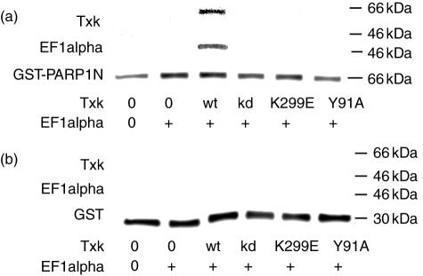
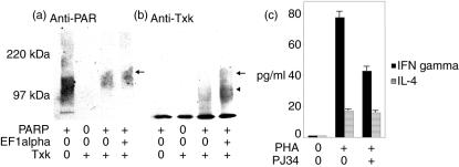
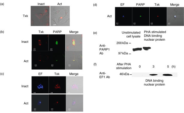
We have found previously that Txk, a member of the Tec family tyrosine kinases, is involved importantly in T helper 1 (Th1) cytokine production. However, how Txk regulates interferon (IFN)-gamma gene transcription in human T lymphocytes was not fully elucidated. In this study, we identified poly(ADP-ribose) polymerase 1 (PARP1) and elongation factor 1alpha (EF-1alpha) as Txk-associated molecules that bound to the Txk responsive element of the IFN-gamma gene promoter. Txk phosphorylated EF-1alpha and PARP1 formed a complex with them, and bound to the IFN-gamma gene promoter in vitro. In particular, the N terminal region containing the DNA binding domain of PARP1 was important for the trimolecular complex formation involving Txk, EF-1alpha and PARP1. Several mutant Txk which lacked kinase activity were unable to form the trimolecular complex. A PARP1 inhibitor, PJ34, suppressed IFN-gamma but not interleukin (IL)-4 production by normal peripheral blood lymphocytes (PBL). Multi-colour confocal analysis revealed that Txk and EF-1alpha located in the cytoplasm in the resting condition. Upon activation, a complex involving Txk, EF-1alpha and PARP1 was formed and was located in the nucleus. Collectively, Txk in combination with EF-1alpha and PARP1 bound to the IFN-gamma gene promoter, and exerted transcriptional activity on the IFN-gamma gene.


Figures

Fig. 5
Fig. 5
Binding activity of the trimolecular complex consisting of Txk, poly(ADP-ribose) polymerase 1 (PARP1) and elongation factor 1α (EF-1α) to interferon (IFN)-γ promoter −53/−39 region. IFN-γ promoter region (core region; −53 to −39) was labelled with digoxigenin (DIG). (a) Nuclear proteins of the PHA stimulated Jurkat cells were collected at the indicated time and were incubated with DIG-labelled oligoDNA in the presence of poly(dI-dC)(dI-dC). Binding of protein complex to IFNγ promoter region was detected after PHA stimulation transiently. (b) We next performed a gel shift assay to determine whether the trimolecular complex involving Txk bound to the IFN-γ promoter region. A mixture of 50 ng GST-PARP1N, 25 ng Txk-wt and 25 ng EF-1α was incubated with the DIG-labelled oligoDNA in the presence of 1 µg poly(dI-dC)(dI-dC). To confirm binding specificity, the protein mixture was incubated with the DIG-labelled probe in the presence of a 10-fold molar excess of unlabelled relevant oligoDNA (−53/−39 probe) (lane 5) and unlabelled Oct-2 competitor oligoDNA (lane 6). The DNA–protein complex was disappeared specifically by introduction of the relevant oligoDNA indicating the specificity of the binding (arrow, specific binding of trimolecular complex to the promoter DNA). Two arrows (below the specific bands) indicate non-specific binding of glutathione-S-transferase (GST)–PARP1N to the probe, which appeared in the presence of GST–PARP1N.
Fig. 1
Fig. 1
Identification of T cell nuclear proteins that bind to interferon (IFN)-γ promoter (−53/−39) region together with Txk. Jurkat cells were transfected with pME18S-Txk-wt (wild-type Txk) expression vector, and were cultured for 48 h. Thereafter, the cells were activated with phytohaemagglutinin (PHA) or kept unstimulated. IFN-γ promoter region (core region, −53/−39; actual synthetic oligo DNA, −56/−36) to which Txk bound was digoxigenin (DIG)-labelled, and was incubated with nuclear proteins of the Txk transfected Jurkat cells in the presence of 5 µg/reaction poly(dI-dC)(dI-dC). The DNA-binding nuclear proteins were recovered by using anti-DIG antibody and appropriate-Dynabeads, followed by magnetic separation. As control DNA, calf thymus DNA was sonicated, approximately 50 base pairs (bp) DNA fragment were recovered by glass beads and treated similarly. The DNA–protein complexes were washed extensively and were loaded onto sodium dodecyl sulphate–polyacrylamide gel electrophoresis (SDS-PAGE). The results shown are representative of three independent experiments with essentially the same result. (a) IFN-γ promoter (−53/−39) region oligoDNA binding proteins were detected by silver staining. A 50-kDa (two arrows) and an 110-kDa protein (one arrow) bound to the IFN-γ promoter region oligoDNA were reproducibly detected. An arrowhead indicates Txk protein. The 110 kDa and 50 kDa proteins were electrotransferred and their sequences determined by proteinase digestion and subsequent high performance liquid chromatography (HPLC) analysis. The peptide sequences of the 110 kDa protein were NREELGFRPEYS and IFPPETSASVAA, indicating human PARP1. Those of the 50 kDa protein were YYVTIIDAPGHR and HINIVVIGHVD, indicating EF-1α. (b) Schematic representation of the domain structure of Txk and its mutants. Wild-type Txk (Txk-wt; amino acids, 1–527; His-Txk-wt ∼63 kDa) contains proline-rich region (PRR), Src homology region 3 (SH3), SH2 and kinase domain. Kinase domain-deleted mutant Txk (Txk-kd; amino acids, 1–387; His-Txk-kDa ∼46 kDa) lacks a vast majority of kinase domain. Txk-Y91A was a point mutant whose autophosphorylation site Y residue was substituted with A. Txk-K299E was a point mutant Txk whose ATP binding K residue was substituted with E.
Fig. 2
Fig. 2
Phosphorylation of poly(ADP-ribose) polymerase 1 (PARP1) and elongation factor 1α (EF-1α) by wild-type Txk. (a) Kinase activity of Txk-wt and kinase domain deleted mutant Txk (Txk-kd) detected by autophosphorylation. Txk-wt and Txk-kd were incubated with adenosine triphosphate (ATP) for 30 min, and were then analysed with immunoblotting. The upper panel was anti-phosphotyrosine (PY20) immunoblotting. The membrane was re-probed with anti-His protein antibody (the lower panel), indicating amounts of Txk in the reactions. In the presence of exogenous ATP Txk-wt phosphorylated itself (∼63 kDa) by its own kinase activity, confirming that Txk-wt has kinase activity (arrow). Txk-kd (∼43 kDa) incubated with ATP for 30 min did not phosphorylate itself, indicating that Txk-kd lacked kinase activity (arrowhead). (b) Schematic representation of the domain structure of PARP1 and its deletion mutants. PARP1 includes the DNA binding domain (amino acids, 1–339) and the automodification domain and catalytic domain (amino acids, 338–1014). Wild-type PARP1 (PARP1-wt; ∼113 kDa), glutathione-S-transferase (GST)-tagged N terminus region of PARP1 (GST–PARP1N; amino acids, 1–339; ∼64 kDa) and GST-tagged C terminus region of GST (GST–PARP1C; amino acids, 338–1014; ∼101 kDa) were produced by Escherichia coli. (c) Phosphorylation of PARP1 by wild-type Txk. GST–PARP1N and Txk-wt were coincubated with ATP for 0–1−2 h. GST-tagged protein was recovered by the addition of glutathione beads, and was analysed with anti-phosphotyrosine immunoblotting, followed by anti-GST protein immunoblotting. In the presence of ATP, Txk-wt phosphorylated GST–PARP1N (arrowhead). Similarly, phosphorylation of GST–PARP1C was evident by co-incubation with Txk for 1 h (arrowhead). (d) Schematic representation of the domain structure of EF-1α. EF-1α includes domains 1, 2 and 3. We prepared both His-tagged wild-type EF-1α (∼51 kDa) and GST-tagged wild-type EF-1α (∼76 kDa) by culturing E. coli. (e) Phosphorylation of EF-1α by Txk-wt. GST-EF-1α was incubated with Txk-wt in the presence of ATP for 0–120 min, and glutathione-binding proteins were recovered. Arrowhead indicates phosphorylated GST-EF-1α.
Fig. 3
Fig. 3
Phosphorylation dependent trimolecular complex formation of poly(ADP-ribose) polymerase 1 (PARP1), elongation factor 1α (EF-1α) and Txk in vitro. (a) Phosphorylation-dependent interaction of unlabelled full-length PARP1 with Txk. Full-length wild-type PARP1 was incubated with Txk-wt and Txk-kd in the presence of adenosine triphosphate (ATP). Txk-wt and Txk-kd, both of which were tagged with poly Histidine, were precipitated by the addition of Ni-NTA resin. After extensive washing, the binding proteins to the beads were analysed by immunoblotting with anti-Txk antibody. The membrane was next analysed with anti-PARP1 antibody. Lower panel indicates amounts of Txk-wt and Txk-kd proteins detected by anti-Txk antibody. Txk-wt co-precipitated PARP1 in the presence of adenosine triphosphate (ATP). Txk-kd did not precipitate PARP1. In the absence of ATP, no binding protein was detected, thus, the results were omitted. (b) Trimolecular complex formation including Txk-wt, EF-1α and PARP1 in the presence of ATP. Txk-wt and EF-1α were incubated with glutathione-S-transferase (GST)–PARP1N (a), GST–PARP1C (b) and GST (c) for 60 min in the presence of ATP. The binding proteins were recovered by the addition of glutathione-sepharose beads. ‘Anti-GST protein’ indicates amounts of GST-tagged protein. EF-1α and Txk-wt that had co-precipitated with GST-tagged protein were detected by anti-EF-1α antibody (upper panel) and anti-Txk (middle panel) antibody, respectively. (i) GST–PARP1N co-precipitated Txk-wt when the two proteins were co-incubated. Furthermore, GST–PARP1N co-precipitated Txk-wt and EF-1α when the three proteins were co-incubated. (ii) GST–PARP1C bound to Txk-wt when the two proteins were co-incubated. (iii) GST protein (∼26 kDa) did not bind either protein. Txk-wt formed the trimolecular complex involving GST–PARP1N and EF-1α, suggesting that Txk bound to EF-1α together with PARP1N (which includes DNA binding domain). (c) To further confirm phosphorylation dependency of the trimolecular complex formation, a mixture of Txk-wt, GST–PARP1N (two arrows) or GST (two arrowheads) and EF-1α was incubated with or without ATP for 60 min, and glutathione-sepharose beads was applied to the mixture. The bead-binding proteins were analysed similarly. EF-1α and Txk-wt bound to PARP1N only in the presence of ATP, suggesting that trimolecular complex formation was phosphorylation-dependent.
Fig. 4
Fig. 4
Phosphorylation dependency of trimolecular complex formation of Txk with poly(ADP-ribose) polymerase 1 (PARP1) and elongation factor 1α (EF-1α). (a) To further confirm phosphorylation dependency of the trimolecular complex formation, a mixture of glutathione-S-transferase (GST)–PARP1N and EF-1α was incubated with Txk-wt or its mutants for 60 min, and glutathione-sepharose beads were applied to the mixture. The binding proteins were analysed similarly. Anti-GST immunoblotting indicated the amounts of GST–PARP1N in the reactions. Anti-Txk and anti-EF-1α indicated the co-precipitated Txk and EF-1α, respectively. EF-1α and Txk-wt bound to PARP1N only when Txk had kinase activity. Mutant Txk which lacked kinase activity did not form a trimolecular complex. (b) As a control protein, GST was analysed similarly.
Fig. 6
Fig. 6
Poly(ADP-ribosyl)ation of Txk by poly(ADP-ribose) polymerase 1 (PARP1). (a,b) In vitro poly(ADP-ribosyl)ation assay was carried out. His-tagged Txk was incubated with or without elongation factor 1α (EF-1α) in the presence of catalytically active full-length PARP1. Thereafter, Txk was recovered as Ni-NTA resin-binding proteins. The precipitated proteins were analysed with immunoblotting. (a) Immunoblotting with anti-poly(ADP-ribose) antibody (anti-PAR) to detect poly(ADP-ribosyl)ated proteins. Arrow indicates the heavily poly(ADP-ribosyl)ated protein, appearing in the presence of Txk, EF-1α and PARP1. (b) The same membrane was re-probed with anti-Txk antibody. Arrow indicates that the precipitated Txk was poly(ADP-ribosyl)ated by PARP1. Arrowhead indicates Txk with less and/or without poly(ADP-ribosyl)ation. (c) Effects of PARP1 inhibitor on the cytokine production of human peripheral blood lymphocytes (PBL). Culture supernatants of phytohaemagglutinin (PHA)-stimulated PBL in the presence/absence of 4000 nM PJ34 were assessed for cytokine production. PJ34 treatment of normal PBL reduced interferon (IFN)-γ production of PBL specifically. Interleukin (IL)-4 production was affected marginally by the PARP1 inhibitor.
Fig. 7
Fig. 7
Multi-colour confocal analysis of molecular complex formation upon activation of Txk, poly(ADP-ribose) polymerase 1 (PARP1) and elongation factor 1α (EF-1α). Cos7 cells were electrotransfected with the combinations of Txk-DsRed monomer, PARP1–GFP and EF-1α-CFP. To achieve activation and phosphorylation of the Txk protein, active Fyn and, as a negative control, inactive Fyn, were transfected simultaneously, and 24 h later the Cos7 cells were analysed. (a) DsRed-Txk was transfected with active and inactive Fyn. DsRed-labelled Txk was present in cytoplasm in the inactivated condition, and the Txk accumulated in the nucleus when active Fyn was co-transfected. (b) GFP-labelled PARP1 and DsRed-Txk were transfected. GFP-labelled PARP1 was located in the nucleus constitutively. In the activated condition, PARP and Txk co-localized in the nucleus. (c) CFP-labelled EF-1α and DsRed-Txk were transfected. A vast majority of CFP-labelled EF-1α and Txk were located in the cytoplasm in the inactive condition. Some of the EF-1α and Txk translocated into nucleus upon activation. EF-1α and Txk co-localized in the nucleus when activated. (d) When CFP-labelled EF-1α, GFP-labelled PARP1 and Ds-Red-labelled Txk were co-transfected with inactive Fyn, Txk and EF-1α located in the cytoplasm, whereas PARP1 located in the nucleus. When active Fyn was used, co-localization of the three molecules was detected in the nucleus. (e, f) Immunoblotting analysis of the IFN-γ promoter (−53/−39) oligoDNA-binding proteins of human T cells. To confirm that Txk binding proteins were PARP1 and EF-1α in the human T cells, Jurkat cells were stimulated with phytohaemagglutinin (PHA) and the nuclear proteins were recovered. The nuclear proteins were allowed to bind to the Txk binding element, and the binding proteins were purified and were analysed using the immunoblotting method. The blots were probed with anti-PARP1 antibody and anti-EF-1α antibody. (e) Nuclear proteins of the PHA-stimulated Jurkat cells for 3 h were probed with the DNA and the binding proteins were analysed. As a control, whole-cell lysate of unstimulated Jurkat cells was included. We confirmed that the 110 kDa protein in the nuclear protein complex from Jurkat cells was PARP1 protein. (f) Nuclear proteins of Jurkat cells with or without PHA stimulation were analysed similarly. The 50 kDa protein in the nuclear protein complex from Jurkat cells were EF-1α.

Similar articles

Cited by

References

    1. Szabo SJ, Kim ST, Costa GL, Zhang X, Fathman CG, Glimcher LH. A novel transcription factor, T-bet, directs Th1 lineage commitment. Cell. 2000;100:655–69. - PubMed
    1. Mullen AC, Hutchins AS, High FA, et al. Hlx is induced by and genetically interacts with T-bet to promote heritable T(H)1 gene induction. Nat Immunol. 2002;3:652–8. - PubMed
    1. Zheng W, Flavell RA. The transcription factor GATA-3 is necessary and sufficient for Th2 cytokine gene expression in CD4 T cells. Cell. 1997;89:587–96. - PubMed
    1. Messi M, Giacchetto I, Nagata K, Lanzavecchia A, Natoli G, Sallusto F. Memory and flexibility of cytokine gene expression as separable properties of human T(H)1 and T(H)2 lymphocytes. Nat Immunol. 2003;4:78–86. - PubMed
    1. Komine O, Hayashi K, Natsume W, et al. The Runx1 transcription factor inhibits the differentiation of naive CD4+ T cells into the Th2 lineage by repressing GATA3 expression. J Exp Med. 2003;198:51–61. - PMC - PubMed

MeSH terms

Substances