Comparative Study
. 2006 Dec 13;26(50):13089-101.
doi: 10.1523/JNEUROSCI.2855-06.2006.

Genetic analysis of Mint/X11 proteins: essential presynaptic functions of a neuronal adaptor protein family

Affiliations
Comparative Study

Genetic analysis of Mint/X11 proteins: essential presynaptic functions of a neuronal adaptor protein family

Angela Ho et al. J Neurosci. .

Abstract

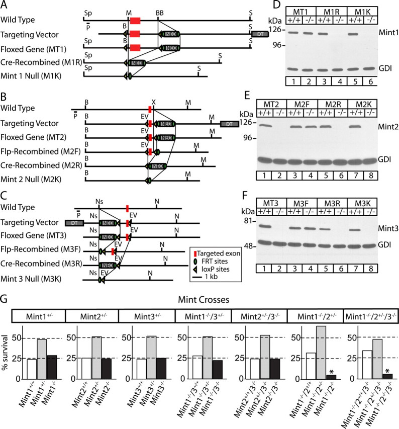
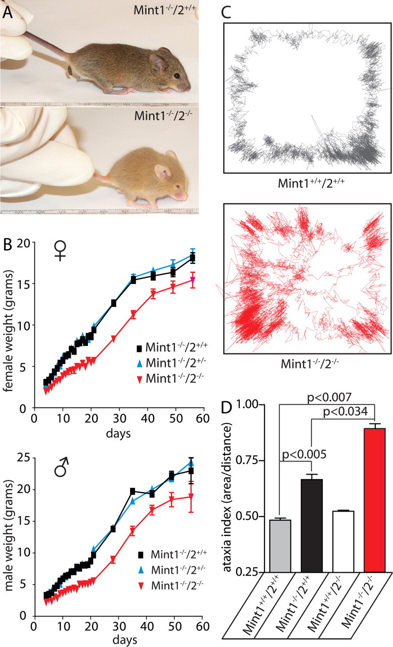
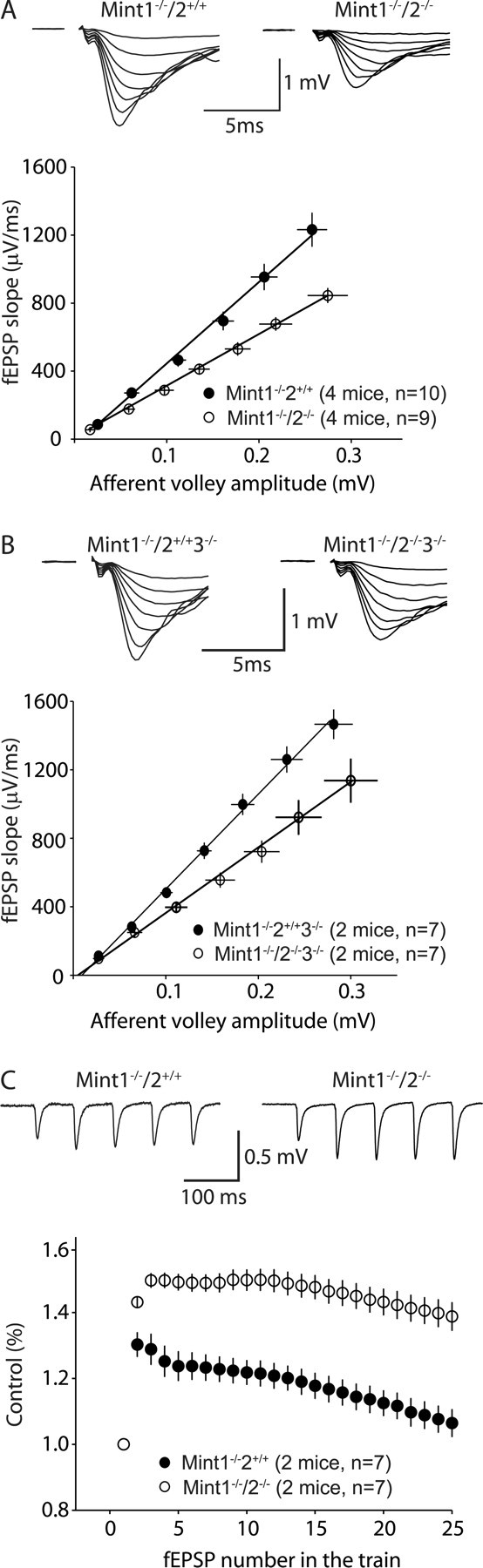
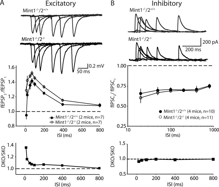
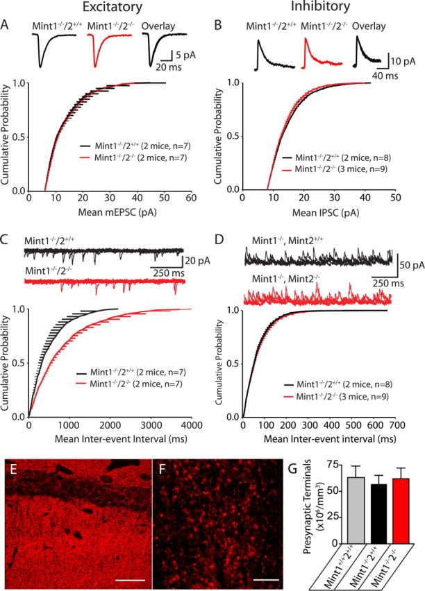
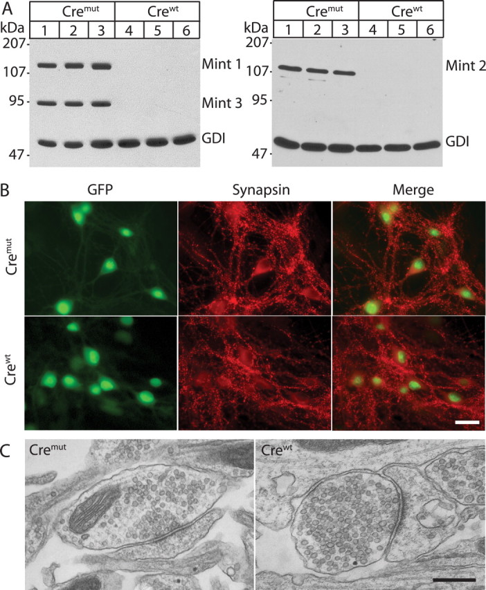
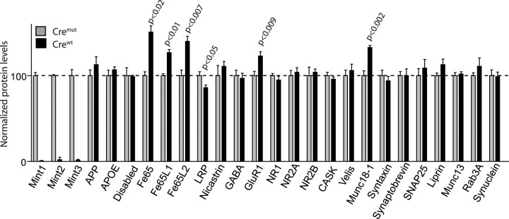
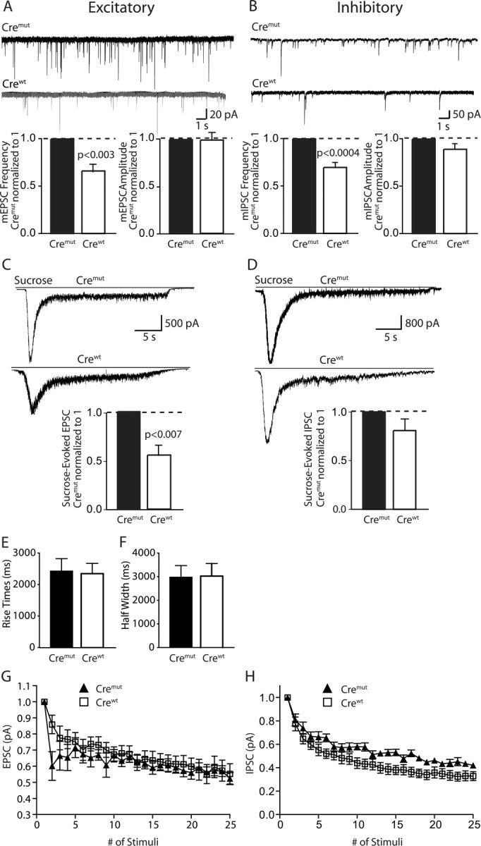
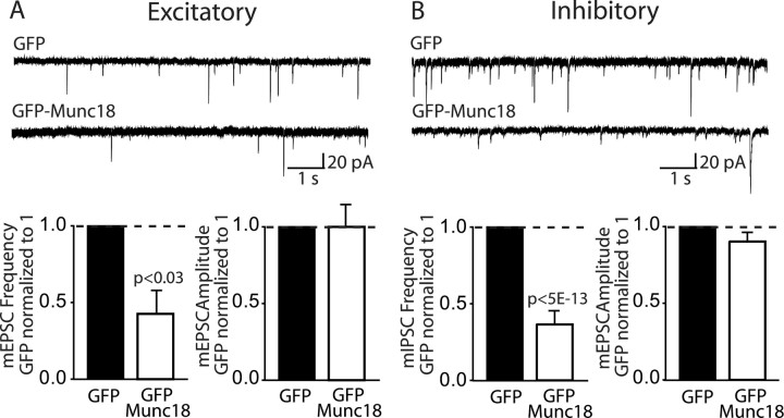
Mints/X11s are adaptor proteins composed of three isoforms: neuron-specific Mints 1 and 2, and the ubiquitously expressed Mint 3. We have now analyzed constitutive and conditional knock-out mice for all three Mints/X11s. We found that approximately 80% of mice lacking both neuron-specific Mint isoforms (Mints 1 and 2) die at birth, whereas mice lacking any other combination of Mint isoforms survive normally. The approximately 20% surviving Mint 1/2 double knock-out mice exhibit a decrease in weight and deficits in motor behaviors. Hippocampal slice electrophysiology uncovered a decline in spontaneous neurotransmitter release, lowered synaptic strength, and enhanced paired-pulse facilitation in Mint-deficient mice, suggesting a decreased presynaptic release probability. Acute ablation of Mint expression in cultured neurons from conditional Mint 1/2/3 triple knock-in mice also revealed a decline in spontaneous release, confirming that deletion of Mints impair presynaptic function. Quantitation of synaptic proteins showed that acute deletion of Mints caused a selective increase in Munc18-1 and Fe65 proteins, and overexpression of Munc18-1 in wild-type neurons also produced a decrease in spontaneous release, suggesting that the interaction of Mints with Munc18-1 may contribute to the presynaptic phenotype observed in Mint-deficient mice. Our studies thus indicate that Mints are important regulators of presynaptic neurotransmitter release that are essential for mouse survival.


Figures

Figure 1.
Figure 1.
Generation and survival of Mint 1, 2, and 3 KO mice. AC, Homologous recombination strategy for Mint 1, 2, and 3, respectively. The targeted exon (red box) was flanked by loxP sites (yellow arrowheads) to allow conditional removal of exon, a NEO for positive selection surrounded by FRT sites (green circles), and a diphtheria toxin (DT) was attached for negative selection. DF, Immunoblot analysis of brain proteins from wild-type (+/+) and homozygous (−/−) mice. AD, Homozygous floxed Mint 1 mutants (MT1) showed normal Mint 1 protein expression in the presence of NEO resistance cassette but were abolished when we crossed these mice with transgenic mice that express cre recombinase in the male germline (M1R). B, C, E, F, For homozygous floxed Mint 2 and 3 mutants (MT2 and MT3, respectively), the presence of the NEO resistance cassette abolished protein expression before floxed exon was excised. However, when NEO was excised by crossing with mice expressing FRT recombinase in the germline, both Mints 2 and 3 expressions returned as expected (M2F, M3F, respectively). In addition, Mints 2 and 3 protein expression were abolished when exon was removed by crossing with mice that express cre recombinase (M2R, M3R, respectively). In addition, we have created Mint KO lines in which protein expression was abolished with excision of both the targeted exon and NEO cassette (M1K, M2K, and M3K). G, Genotype analysis of offspring obtained from single Mint 1, 2, or 3 heterozygous matings show normal Mendelian ratio of genotypes. Genotype analysis of offspring obtained from Mint 1−/−/3+/− or Mint 2+/−/3−/− matings revealed normal Mendelian ratio of genotypes. However, interbreeding of Mint 1−/−/2+/− animals resulted in a Mendelian ratio of 5% instead of the expected 25% in double Mint 1/2 offsprings. Triple KO of all three Mints showed a Mendelian ratio similar to Mint 1/2 double KO mice. Sp, Spe; P, probe; M, Mfe1; BB, BstB1; B, BamH1; S, Sal1; EV, EcoRV; X, Xho1; Ns, Nsi1; N, Not1.
Figure 2.
Figure 2.
Histological examination of newborn Mint mutant mice. A, B, Mint 1/2 double KO mice revealed normal brain development and showed no obvious palate abnormalities by hematoxylin and eosin (H&E) stain compared with Mint 1 single KO littermates. C, Nissl-stained brain sections showed normal development and formation of the cortex, caudate–putamen, thalamus, and hippocampus in Mint 1 single and Mint 1/2 double KO mice. D, Single deletion of Mint 1 stained with Mint 2 revealed normal distribution of expression in CA3 region of the hippocampus, and as expected, all Mint 2 staining was abolished in Mint 1/2 double KO mice. E, Immunostaining of APP showed normal and distinct localization at the CA3 region of the hippocampus in Mint 1 KO mice, and this was indistinguishable in Mint 1/2 double KOs. OE, Olfactory epithelium; B, brain; P, palate; T, tongue; CTX, cerebral cortex; CPu, caudate–putamen; TH, thalamus; HC, hippocampus; CC, central canal; CB, cerebellum; H, hilus. Scale bars: B, 150 μm; (in E) D, E, 50 nm.
Figure 3.
Figure 3.
Overall brain structure and morphology of adult Mint KOs. A, Nissl brain sections of Mint 1−/−/2+/+ (left) and Mint 1−/−/2−/− double KO mice (right) at 6–8 weeks of age showed normal brain architecture. CTX, Cerebral cortex; CPu, caudate–putamen; TH, thalamus; OB, olfactory bulb; HC, hippocampus; CB, cerebellum. B, C, Mint 2 immunostaining was abolished in Mint 1−/−/2−/− double KO mice (right) as expected in the hippocampus (B) and cerebellum (C) compared with Mint 1−/−/2+/+ littermate control mice. DG, Dentate gyrus; ML, molecular layer; PL, Purkinje cell layer; GCL, granule cell layer. D, E, Immunostaining for APP (D) and Rab3A (E) displayed normal and distinct localization in the hippocampus in Mint 1−/−/2+/+ and Mint 1−/−/2−/− double KO littermate mice. Scale bar: (in E) BE, 100 μm.
Figure 4.
Figure 4.
Abnormal weight and motor behavior of adult Mint 1/2 double KO mice. A, Photograph of Mint 1 single KO and Mint 1/2 double KO mice at 21 d of age. B, Plot of average weight of wild-type, heterozygous, and homozygous Mint 2 KO mice in a Mint 1 KO background as a function of age for littermate female (♀) and male mice (♂). Data shown represent means ± SEMs. C, Locomotor sample traces of wild-type (Mint 1+/+/2+/+; gray) and Mint 1−/−/2−/− double KO mice (red), which display the position of the mouse as it moves within the force plate as represented by the box within 6 min. D, Force plate analysis revealed a significant increase in ataxia indices (area covered in 6 min/net distance traveled) of Mint 1/2 double KO mice compared with wild type. Single Mint 1 KO mice exhibited a slight increase, whereas single Mint 2 KO mice displayed normal locomotive movements. Data shown represent means ± SEMs.
Figure 5.
Figure 5.
Synaptic transmission deficits at Schaffer collateral/CA1 pyramidal cell synapses of Mint 1/2 double KO mice. A, Synaptic strength analysis using input–output measurements. The fEPSP slope is plotted as a function of the afferent volley amplitude (in millivolts). Linear fit slopes are significantly different between Mint 1−/−/2−/− double KO mice, which displayed a decrease in the magnitude of synaptic responses compared with Mint 1−/−/2+/+ single KO littermate controls. B, Input–output measurements illustrating Mint 1−/−2−/−3−/− triple KO mice showed a similar decrease in the magnitude of synaptic responses compared with Mint 1−/−2−/− double KO mice shown in A. C, Response to 14 Hz stimulus trains. Control synaptic responses (Mint 1−/−/2+/+) exhibited initial facilitation that slowly decreased, in contrast to Mint 1−/−/2−/− double KO mice, which displayed extended facilitation.
Figure 6.
Figure 6.
Excitatory and inhibitory synaptic responses to paired-pulse stimulation of Mint 1/2 double KO mice. A, Mint 1−/−/2−/− double KO mice display an enhanced increase in paired-pulse facilitation compared with Mint 1−/−/2+/+ single KO mice. A, Bottom, Ratio of the paired-pulse facilitation values between Mint 1−/−/2−/− to Mint 1−/−/2+/+ KO mice at excitatory synapses. B, Paired-pulse depression of inhibitory synaptic currents did not exhibit any difference between Mint 1−/−/2+/+ single and Mint 1−/−/2−/− double KO mice. B, Bottom, Ratio of paired-pulse responses at inhibitory synapses between Mint 1−/−/2−/− to Mint 1−/−/2+/+ KO mice. ISI, Interstimulus interval.
Figure 7.
Figure 7.
Deletion of Mints 1 and 2 decreases spontaneous release at excitatory, but not inhibitory synapses onto CA1 pyramidal neurons. AB, Cumulative probability plot of mEPSC (A) and mIPSC (B) amplitudes for Mint 1−/−/2+/+ and Mint 1−/−/2−/− KO neurons did not show any significant differences between the genotypes. C, Meanwhile, there was a statistically significant decline in the cumulative probability plot of mEPSC frequency in Mint 1−/−/2−/− double KO compared with Mint 1−/−/2+/+ mice (*p = 0.0024). D, No changes were detected in the cumulative probability plot of mIPSC frequency in Mint 1−/−/2−/− double KO compare with Mint 1−/−/2+/+ mice. E, F, Representative confocal image of the CA1 region of the hippocampus immunostained for synaptophysin. Scale bars: E, 47.62 μm; F, 9.52 μm. G, Quantitative assessment of presynaptic terminals in the CA1 subfields of the hippocampus (n = 3 mice per group and genotype).
Figure 8.
Figure 8.
Acute deletion of all three Mint proteins in neuronal cultures infected with lentiviral cre recombinase (crewt) or cremut (vector carrying deletion of cre recombinase). Neurons were cultured from Mint triple-floxed newborn pups and infected with either lentiviral crewt or cremut at 4 DIV for 48 h. Lysates were collected at 14 DIV and subjected for SDS-PAGE and immunoblot for all Mint 1, 2, and 3 with GDP dissociation inhibitor (GDI), a ubiquitously expressed protein used as internal loading control. A, Neuronal cultures infected with cremut virus showed normal expression of all three Mint proteins, whereas lentiviral infection with crewt efficiently deleted all three Mint protein expressions. B, Neuronal cultures transfected with lentiviral cremut or crewt displayed normal synaptic staining with the synaptic marker synapsin. Both lentiviral vectors contain nuclear localization signal, and a GFP tag that labels cell nuclei of infected neurons exclusively. C, Synaptic structure of neuronal cultures transfected with lentiviral cremut and crewt showed normal presynaptic nerve terminal clustered with synaptic vesicles at the active zone with a filamentous postsynaptic density. Scale bars: B, 30 μm; C, 400 nm.
Figure 9.
Figure 9.
Effect of Mint-deficient neurons on protein level expression. Levels of selected brain proteins in Mint-deficient neurons (crewt; black bars) and control (cremut; gray bars) at 13–15 DIV. As expected, we efficiently abolished all three Mint expressions in neurons that were treated with crewt compared with cremut control neurons. We found a selective increase in Fe65 family of adaptor proteins (Fe65, Fe65L1, and Fe65L2), postsynaptic receptor GluR1, and synaptic protein Munc18-1. Meanwhile, a small significant decrease in LRP protein was observed in Mint-deficient neurons. APOE, Apolipoprotein E; NR1, NMDA receptor subunit 1; SNAP25, soluble N-ethylmaleimide-sensitive factor attached protein 25.
Figure 10.
Figure 10.
Altered synaptic responses in Mint-deficient neurons. A, B, Sample traces showing miniature excitatory and inhibitory recordings of cremut and crewt at 13–15 DIV. Bar graphs of the quantification revealed a significant decrease in miniature frequency but not amplitude in neurons transfected with crewt that abolished all three Mint proteins. C, D, Sample traces showing sucrose-evoked excitatory and inhibitory recordings of cremut and crewt at 13–15 DIV. Bar graphs showed a significant decrease in sucrose-evoked response in excitatory but not inhibitory synapses of Mint deficient neurons. Data shown represent absolute mean EPSP and IPSP amplitudes. E, F, Plots of 10–90% rise times and half-width showed no changes. G, H, Traces of synaptic depression responses recorded from excitatory and inhibitory neurons, respectively, during a 10 Hz stimulation.
Figure 11.
Figure 11.
Munc18-1 overexpression decreases spontaneous miniature frequency in excitatory and inhibitory synapses. A, B, Sample traces showing miniature excitatory and inhibitory recordings of GFP and GFP–Munc18 at 13–15 DIV. Bar graphs showed a significant decrease in miniature frequency but not amplitude in both excitatory and inhibitory synapses in Munc18-1 overexpressed neurons compared with GFP alone.

Similar articles

Cited by

References

    1. Biederer T, Südhof TC. Mints as adaptors: direct binding to neurexins and recruitment of munc18. J Biol Chem. 2000;275:39803–39806. - PubMed
    1. Biederer T, Cao X, Südhof TC, Liu X. Regulation of APP-dependent transcription complexes by Mint/X11s: Differential functions of mint isoforms. J Neurosci. 2002;22:7340–7351. - PMC - PubMed
    1. Borg JP, Ooi J, Levy E, Margolis B. The phosphotyrosine interaction domains of X11 and FE65 bind to distinct sites on the YENPTY motif of amyloid precursor protein. Mol Cell Biol. 1996;16:6229–6241. - PMC - PubMed
    1. Borg JP, Straight SW, Kaech SM, de Taddeo-Borg M, Kroon DE, Karnak D, Turner RS, Kim SK, Margolis B. Identification of an evolutionarily conserved heterotrimeric protein complex involved in protein targeting. J Biol Chem. 1998;273:31633–31636. - PubMed
    1. Borg JP, Lopez-Figueroa MO, de Taddeo-Borg M, Kroon DE, Turner RS, Watson SJ, Margolis B. Molecular analysis of the X11-mLin-2/CASK complex in brain. J Neurosci. 1999;19:1307–1316. - PMC - PubMed

Publication types

MeSH terms