Comparative Study
. 2006 Dec 13;26(50):12904-13.
doi: 10.1523/JNEUROSCI.2531-06.2006.

Suppression of microglial inflammatory activity by myelin phagocytosis: role of p47-PHOX-mediated generation of reactive oxygen species

Affiliations
Comparative Study

Suppression of microglial inflammatory activity by myelin phagocytosis: role of p47-PHOX-mediated generation of reactive oxygen species

Yang Liu et al. J Neurosci. .

Abstract

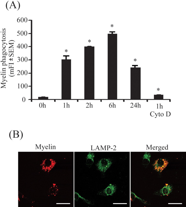
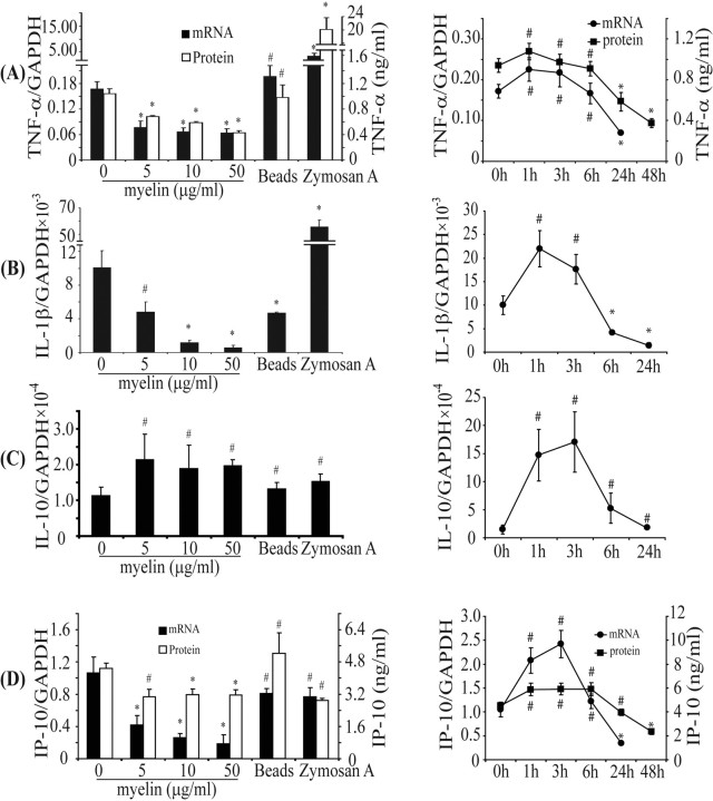
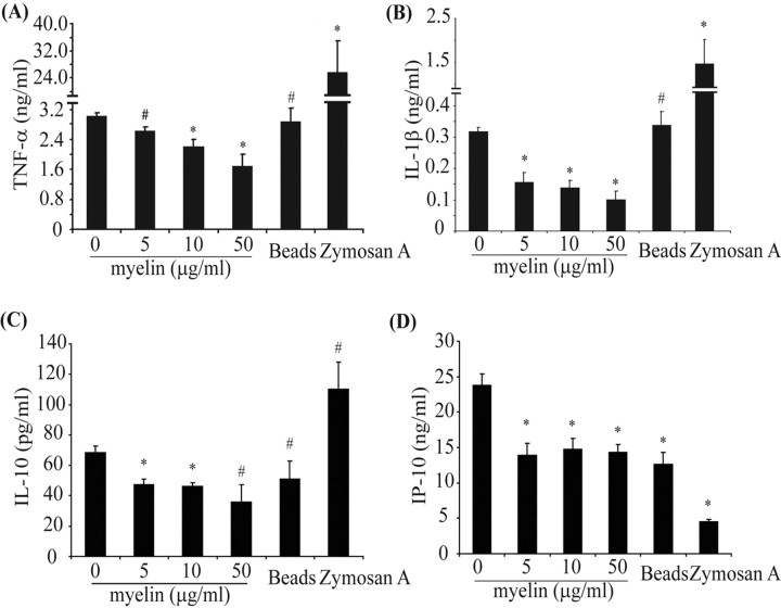
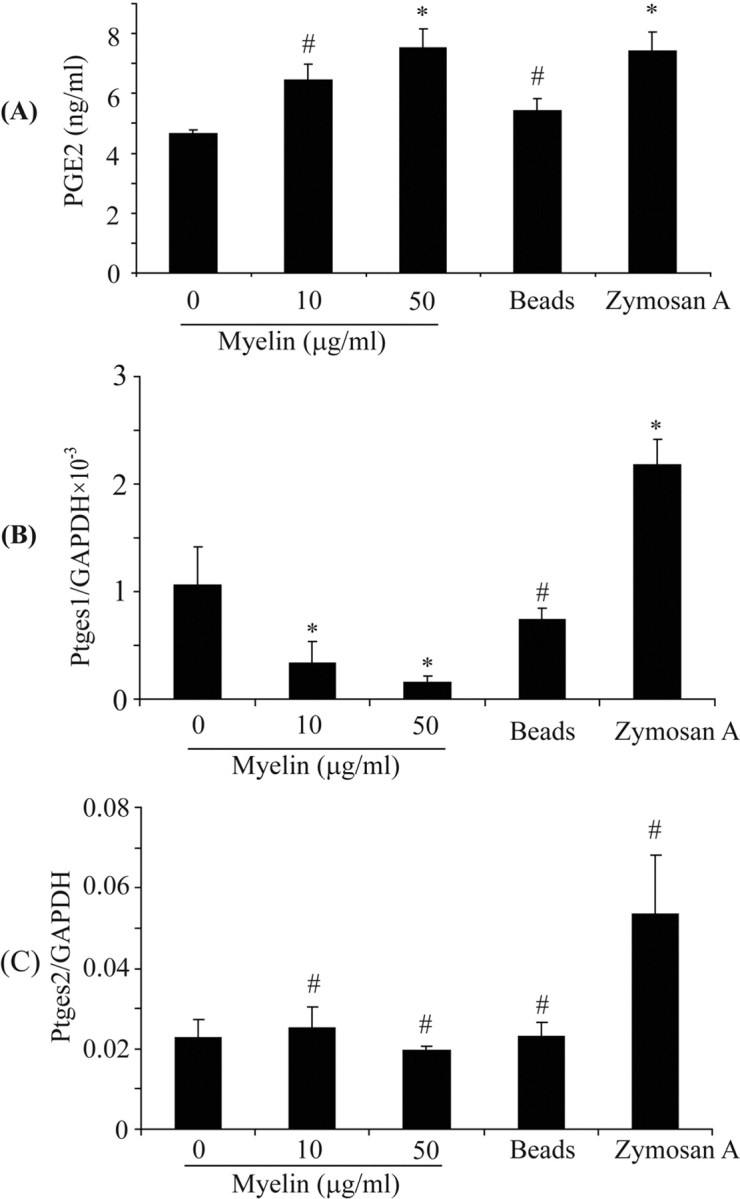
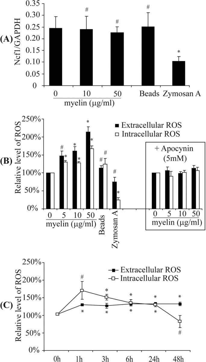
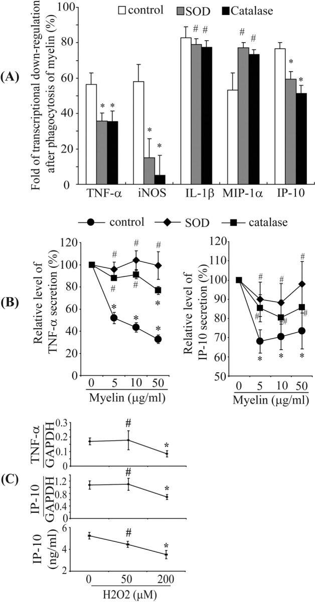
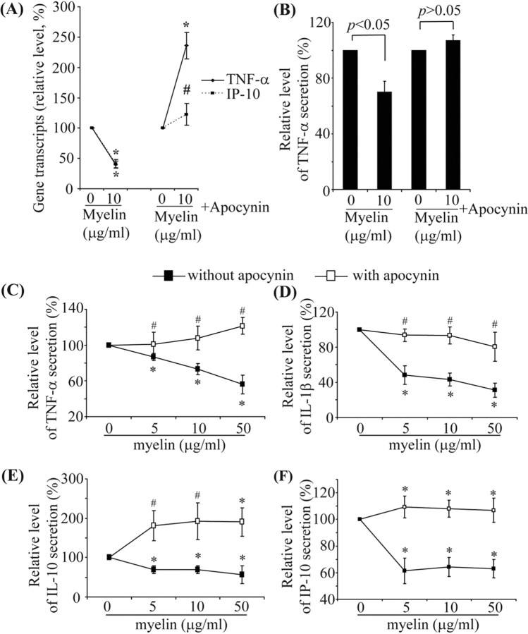
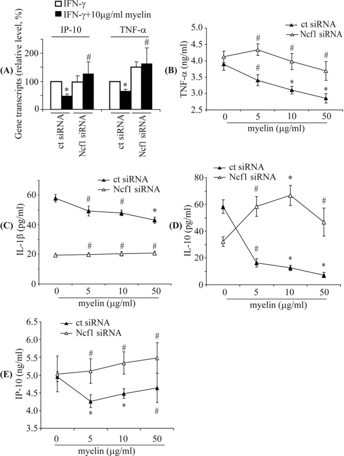
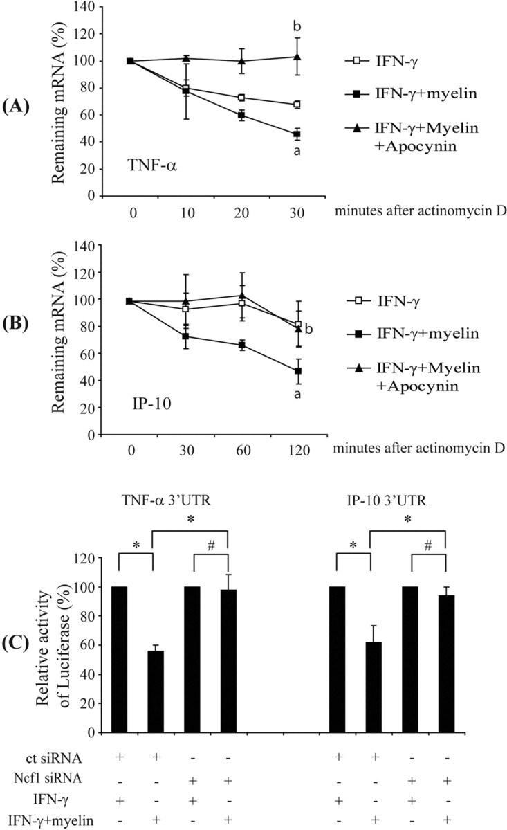
Multiple sclerosis (MS) is pathologically characterized by inflammatory demyelination and neuronal injury. Although phagocytosis of myelin debris by microglia and macrophages in acute MS lesions is well documented, its pathophysiological significance is unclear. Using real-time quantitative PCR, flow cytometry, ELISA, and reactive oxygen species (ROS) measurement assays, we demonstrated that phagocytosis of myelin modulates activation of microglial cells prestimulated by interferon-gamma (IFN-gamma) or a combination of IFN-gamma and lipopolysaccharide with a biphasic temporal pattern, i.e., enhanced production of proinflammatory mediators during the first phase (< or = 6 h), followed by suppression during the second (6-24 h) phase. In this second phase, myelin phagocytosis leads to an enhanced release of prostaglandin E2 and ROS in microglia, whereas the production of anti-inflammatory cytokines (particularly interleukin-10) remains unchanged. Suppression of inflammatory microglial activation by myelin phagocytosis was reversed by treatment with superoxide dismutase and catalase, by inhibition of the NADPH-oxidase complex, or by specific knockdown of the NADPH-oxidase-required adaptor p47-phagocyte oxidase (PHOX). Furthermore, we observed that myelin phagocytosis destabilized tumor necrosis factor-alpha and interferon-induced protein-10 mRNA through an adenine-uridine-rich elements-involved mechanism, which was reversed by blocking the function of NADPH-oxidase complex. We conclude that phagocytosis of myelin suppresses microglial inflammatory activities via enhancement of p47-PHOX-mediated ROS generation. These results suggest that intervention in ROS generation could represent a novel therapeutic strategy to reduce neuroinflammation in MS.


Figures

Figure 1.
Figure 1.
Microglia readily phagocytose myelin. Primary microglia were plated on 24-well plates and treated with Cy3-labeled myelin for 0, 1, 2, 6, and 24 h. A, The mean fluorescence intensity (mFI) of Cy3 in microglia was analyzed by flow cytometry. Microglial internalization reached its maximum ∼6 h after myelin administration. Myelin internalization was blocked by 5 μm cytochalasin D (Cyto D) as showed by mean fluorescence intensity of Cy3 after 1 h (all of the experiments were independently repeated 3 times with the same trends; one-way ANOVA vs that at zero incubation time point, *p < 0.05; n = 3 per group). Under confocal microscopy, myelin (shown in red) colocalized (shown in yellow) with LAMP-2, a specific marker of lysosomes (shown in green), within 2 h after myelin administration. Scale bars, 10 μm.
Figure 2.
Figure 2.
Myelin phagocytosis downregulates transcription and secretion of proinflammatory mediators in IFN-γ-activated microglia. Primary microglia were activated by IFN-γ and then treated in the presence of IFN-γ with (1) myelin at 0, 5, 10, and 50 μg/ml for 24 h, or (2) 10 μg/ml myelin for 0, 1, 3, 6, 24, and 48 h. Total RNA was isolated, and transcripts of TNF-α (A), IL-1β (B), IL-10 (C), and IP-10 (D) were measured using real-time quantitative PCR. Release of TNF-α and IP-10 in the culture medium was detected with ELISA. After 24 h, transcription of TNF-α (A), IL-1β (B), or IP-10 (D) and secretion of TNF-α (A) or IP-10 (D) were dose-dependently downregulated by myelin phagocytosis. In contrast, transcription of IL-10 (C) was slightly enhanced by myelin. After treatment with 10 μg/ml myelin, transcription of TNF-α (A), IL-1β (B), IL-10 (C), or IP-10 (D) and secretion of TNF-α (A) or IP-10 (D) were initially (≤6 h) upregulated and later (24 h and thereafter) downregulated. Beads and zymosan A were used as positive controls of phagocytosis. After 24 h, beads did not significantly affect inflammatory response except by downregulating IL-1β (B); zymosan A strongly increased production of TNF-α (A) and IL-1β (B) (all of the experiments were independently repeated at least 3 times with the same trends; one-way ANOVA vs IFN-γ-activated control, *p < 0.05, #p > 0.05; n = 3–10 per group).
Figure 3.
Figure 3.
Myelin phagocytosis decreases release of inflammatory mediators from IFN-γ- and LPS-activated microglia. Primary microglia preactivated by a combination of IFN-γ and LPS were treated with myelin at 0, 5, 10, and 50 μg/ml in the presence of preactivators for 24 h. AC, Release of cytokines into the supernatant was determined by ELISA. The release of TNF-α (A), IL-1β (B), and IL-10 (C) was downregulated by myelin in a dose-dependent manner. D, IP-10 secretion was significantly reduced but not in a dose-dependent manner (all of the experiments were independently repeated at least 3 times with the same trends; one-way ANOVA vs control activated by IFN-γ plus LPS, *p < 0.05, #p > 0.05; n = 3–8 per group).
Figure 4.
Figure 4.
Myelin phagocytosis induces production of PGE2. IFN-γ-preactivated primary microglia were treated with myelin at 0, 10, and 50 μg/ml in the presence of IFN-γ for 24 h. Release of PGE2 as well as gene transcription of Ptges1 and Ptges2 were determined by ELISA and real-time PCR, respectively. A, The release of PGE2 detected by ELISA was significantly enhanced in relation to the myelin concentrations. B, C, The transcription of Ptges1 was downregulated by myelin in a dose-dependent manner (B) and that of Ptges2 was not significantly changed (C) (all of the experiments were independently repeated at least 4 times with the same trends; one-way ANOVA vs IFN-γ-activated control, *p < 0.05, #p > 0.05; n = 4–5 per group).
Figure 5.
Figure 5.
Myelin phagocytosis induces production of reactive oxygen species. IFN-γ-preactivated primary microglia were treated in the presence of IFN-γ with myelin at 0, 5, 10, and 50 μg/ml for 24 h or with 10 μg/ml myelin for 0, 1, 3, 6, 24, and 48 h. A, After 24 h, the transcription of Ncf1, encoding p47–PHOX, the main adaptor of NADPH–oxidase complex, was not changed. B, The extracellular and intracellular ROS were significantly enhanced relative to the concentration of myelin used as determined by measuring SOD-inhibitible reduction of WST-1 and luminol chemiluminescence assay, respectively. Apocynin at 5 mm was able to block the production of ROS caused by treatment of myelin. C, Production of both extracellular and intracellular ROS caused by phagocytosis of 10 μg/ml myelin was maintained at significantly higher level for >24 h (all of the experiments were independently repeated at least 4 times with the same trends; one-way ANOVA vs control without myelin treatment, *p < 0.05, #p > 0.05; n = 4–6 per group).
Figure 6.
Figure 6.
Reactive oxygen species are involved in downregulation of inflammatory activities. IFN-γ-preactivated primary microglia were treated with IFN-γ and myelin at 0, 5, 10, and 50 μg/ml in the presence or absence of 500 U/ml SOD or 1000 U/ml catalase for 24 h. Total RNA was isolated, and transcripts of inflammatory genes were measured using real-time quantitative PCR. Release of TNF-α and IP-10 in the culture medium was detected with ELISA. A, After 24 h, the folds of transcriptional downregulation of TNF-α, iNOS, and IP-10 were significantly reduced by cotreatment of SOD or catalase, but the downregulation of IL-1β or MIP-1α transcription was not altered (all of the experiments were independently repeated at least 5 times with the same trends; two-independent-samples t test vs control without cotreatment, *p < 0.05, #p > 0.05; n = 5–10 per group). B, Secretion of TNF-α or IP-10 was dose-dependently downregulated by myelin phagocytosis, which could be reversed by SOD and catalase (all of the experiments were independently repeated 4 times with the same trends; one-way ANOVA vs control without myelin treatment, *p < 0.05, #p > 0.05; n = 4 per group). C, When IFN-γ-activated primary microglia were directly treated with 50 and 200 μm H2O2 for 24 h, the transcription of TNF-α or IP-10 and secretion of IP-10 were downregulated in a dose-dependent manner (all of the experiments were independently repeated 4 times with the same trends; one-way ANOVA vs control without H2O2 treatment, *p < 0.05, #p > 0.05; n = 4–5 per group).
Figure 7.
Figure 7.
Inhibition of NADPH–oxidase reverses downregulation of microglial transcription and secretion of inflammatory mediators by myelin phagocytosis. AF, Primary microglia were preactivated by IFN-γ (A, B) or a combination of IFN-γ and LPS (CF) and then treated with myelin (0, 5, 10, or 50 μg/ml) and the preactivator in the presence or absence of 5 mm apocynin for 24 h. Transcripts and secretion of inflammatory mediators were measured by quantitative PCR and ELISA, respectively. In the absence of apocynin treatment, transcription of TNF-α and IP-10 (A) and secretion of TNF-α (B) in IFN-γ-activated microglia were significantly downregulated; apocynin treatment reversed the downregulation of TNF-α and IP-10. Similarly, in IFN-γ plus LPS-activated microglia, release of TNF-α (C), IL-1β (D), IL-10 (E), and IP-10 (F) were significantly downregulated by myelin treatments; apocynin treatment reversed the downregulation of cytokines and IP-10 (all of the experiments were independently repeated at least 3 times with the same trends; one-way ANOVA vs control without myelin treatment, *p < 0.05, #p > 0.05; n = 3–8 per group).
Figure 8.
Figure 8.
Knockdown of p47–PHOX reverses downregulation of transcription and secretion of inflammatory mediators by myelin phagocytosis in siRNA-transfected BV-2 cells. BV-2 cells were transfected with control siRNA (ct siRNA) or siRNA targeting mouse Ncf1 (Ncf1 siRNA), activated by IFN-γ and then administrated with 10 μg/ml myelin in the presence of IFN-γ for 24 h. Transcripts of TNF-α and IP-10 were measured by quantitative PCR. As seen in A, in ct siRNA-transfected cells, TNF-α and IP-10 were significantly downregulated during challenge with 10 μg/ml myelin, but after transfection with Ncf1 knockdown siRNA, no decreased transcription of TNF-α and IP-10 was observed (all of the experiments were independently repeated 3 times with the same trends; two-independent-samples t test vs control without myelin treatment, *p < 0.05, #p > 0.05; n = 3 per group). To detect secretion of inflammatory mediators, transfected BV-2 cells were activated by IFN-γ and LPS and then administrated with 0, 5, 10, and 50 μg/ml myelin in the presence of preactivators for 24 h. Secretions were determined by ELISA. As seen in the BE, myelin phagocytosis significantly reduced release of TNF-α, IL-1β, IL-10, and IP-10 from ct siRNA-transfected BV-2 cells but not from Ncf1 siRNA-transfected BV-2 cells (all of the experiments were independently repeated at least 3 times with the same trends; one-way ANOVA vs control without myelin treatment, *p < 0.05, #p > 0.05; n = 3–8 per group).
Figure 9.
Figure 9.
Myelin phagocytosis impairs TNF-α and IP-10 mRNA stability in microglia through an AREs-involved mechanism. IFN-γ-preactivated primary microglia were treated with 10 μg/ml myelin in the presence or absence of 5 mm apocynin for 3 h before incubation with 10 μg/ml actinomycin D. TNF-α and IP-10 transcripts were measured by quantitative RT-PCR. Lifetime of transcripts were determined by plotting of the percentage of remaining mRNA versus time after treatment with actinomycin D. A, B, Lifetime of TNF-α and IP-10 transcripts after treatment with myelin and IFN-γ were significantly shortened compared with treatment with only IFN-γ. Apocynin treatment could prolong the lifetime of TNF-α and IP-10 transcripts (all of the experiments were independently repeated 4 times with the same trends; two-independent-samples t test, ap < 0.05 vs IFN-γ treatment; bp < 0.05 vs cotreatment of IFN-γ and myelin; n = 4 per group). BV-2 cells were cotransfected with 0.15 μg of luciferase reporter genes carrying various 3′ UTR of gene encoding TNF-α or IP-10, 0.05 μg of pcDNA3.1/His/LacZ, and 10 pmol of double-stranded siRNA targeting mouse Ncf1 sequence (Ncf1 siRNA) or a nonsense sequence (ct siRNA). Reporter activity in cell lysates of BV-2 cells was measured after IFN-γ preactivation and myelin treatment. The ratio of luciferase activity to β-galactosidase activity was calculated. C, Reporter activity of BV-2 cells transfected with siRNA targeting a nonsense sequence but not with siRNA targeting Ncf1 was reduced by myelin treatment (all of the experiments were independently repeated 4 times with the same trends; two-independent-samples t test, *p < 0.05, #p > 0.05; n = 4 per group).

Similar articles

Cited by

References

    1. Akiyama N, Shimma N, Takashiro Y, Hatori Y, Hirabayashi T, Horie S, Saito T, Murayama T. Decrease in cytosolic phospholipase A2alpha mRNA levels by reactive oxygen species via MAP kinase pathways in PC12 cells: effects of dopaminergic neurotoxins. Cell Signal. 2005;17:597–604. - PubMed
    1. Bauer J, Sminia T, Wouterlood FG, Dijkstra CD. Phagocytic activity of macrophages and microglial cells during the course of acute and chronic relapsing experimental autoimmune encephalomyelitis. J Neurosci Res. 1994;38:365–375. - PubMed
    1. Blasi E, Barluzzi R, Bocchini V, Mazzolla R, Bistoni F. Immortalization of murine microglial cells by a v-raf/v-myc carrying retrovirus. J Neuroimmunol. 1990;27:229–237. - PubMed
    1. Boven LA, Van Meurs M, Van Zwam M, Wierenga-Wolf A, Hintzen RQ, Boot RG, Aerts JM, Amor S, Nieuwenhuis EE, Laman JD. Myelin-laden macrophages are anti-inflammatory, consistent with foam cells in multiple sclerosis. Brain. 2006;129:517–526. - PubMed
    1. Chen JW, Murphy TL, Willingham MC, Pastan I, August JT. Identification of two lysosome membrane glycoproteins. J Cell Biol. 1985;101:85–95. - PMC - PubMed

Publication types