. 2007 Jan 10;26(1):144-57.
doi: 10.1038/sj.emboj.7601478. Epub 2006 Dec 7.

The human synMuv-like protein LIN-9 is required for transcription of G2/M genes and for entry into mitosis

Affiliations

The human synMuv-like protein LIN-9 is required for transcription of G2/M genes and for entry into mitosis

Lisa Osterloh et al. EMBO J. .

Abstract

Regulated gene expression is critical for the proper timing of cell cycle transitions. Here we report that human LIN-9 has an important function in transcriptional regulation of G2/M genes. Depletion of LIN-9 by RNAi in human fibroblasts strongly impairs proliferation and delays progression from G2 to M. We identify a cluster of G2/M genes as direct targets of LIN-9. Activation of these genes is linked to an association between LIN-9 and B-MYB. Chromatin immunoprecipitation assays revealed binding of both LIN-9 and B-MYB to the promoters of G2/M regulated genes. Depletion of B-MYB recapitulated the biological outcome of LIN-9 knockdown, including impaired proliferation and reduced expression of G2/M genes. These data suggest a critical role for human LIN-9, together with B-MYB, in the activation of genes that are essential for progression into mitosis.


Figures
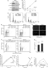
Figure 1
Figure 1
LIN-9 is required for entry into mitosis. (A) LIN-9 protein in hTERT immortalized primary BJ fibroblasts (BJ-ET) and in SHEP cells infected with the indicated retroviral shRNA vectors was analyzed by immunoprecipitation followed by immunoblotting. β-Tubulin was detected by immunoblotting. (B) Control and LIN-9 depleted BJ-ET cells were passaged according to a 3T3-like protocol. At every passage, the increase in cell number was determined and the cumulative growth was calculated. (C) FACS analysis of control and LIN-9 depleted BJ-ET cells. A representative experiment is shown. (D) Control cells and LIN-9 depleted cells were labeled with BrdU and analyzed by FACS to determine the fraction of cells in each phase of the cell cycle. Data are from three independent experiments. (E) Phospho-histone H3 positive cells (control infected BJ cells and LIN-9 depleted BJ cells) were identified by immunostaining. Data are from three independent experiments. (F) Control infected cells or LIN-9 depleted BJ-ET cells were pulsed for 1 h with BrdU and analyzed by FACS immediately or 9 h later. The fraction of BrdU-positive cells that progressed to the next G1 phase within the 9 h pulse was determined. Data are from three independent experiments. (G) S-phase length was determined by BrdU-pulse chase labeling as described by Begg et al (1985). Data are from five independent experiments. (H) SHEP cells were grown to confluence to induce cell cycle arrest in G1 and then released into the cell cycle by replating. At 18 h later (at which time most of the cells were in S-phase), they were pulsed with BrdU. At the start of the experiment, 66.25% (control cells) and 70.28% (LIN-9 depleted cells) were BrdU positive. BrdU was washed away and movement of BrdU positive cells into G2/M (left panel) or into the G1 phase of the next cell cycle (right panel) was followed by FACS. A representative experiment is shown. (I) Mitotic index of LIN-9 depleted SHEP cells and control infected SHEP cells following exposure to nocodazole. A representative experiment is shown.
Figure 2
Figure 2
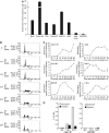
LIN-9 is required for transcription of a cluster of G2/M specific genes. Percentage (A) and list (B) of downregulated G2/M genes in LIN-9 depleted BJ-ET cells as determined by cDNA microarrays. (C) Quantitative RT–PCRs of LIN-9 target genes in LIN-9 depleted cells relative to control infected BJ-ET cells. Expression was normalized to S14. (D) Western blot analysis of G2/M (cyclin A, cyclin B, cdc2, BIRC5), G1 (cyclin E, cyclin D1) and noncycling control proteins (CDK2 and β-tubulin) in control cells and LIN-9 depleted BJ-ET cells. (E) Cyclin B1-dependent kinase activity towards histone H1 in lysates of control and LIN-9 depleted BJ-ET cells. Immunoblotting of the same lysates with an anti-β-tubulin antibody served as a control. (F) Inhibition of G2/M transcription following suppression of LIN-9 is independent on p53. BJ-ET cells were first infected with a p53-specific retroviral shRNA vector or with a control vector. After selection, cells were transfected with control oligonucleotides or LIN-9 specific RNAi oligonucleotides. After 72 h, gene expression was determined by quantitative RT–PCR.
Figure 3
Figure 3
LIN-9 binds to promoters of G2/M genes. (A) Binding of LIN-9 to the indicated promoters was analyzed by ChIP with chromatin isolated from BJ-ET cells enriched in S/G2/M. As a control, chromatin was immunoprecipitated with nonspecific IgG. Bound DNA was analyzed by quantitative real-time PCR. Enrichment compared with Input DNA was calculated. As controls, the GAPDH2 promoter and a region 6 kb downstream of the cdc2 promoter were analyzed. The promoters and the location of PCR amplicons are schematically shown in Supplementary Figure S3. (B) FACS analysis of T98G cells synchronized in G0 by serum starvation and released into the cell cycle by the addition of serum. (C) Gene expression in synchronized T98G cells was determined by real-time RT–PCR. Expression levels were normalized to GAPDH. (D) Binding of E2F4 and LIN-9 to the cyclin B1 and GAPDH2 promoter in G0 and S phase. Chromatin was isolated from serum starved T98G cells and at 20 h after addition of serum. Chromatin was immunoprecipitated with antisera specific for LIN-9 or E2F4. Nonspecific IgG served as control.
Figure 4
Figure 4
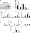
LIN-9 switches during cell cycle re-entry from E2F4/p130 to B-MYB. (A) Lysates from quiescent and S-phase T98G cells were immunoprecipitated with nonspecific IgG (control) or with E2F3, E2F4, B-MYB or LIN-9-specific antisera. Immunoprecipitations were immunoblotted for p130, E2F3, E2F4 and B-MYB. (B) Binding of B-MYB to promoters of G2/M genes was analyzed by ChIP with chromatin isolated from BJ-ET cells synchronized in S/G2/M. For details see legend of Figure 3A. (C) Binding of LIN-9, B-MYB and E2F4 to the promoters of E2F-regulated G2/M genes (Ubch10, Birc5, Plk1), G1/S genes (cdc6, RR1) or to the GAPDH2 promoter (control) in G0 and S phase was analyzed by ChIP. Chromatin was isolated from serum starved T98G cells (quiescent) and at 20 h after addition of serum (S-phase). Chromatin was immunoprecipitated with antisera specific for LIN-9 or E2F4 and B-MYB. Nonspecific IgG served as control. (D) Re-ChIP experiments with chromatin from T98G cells. S-phase chromatin was immunoprecipitated with IgG or B-MYB or LIN-9 antibodies followed by elution with DTT and reprecipitation (Re-ChIP) with reciprocal antibodies. Data are normalized to 1% input and are expressed relative to IgG (fold enrichment).
Figure 5
Figure 5
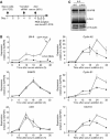
LIN-9 is required for activation of G2/M genes but not for their repression in quiescent cells. (A) Experimental design and time frame. (B) Gene expression during the cell cycle in control depleted cells and LIN-9 depleted T98G cells was analyzed by real-time RT–PCR and normalized to GAPDH. (C) Protein levels of B-MYB and LIN-9 in LIN-9 depleted and control-depleted, S-phase synchronized T98G cells. β-Tubulin served as a control. The asterisk denotes the heavy chain.
Figure 6
Figure 6
LIN-9 interacts with the C-terminus of B-MYB. (A) Lysates of HeLa cells were immunoprecipitated and immunoblotted with the indicated antisera (Pre=preimmunserum). (B) Association between LIN-9 and B-MYB in control transfected cells and in LIN-9 depleted HeLa cells. (C) Lysates of HeLa cells expressing HA-LIN-9 and flag-B-MYB mutants (see D) were immunoprecipitated with flag-antiserum and immunoblotted with an anti-HA-antibody. Ten percent of total lysate was immunoblotted (Input). HDAC2 was used as positive control because B-MYB has been shown to interact with N-CoR and SMRT corepressor that associate with HDACs (Masselink et al, 2001; Li and McDonnell, 2002) (D) Scheme of B-MYB mutants (adopted from Johnson et al, 2002) and summary of binding data. Asterisks indicate the location of the mutated sites in 15mut.
Figure 7
Figure 7
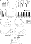
B-MYB is required for the expression of G2/M genes and for the transition through G2/M. (A) Immunoblot of B-MYB and β-tubulin using extracts from HeLa cells transfected with control or the B-MYB shRNA plasmid. (B) Growth of B-MYB depleted BJ-ET cells was analyzed as described in Figure 1B. (C) BrdU-FACS analysis of BJ-ET cells infected with the indicated shRNA viruses or with a control virus. The absolute change in G1, S and G2/M cells compared to control infected cells is shown. Data are from three different experiments. Error bars represent standard deviation. (D) Mitotic cells were identified in control infected BJ-ET cells and B-MYB depleted BJ-ET cells by immunostaining using a phospho-histone H3 antibody. Data are from three independent experiments. Error bars represent standard deviation (E) Mitotic index of B-MYB depleted SHEP cells and control infected SHEP cells following exposure to nocodazole. (F) Real-time RT–PCR analysis of the indicated genes in B-MYB depleted and control infected BJ-ET cells. Expression was normalized to S14. (G). B-MYB and/or LIN-9 were depleted in starved T98G cells by RNAi as described in Figure 5. Gene expression following re-addition of serum was analyzed by real-time RT–PCR. Expression levels were normalized to GAPDH. (H) Model for the role of LIN-9 and B-MYB in the regulation of transcription at G2/M.

Similar articles

Cited by

References

    1. Badie C, Itzhaki JE, Sullivan MJ, Carpenter AJ, Porter AC (2000) Repression of CDK1 and other genes with CDE and CHR promoter elements during DNA damage-induced G(2)/M arrest in human cells. Mol Cell Biol 20: 2358–2366 - PMC - PubMed
    1. Beall EL, Bell M, Georlette D, Botchan MR (2004) Dm-myb mutant lethality in Drosophila is dependent upon mip130: positive and negative regulation of DNA replication. Genes Dev 18: 1667–1680 - PMC - PubMed
    1. Beall EL, Manak JR, Zhou S, Bell M, Lipsick JS, Botchan MR (2002) Role for a Drosophila Myb-containing protein complex in site-specific DNA replication. Nature 420: 833–837 - PubMed
    1. Begg AC, McNally NJ, Shrieve DC, Karcher H (1985) A method to measure the duration of DNA synthesis and the potential doubling time from a single sample. Cytometry 6: 620–626 - PubMed
    1. Carvalho A, Carmena M, Sambade C, Earnshaw WC, Wheatley SP (2003) Survivin is required for stable checkpoint activation in taxol-treated HeLa cells. J Cell Sci 116: 2987–2998 - PubMed

Publication types