. 2006 Dec 1;20(23):3255-68.
doi: 10.1101/gad.1495506.

Cucumber mosaic virus-encoded 2b suppressor inhibits Arabidopsis Argonaute1 cleavage activity to counter plant defense

Affiliations

Cucumber mosaic virus-encoded 2b suppressor inhibits Arabidopsis Argonaute1 cleavage activity to counter plant defense

Xiuren Zhang et al. Genes Dev. .

Abstract

RNA silencing refers to small regulatory RNA-mediated processes that repress endogenous gene expression and defend hosts from offending viruses. As an anti-host defense mechanism, viruses encode suppressors that can block RNA silencing pathways. Cucumber mosaic virus (CMV)-encoded 2b protein was among the first suppressors identified that could inhibit post-transcriptional gene silencing (PTGS), but with little or no effect on miRNA functions. The mechanisms underlying 2b suppression of RNA silencing are unknown. Here, we demonstrate that the CMV 2b protein also interferes with miRNA pathways, eliciting developmental anomalies partially phenocopying ago1 mutant alleles. In contrast to most characterized suppressors, 2b directly interacts with Argonaute1 (AGO1) in vitro and in vivo, and this interaction occurs primarily on one surface of the PAZ-containing module and part of the PIWI-box of AGO1. Consistent with this interaction, 2b specifically inhibits AGO1 cleavage activity in RISC reconstitution assays. In addition, AGO1 recruits virus-derived small interfering RNAs (siRNAs) in vivo, suggesting that AGO1 is a major factor in defense against CMV infection. We conclude that 2b blocks AGO1 cleavage activity to inhibit miRNA pathways, attenuate RNA silencing, and counter host defense. These findings provide insight on the molecular arms race between host antiviral RNA silencing and virus counterdefense.


Figures
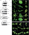
Figure 1.
Figure 1.
CMV 2b causes developmental anomalies in Arabidopsis. (A) CMV (FNY strain) infection enhances accumulation of AGO1 (panel a) and miR168 and miR168* (panel b). Samples were collected 7 or 12 d post-inoculation (dpi). Control plants were treated with buffer only (Mock). The numbers below each lane refer to expression levels relative to WT, after normalization against the loading controls (25S or 5S rRNAs). (B) FNY2b transcript and protein levels in 35S-FNY2b lines and CMV (FNY strain)-infected plants. Polyclonal antibody against FNY2b was used. (Top panels, lanes 1–3) For CMV infection experiment, samples were collected 7 or 12 dpi. Transgenic line numbers are given on top. A cross-reacting band (asterisk) was used as a loading control. (C) Morphological phenotypes of 35S-FNY2b transgenic lines. Photographs were taken for 4-wk-old seedlings, their fourth true leaves, and adult flowers of WT Col-0 (panels ac) and representative 35S-FNY2b lines (#1, #3, and #5), and for 3-wk-old seedlings, second true leaves, and adult flowers of ago1-27 (panels m–o). (Panels d–f) 35S-FNY2b-1. (Panels g–i) 35S-FNY2b-3. (Panels j–l) 35S-FNY2b-5. Bars: panels a,d,g,j,m, 10 mm; panels b,e,h,k,n, 2 mm; panels c,f,i,l,o, 1 mm. (D) 35S-FNY2b, a series of ago1 mutant alleles and WT Col-0 seedlings at the cotyledon stage. (Panel a) WT Col-0. (Panel b,c) Extremely severe 35S-FNY2b lines with unexpanded cotyledons. (Panels df) 35S-FNY-3, 35S-FNY-4 and 35S-FNY-5, respectively. (Panels gi) ago1-36, ago1-25, and ago1-27. Bars, 3 mm.
Figure 2.
Figure 2.
Accumulation of small RNAs and their * strands and target mRNAs in transgenic Arabidopsis plants expressing CMV 2b. (A) Northern blot analysis of high-molecular-weight RNAs. Transgenic line numbers are given on top. Blots were hybridized with the indicated gene-specific probes. A non-small-RNA-targeted mRNA (At2g24430) was used as a loading control. (B,C) Low-molecular-weight RNA blot. Duplicate blots were hybridized to the oligonucleotide probes complementary to the small RNAs, and their star strands are indicated. The numbers below each lane in AC refer to expression levels relative to WT, after normalization against the loading control. For DCL1 in A, only the full-length transcript (∼6.2 kb) is targeted by miR162, and its accumulation level was calculated and is shown below.
Figure 3.
Figure 3.
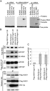
CMV 2b suppresses RNA silencing by a mechanism different from that used by P19. (A) Binding of ss and ds siRNAs and ss mRNA in vitro. Complexes were analyzed using native PAGE. (B) Analysis of miRNA and miRNA* in RISC of 2b-expressing plants. RNA were extracted from flowers or Flag-AGO1 immunoprecipitates of transgenic plants (two independent lines, #1 and #10) harboring 35S-FNY2b in a Flag-AGO1/ago1-36 background (Baumberger and Baulcombe 2005) and Flag-AGO1/ago1-36 control plants (Flag-AGO1/ago1–36). For middle panels, each lane contained small RNAs associated with Flag-AGO1 immunoprecipitated from 0.2 g of flowers. (Bottom panels) The input and immunoprecipitates of Flag-AGO1 and FNY2b were analyzed by Western blot assays in the same samples for small RNA blots. A cross-reacting band (*) was used as a loading control. (C) The relative ratio of miR165* and miR165 is increased in Flag-AGO1 immunoprecipitates from 35S-FNY2b-overexpressing lines compared with those from control plants. The mean signal values of miR165* and miR165 levels in 35S-FNY2b were calculated relative to those in control plants (Flag-AGO1/ago1-36), where the ratio was arbitrarily assigned a value of 1. Standard errors represent at least four experiments.
Figure 4.
Figure 4.
Specific interaction of CMV 2b and AGO1 in vivo. (A,B) Coimmunoprecipitation of FNY2b and AGO1 transiently expressed in N. benthamiana. Proteins were tagged either with 6myc or 3HA. Total crude protein extracts (Input) were immunoprecipitated (IP) with polyclonal antibody to myc or MBP. Western blots were analyzed with a monoclonal antibody to myc to detect myc-tagged proteins (top panels), and a monoclonal antibody to HA to detect coimmunoprecipitated HA-tagged proteins (bottom panels). Arrowheads (◀) indicate transiently expressed proteins. (C) Interaction of FNY2b and AGO1 proteins in Arabidopsis plants. Two-week-old seedlings expressing 35-FNY2b-3HA/XVE-1-6myc-AGO1 were treated overnight with 50 μM MG132 in the absence (lanes1,3) or presence (lanes 2,4,5) of β-estradiol (25 μM) to induce 6myc-AGO1 expression. Total protein extracts were immunoprecipitated (IP) with polyclonal antibody to myc or MBP. Western blots were analyzed with a monoclonal antibody to myc to detect 6myc-AGO1 (top panel), and a monoclonal antibody to HA to detect coimmunoprecipitated FNY2b-3HA (bottom panel). A monoclonal antibody to phyB was used as a negative control to detect the presence of phyB. (D) CMV-encoded 2b suppressor is incorporated into the AGO1 complex in Arabidopsis plants during viral infections. Flag-AGO1/ago1-36 plants at flowering stages were treated with buffer only (mock) or infected with the CMV FNY strain. Total protein extracts from flowers were immunoprecipitated (IP) with monoclonal antibody to Flag. Western blots were analyzed with the same antibody to detect Flag-AGO1 (top panel), and a polyclonal antibody to FNY2b to detect coimmunoprecipitated FNY2b (middle panel). A polyclonal antibody to CMV CP was used as a negative control to detect the presence of coat protein. (AD) In all panels, an asterisk (*) indicates a cross-reacting band in the input fraction, which served as a negative control since it was absent in the IP fractions. Double asterisks (**) indicate the heavy or light chains of the protein A-conjugated antibody.
Figure 5.
Figure 5.
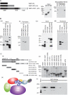
CMV 2b directly interacts with AGO1 in vitro. (A) In vitro pull-down assays of 6His-SUMO-FNY2b with three bait proteins: MBP (M), MBP-AIP2 (AI), and MBP-AGO1 (AG). (Left panel) A schematic diagram of the three bait proteins. (Middle panel) The input of the bait proteins. (Right panel) The input and output of 6His-SUMO-FNY2b. Western blots were analyzed with monoclonal antibodies to MBP (middle panel) or to 6His (right panel). (B). In vitro pull-down assays of several 6His- or GST-tagged proteins by MBP-AGO1. (Panels a,b) The input and output of the indicated 6His-tagged target proteins, respectively. (Panels c,d) The input and output fraction of the indicated GST-tagged target proteins, respectively. Western blots were analyzed with monoclonal antibodies to 6His (panels a,b) or to GST (panels c,d). (C) CMV 2b interacts with one face of AGO1. (Panel a) A schematic diagram of full-length and truncated forms of AGO1. The numbers refer to the amino acid residues in the WT AGO1 protein. Locations of the PAZ and PIWI domains are shown. All bait proteins were tagged with MBP at the N terminus. (Panel b) Coomassie brilliant blue G250 staining of the bait proteins, showing their mobility. The major band representing the bait protein is indicated with arrowheads (◀). (Panel c) The output of pulled-down GST-FNY2b. Western blots were analyzed with a monoclonal antibody to GST. All MBP-tagged bait proteins and 6His- and GST-tagged target proteins were purified from Escherichia coli using amylose resins, nitrilotriacetate resins, and glutathione resins, respectively. In all assays, 2 μg of target proteins were pulled down with the indicated bait proteins (2 μg each) using amylose resins. (Panel d) A cartoon showing binding of CMV 2b to one surface of the PAZ-containing module of AGO1, which harbors ss small RNA and its target mRNA-binding groove. The predicted domains of poly-Q (Q), N terminus (N), Linker 1 (L1), Mid, PAZ, and PIWI are shown.
Figure 6.
Figure 6.
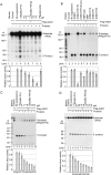
CMV 2b inhibits AGO1 slicer activity in RISC reconstituted in vitro. (A) FNY2b blocks the activity of RISC reconstituted in vitro. (Lanes 39) Immunoprecipitates (IPs) containing Flag-AGO1 were prepared from inflorescences of Flag-AGO1/AGO1-36 plants using a monoclonal antibody to Flag. (Lane 2) Similar immunoprecipitates prepared from inflorescences of Flag-AGO1/AGO1-36 plants using a monoclonal antibody to 6His were used as negative controls. Immunoprecipitates were incubated with the indicated suppressor protein (in micromolar concentration; lanes 49) or without any suppressor protein (lane 3, buffer only, containing the same ionic concentrations) before mixing with the ss siRNA complementary to the target PDS mRNA. Reconstituted RISC was tested for cleavage activity by incubation with a 32P-cap-labeled PDS mRNA. RNAs recovered from the reaction mix were fractionated on 12% denaturing gels. The positions of intact substrates, 5′ cleavage products, and RNA markers are shown. (B) FNY2b inhibits cleavage activity of Flag-AGO1. Immunoprecipitates containing Flag-AGO1 were incubated without (buffer only, lane 3) or with the indicated proteins (lanes 411) before addition of 32P-cap-labeled in vitro transcripts of At4g29770. (Top panel, lane 2) Immunoprecipitates of WT Col-0 inflorescences using Flag antibody were used as a negative control. The final concentration of the indicated suppressors and control proteins was 5 μM except MBP and MBP-Q2b, which were 5 μg (∼1.5 and 1 μM, respectively). RNAs recovered from the reaction mix were analyzed as in A. (C,D) Inhibition of AGO1 cleavage activity is proportional to the FNY2b amount. Immunoprecipitates containing Flag-AGO1 were incubated without (buffer only, lane 3) or with the indicated suppressors at different concentrations before mixing with 32P-cap-labeled in vitro transcripts of At4g29770 (C) and PHAVOLUTA (PHV) (D). (Lane 2) Control immunoprecipitates were prepared from WT Col-0 inflorescences using antibody to Flag. RNAs recovered from the reaction mix were analyzed as in A. (A–D) In all the four panels, the experiments were done at least three times. (Lane 3) In each experiment, the cleavage efficiency was normalized to that obtained with the buffer only. (Bottom panels) Mean values of the relative cleavage efficiency are shown along with standard error.
Figure 7.
Figure 7.
AGO1 recruits viral siRNAs. Flag-AGO1/ago1-36 plants were infected with the CMV (FNY strain) (A), the CMV (NT9 strain) (B), and TYMV (C), or with buffer (Mock). Inflorescence tissues were collected 7 dpi for immunoprecipitation with Flag or His antibody except for CMV (FNY) experiments in which samples were harvested at 7 and 12 dpi. Small RNAs were extracted directly from flowers (crude extract) or from Flag-AGO1 immunoprecipitates and control immunoprecipitates obtained with an antibody to 6His (IP). Viral siRNAs were detected by hybridization with gene-specific probes (Materials and Methods). The same membrane was hybridized to a 5S rRNA probe as a control, indicating no small RNA contamination in immunoprecipitate fractions. miR165 was used as a positive control of AGO1–small RNA coimmunoprecipitation. For the crude extract, each lane contained 5 μg of total RNA. The immunoprecipitate samples were derived from 0.3 g of flowers. Two-thirds of the samples were used for siRNA recovery and the remainder for Western blots to monitor Flag-AGO1 levels. Western blot analyses were performed using antibodies to Flag. A cross-reacting band (*) served as a loading control.

Similar articles

Cited by

References

    1. Allen, E., Xie, Z.X., Gustafson, A., Carrington, J. microRNA-directed phasing during trans-acting siRNA biogenesis in plants. Cell. 2005;121:207–221. - PubMed
    1. Anandalakshmi, R., Pruss, G., Ge, X., Marathe, R., Mallory, A., Smith, T., Vance, V. A viral suppressor of gene silencing in plants. Proc. Natl. Acad. Sci. 1998;95:13079–13084. - PMC - PubMed
    1. Bartel, D.P. MicroRNAs: Genomics, biogenesis, mechanism, and function. Cell. 2004;116:281–297. - PubMed
    1. Baulcombe, D. RNA silencing in plants. Nature. 2004;431:356–363. - PubMed
    1. Baumberger, N., Baulcombe, D. Arabidopsis ARGONAUTE1 is an RNA slicer that selectively recruits miRNAs and short interfering RNAs. Proc. Natl. Acad. Sci. 2005;102:11928–11933. - PMC - PubMed

Publication types

MeSH terms