. 2006 Dec;18(12):3386-98.
doi: 10.1105/tpc.106.047605. Epub 2006 Dec 8.

Arabidopsis DCP2, DCP1, and VARICOSE form a decapping complex required for postembryonic development

Affiliations

Arabidopsis DCP2, DCP1, and VARICOSE form a decapping complex required for postembryonic development

Jun Xu et al. Plant Cell. 2006 Dec.

Abstract

mRNA turnover in eukaryotes involves the removal of m7GDP from the 5' end. This decapping reaction is mediated by a protein complex well characterized in yeast and human but not in plants. The function of the decapping complex in the development of multicellular organisms is also poorly understood. Here, we show that Arabidopsis thaliana DCP2 can generate from capped mRNAs, m7GDP, and 5'-phosphorylated mRNAs in vitro and that this decapping activity requires an active Nudix domain. DCP2 interacts in vitro and in vivo with DCP1 and VARICOSE (VCS), an Arabidopsis homolog of human Hedls/Ge-1. Moreover, the interacting proteins stimulate DCP2 activity, suggesting that the three proteins operate as a decapping complex. Consistent with their role in mRNA decay, DCP1, DCP2, and VCS colocalize in cytoplasmic foci, which are putative Arabidopsis processing bodies. Compared with the wild type, null mutants of DCP1, DCP2, and VCS accumulate capped mRNAs with a reduced degradation rate. These mutants also share a similar lethal phenotype at the seedling cotyledon stage, with disorganized veins, swollen root hairs, and altered epidermal cell morphology. We conclude that mRNA turnover mediated by the decapping complex is required for postembryonic development in Arabidopsis.


Figures

Figure 1.
Figure 1.
Schemes of Arabidopsis (At) DCP2, DCP1, VCS, and VCR Aligned with Their Homologs in S. cerevisiae (Sc) and Homo sapiens (h). Conserved domains are boxed and labeled. The number of amino acid (aa) residues in each protein is shown along with the degree of sequence similarity. (A) DCP2. Gray bar, N-terminal domain; black bar, Nudix domain. (B) DCP1. Gray bar, EVH1/WH1, a member of the PH domain superfamily (She et al., 2004). (C) VCS and VCR. WD40, nuclear localization signal (NLS), S domain, and ψ(X2-3) repeats are indicated below (Yu et al., 2005).
Figure 2.
Figure 2.
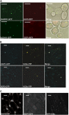
Subcellular Localization of DCP2, DCP1, and VCSc. Single focal planes for each sample are shown. (A) Subcellular localization of DCP2-RFP and DCP1-RFP compared with Sc DHH1-GFP in yeast P bodies. RFP fluorescence is pseudocolored in red, and GFP is pseudocolored in green. Bars = 5 μm. (B) Subcellular localization of DCP1-CFP and DCP2-CFP with YFP-VCSc in Nicotiana epidermal cells. CFP fluorescence is pseudocolored in blue, and YFP is pseudocolored in yellow. Bars = 20 μm. (C) Subcellular localization of DCP2-CFP, DCP1-CFP, and YFP-VCSc in root cells of Arabidopsis transgenic plants 3 d after germination carrying individual transgenes. Bars = 20 μm.
Figure 3.
Figure 3.
Protein Interactions among DCP2, DCP1, and VCS. (A) Self-association of VCS and its interaction with DCP2 and DCP1 in vivo. Coimmunoprecipitation (IP) was performed with extracts prepared from the wild type (lanes 1 and 3) and various Arabidopsis transgenic lines (lanes 2 and 4). Panels a and b, 35S-myc-DCP2 and XVE-YFP-DCP1 coexpression lines. Panel c, 35S-YFP-VCSc line. Panel d, 35S-eIF4E-CFP line. Extracts were immunoprecipitated with anti-VCS antibody (a to d). Immunoprecipitates were then analyzed by protein gel blotting using antibodies against myc (a and e) and YFP/CFP (b to d and f). For panels e and f, transgenic plants carrying 35S-myc-DCP2 and XVE-YFP-DCP1 were used. Plants were either not treated (lanes 5 and 7) or induced using β-estradiol (20 μM) for 6 d (lanes 6 and 8). Extracts were immunoprecipitated with anti-myc antibody. (B) In vitro interactions between DCP2 and DCP1 (a) and interactions between DCP2 and various VCS domains (b). Proteins were purified from E. coli, and amylose resin was used in the pull-down assay. Proteins input are indicated above each lane (a and b), and different portions of VCS used in the assays are illustrated in panel c. The presence of GST fusion proteins was detected by anti-GST monoclonal antibody on protein gel blots. (C) A proposed model of the DCP2/DCP1/VCS complex based on in vivo and in vitro interaction data. aa, amino acids.
Figure 4.
Figure 4.
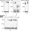
Decapping Activity of DCP2 in Vitro. (A) Decapping assay of the MBP-DCP2 fusion protein. Amounts of protein in each reaction mix are shown at top. Migration positions of m7Gppp and m7Gpp are indicated at left. (B) Comparison of decapping products generated from MBP-DCP2 (lanes 1 and 3) and MBP-hDCP2 (lanes 2 and 4) with and without NDPK treatment. (C) Decapping activity of MBP-DCP2 and its mutants. EQ, mutant MBP-DCP2 bearing the E158Q mutation in the Nudix domain; RFAA, mutant MBP-DCP2 bearing the R30A and F31A mutations. (D) Decapping activity of MBP-DCP2 with or without the presence of MBP-DCP1 (lanes 1 and 4 to 6) and MBP-VCSc (lanes 2 and 7 to 9). In lane 10, GST-At3g20650 (10 pmol) was added as a negative control. Amounts of protein added are indicated above each lane.
Figure 5.
Figure 5.
Phenotypical Analysis of Mutants Deficient in DCP2, DCP1, and VCS. (A) Schemes of T-DNA insertion lines and alleles of each locus. Gray boxes indicate sequences corresponding to mRNA sequences. Locus numbers, open reading frames (first ATG and stop), and allele names are shown. (B) Six-day-old seedlings of the wild type and various mutant alleles (listed below). col, Columbia; WS, Wassilewskija. Bars = 1 mm. (C) Light microscopy images of 6-d-old wild-type and three mutant seedlings after clearing. Bars = 1 mm. (D) Detailed morphological phenotypes of 6-d-old wild-type and three mutant plants. Top panel, scanning electron microscopic images of cotyledon epidermis. Bars = 20 μm. Middle panel, scanning electron microscopic images of shoot apical meristems. Bars = 50 μm. Bottom panel, light microscopy images of root hairs. Bars = 50 μm.
Figure 6.
Figure 6.
Expression Profiles of DCP2, DCP1, and VCS in Arabidopsis Tissues and Different Mutants. (A) GUS staining of 6-d-old seedlings of ProDCP2-GUS (panels a, c, d, and e) and ProDCP1-GUS (panels b, f, g, and h) transgenic plants. Vascular cells (panels a, b, c, and f; bars = 1 mm), guard cells and epidermal cells (panels d and g; bars = 50 μm) on leaves and root hairs (panels e and h; bars = 50 μm) are shown. (B) RNA gel blot analysis of DCP2, DCP1, and VCS expression in Arabidopsis tissues and in different organs of wild-type plants and in decapping-deficient mutants. Specific bands are indicated at right along with estimated sizes. Ethidium bromide–stained RNA gel lanes are shown at bottom as loading controls. The seedling samples were collected from progeny of the DCP2+/− heterozygote line that did not display a phenotype, which includes wild-type seedlings as well as DCP2+/− heterozygotes. Note that the high molecular mass RNA species detected in dcp2-1 were attributable to the insertion of T-DNA (see Figure 5A). This band was also detected in the control sample because of the presence of DCP2+/− heterozygotes. (C) Transcript levels of VCS (top panel) and Actin2 (bottom panel) in a control sample and vcs-6 as determined by RT-PCR.
Figure 7.
Figure 7.
Transcript Accumulation in Three Decapping-Deficient Mutants. Six-day-old seedlings of each mutant were sampled, and seedlings without a phenotype from progeny of DCP2+/− heterozygotes served as the control. (A) Transcript levels of EXPL1 (top panels) and Actin2 (middle panels) in control and decapping-deficient mutants at different time points after cordycepin treatment. The estimated half-life (t1/2) is shown at right. Ethidium bromide–stained RNA gel lanes (bottom panels) are shown as loading controls. (B) RACE-PCR (see Methods) shows accumulation of capped EXPL1 transcript in decapping mutants and in the wild type treated with cycloheximide (CHX). (C) Semiquantitative RT-PCR to examine transcript abundance in 6-d-old seedlings of the wild type and decapping-deficient mutants. Ethidium bromide–stained cDNA bands corresponding to specific transcripts (listed at right) were confirmed by sequencing. Using templates from reaction mix without the RT step during cDNA synthesis produced no bands, suggesting no genomic DNA contamination in the samples used for RT-PCR (bottom panel). (D) Quantitative representation of the RT-PCR results from (C). Each column represents the mean value from three independent experiments normalized with respect to the background of the gel. se bars are shown. (E) Accumulation of SEN1, EXPL1, and eIF4A transcripts after cordycepin (Cor) and cycloheximide (CHX) treatments. Specific bands are labeled at right, and an ethidium bromide–stained RNA gel (bottom panel) is shown as a loading control. Samples and time course after cycloheximide treatment are shown above each lane.

Similar articles

Cited by

References

    1. Andrei, M.A., Ingelfinger, D., Heintzmann, R., Achsel, T., Rivera-Pomar, R., and Luhrmann, R. (2005). A role for eIF4E and eIF4E-transporter in targeting mRNPs to mammalian processing bodies. RNA 11 717–727. - PMC - PubMed
    1. Baima, S., Possenti, M., Matteucci, A., Wisman, E., Altamura, M.M., Ruberti, I., and Morelli, G. (2001). The Arabidopsis ATHB-8 HD-Zip protein acts as a differentiation-promoting transcription factor of the vascular meristems. Plant Physiol. 126 643–655. - PMC - PubMed
    1. Beelman, C.A., Stevens, A., Caponigro, G., LaGrandeur, T.E., Hatfield, L., Fortner, D.M., and Parker, R. (1996). An essential component of the decapping enzyme required for normal rates of mRNA turnover. Nature 382 642–646. - PubMed
    1. Behm-Ansmant, I., Rehwinkel, J., Doerks, T., Stark, A., Bork, P., and Izaurralde, E. (2006). mRNA degradation by miRNAs and GW182 requires both CCR4:NOT deadenylase and DCP1:DCP2 decapping complexes. Genes Dev. 20 1885–1898. - PMC - PubMed
    1. Beliakova-Bethell, N., Beckham, C., Giddings, T.H.J., Winey, M., Parker, R.O.Y., and Sandmeyer, S. (2006). Virus-like particles of the Ty3 retrotransposon assemble in association with P-body components. RNA 12 94–101. - PMC - PubMed

Publication types

MeSH terms