A system for quantifying dynamic protein interactions defines a role for Herceptin in modulating ErbB2 interactions
- PMID: 17148612
- PMCID: PMC1748177
- DOI: 10.1073/pnas.0605218103
A system for quantifying dynamic protein interactions defines a role for Herceptin in modulating ErbB2 interactions
Abstract
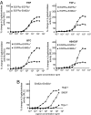
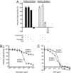
The orphan receptor tyrosine kinase ErbB2 is activated by each of the EGFR family members upon ligand binding. However, difficulties monitoring the dynamic interactions of the membrane receptors have hindered the elucidation of the mechanism of ErbB2 activation. We have engineered a system to monitor protein-protein interactions in intact mammalian cells such that different sets of protein interactions can be quantitatively compared. Application of this system to the interactions of the EGFR family showed that ErbB2 interacts stably with the EGFR and ErbB3, but fails to spontaneously homooligomerize. The widely used anti-cancer antibody Herceptin was found to effectively inhibit the interaction of the EGFR and ErbB2 but not to interfere with the interaction of ErbB2-ErbB3. Treatment of cells expressing EGFR and ErbB2 with Herceptin results in increased EGFR homooligomerization in the presence of EGF and a subsequent rapid internalization and down-regulation of the EGFR. In summary, the protein interaction system described here enabled the characterization of ErbB2 interactions within the biological context of the plasma membrane and provides insight into the mechanism of Herceptin action on cells overexpressing ErbB2.
Conflict of interest statement
Conflict of interest statement: H.M.B. is a major stockholder in a company that might have a gain or loss financially through publication of this paper. T.S.W. and H.M.B. are inventors of the technology described in this article; a patent is pending.
Figures


Similar articles
-
Epidermal growth factor receptor coexpression modulates susceptibility to Herceptin in HER2/neu overexpressing breast cancer cells via specific erbB-receptor interaction and activation.Exp Cell Res. 2005 Apr 1;304(2):604-19. doi: 10.1016/j.yexcr.2004.12.008. Epub 2005 Jan 21. Exp Cell Res. 2005. PMID: 15748904
-
Growth stimulation of non-small cell lung cancer cell lines by antibody against epidermal growth factor receptor promoting formation of ErbB2/ErbB3 heterodimers.Mol Cancer Res. 2007 Apr;5(4):393-401. doi: 10.1158/1541-7786.MCR-06-0303. Mol Cancer Res. 2007. PMID: 17426253
-
Activation of ErbB2 by overexpression or by transmembrane neuregulin results in differential signaling and sensitivity to herceptin.Cancer Res. 2005 Aug 1;65(15):6801-10. doi: 10.1158/0008-5472.CAN-04-4023. Cancer Res. 2005. PMID: 16061662
-
The ErbB/HER family of protein-tyrosine kinases and cancer.Pharmacol Res. 2014 Jan;79:34-74. doi: 10.1016/j.phrs.2013.11.002. Epub 2013 Nov 20. Pharmacol Res. 2014. PMID: 24269963 Review.
-
The neuregulin-I/ErbB signaling system in development and disease.Adv Anat Embryol Cell Biol. 2007;190:1-65. Adv Anat Embryol Cell Biol. 2007. PMID: 17432114 Review.
Cited by
-
Trastuzumab: updated mechanisms of action and resistance in breast cancer.Front Oncol. 2012 Jun 18;2:62. doi: 10.3389/fonc.2012.00062. eCollection 2012. Front Oncol. 2012. PMID: 22720269 Free PMC article.
-
Molecular imaging of epidermal growth factor receptor kinase activity.Anal Biochem. 2011 Oct 1;417(1):57-64. doi: 10.1016/j.ab.2011.05.040. Epub 2011 May 30. Anal Biochem. 2011. PMID: 21693098 Free PMC article.
-
Dynamic analysis of the epidermal growth factor (EGF) receptor-ErbB2-ErbB3 protein network by luciferase fragment complementation imaging.J Biol Chem. 2013 Oct 18;288(42):30773-30784. doi: 10.1074/jbc.M113.489534. Epub 2013 Sep 6. J Biol Chem. 2013. PMID: 24014028 Free PMC article.
-
Interaction of antibodies with ErbB receptor extracellular regions.Exp Cell Res. 2009 Feb 15;315(4):659-70. doi: 10.1016/j.yexcr.2008.10.008. Epub 2008 Oct 22. Exp Cell Res. 2009. PMID: 18992239 Free PMC article. Review.
-
Trastuzumab Blocks the Receiver Function of HER2 Leading to the Population Shifts of HER2-Containing Homodimers and Heterodimers.Antibodies (Basel). 2021 Feb 4;10(1):7. doi: 10.3390/antib10010007. Antibodies (Basel). 2021. PMID: 33557368 Free PMC article.
References
-
- Slamon DJ, Clark GM, Wong SG, Levin WJ, Ullrich A, McGuire WL. Science. 1987;235:177–182. - PubMed
-
- Gschwantler-Kaulich D, Hudelist G, Koestler WJ, Czerwenka K, Mueller R, Helmy S, Ruecklinger E, Kubista E, Singer CF. Oncol Rep. 2005;14:305–311. - PubMed
-
- DiGiovanna MP, Stern DF, Edgerton SM, Whalen SG, Moore D, II, Thor AD. J Clin Oncol. 2005;23:1152–1160. - PubMed
-
- Romond EH, Perez EA, Bryant J, Suman VJ, Geyer CE, Jr, Davidson NE, Tan-Chiu E, Martino S, Paik S, Kaufman PA, et al. N Engl J Med. 2005;353:1673–1684. - PubMed
-
- Piccart-Gebhart MJ, Procter M, Leyland-Jones B, Goldhirsch A, Untch M, Smith I, Gianni L, Baselga J, Bell R, Jackisch C, et al. N Engl J Med. 2005;353:1659–1672. - PubMed
Publication types
MeSH terms
Substances
Grants and funding
- R37 AG009521/AG/NIA NIH HHS/United States
- AG 020961/AG/NIA NIH HHS/United States
- R01 AG024987/AG/NIA NIH HHS/United States
- HD 018179/HD/NICHD NIH HHS/United States
- T32 AG 0259/AG/NIA NIH HHS/United States
- AF 051678/AF/ACF HHS/United States
- AG 009521/AG/NIA NIH HHS/United States
- T32 GM008412/GM/NIGMS NIH HHS/United States
- R01 AG020961/AG/NIA NIH HHS/United States
- R01 AG009521/AG/NIA NIH HHS/United States
- AG 024987/AG/NIA NIH HHS/United States
- R01 HD018179/HD/NICHD NIH HHS/United States
- T32 AG000259/AG/NIA NIH HHS/United States
- T32 GM 08412/GM/NIGMS NIH HHS/United States
LinkOut - more resources
Full Text Sources
Other Literature Sources
Molecular Biology Databases
Research Materials
Miscellaneous

