. 2007 Jan;8(1):92-100.
doi: 10.1038/ni1414. Epub 2006 Dec 3.

HLA-DM targets the hydrogen bond between the histidine at position beta81 and peptide to dissociate HLA-DR-peptide complexes

Affiliations

HLA-DM targets the hydrogen bond between the histidine at position beta81 and peptide to dissociate HLA-DR-peptide complexes

Kedar Narayan et al. Nat Immunol. 2007 Jan.

Abstract

The peptide editor HLA-DM (DM) mediates exchange of peptides bound to major histocompatibility (MHC) class II molecules during antigen processing; however, the mechanism by which DM displaces peptides remains unclear. Here we generated a soluble mutant HLA-DR1 with a histidine-to-asparagine substitution at position 81 of the beta-chain (DR1betaH81N) to perturb an important hydrogen bond between MHC class II and peptide. Peptide-DR1betaH81N complexes dissociated at rates similar to the dissociation rates of DM-induced peptide-wild-type DR1, and DM did not enhance the dissociation of peptide-DR1betaH81N complexes. Reintroduction of an appropriate hydrogen bond (DR1betaH81N betaV85H) restored DM-mediated peptide dissociation. Thus, DR1betaH81N might represent a 'post-DM effect' conformation. We suggest that DM may mediate peptide dissociation by a 'hit-and-run' mechanism that results in conformational changes in MHC class II molecules and disruption of hydrogen bonds between betaHis81 and bound peptide.


Figures

Figure 1
Figure 1
DR1βH81N binds peptides to form complexes similar to peptide–DR1WT. (a) Size-separation profiles of soluble recombinant DR1WT and DR1βH81N molecules in PBS analyzed on a column equilibrated in PBS. The main peak corresponding to the αβ heterodimer (arrow) appears as expected at about 36 min for both molecules. (b) SDS stability of peptide–DR1βH81N and peptide–DR1WT complexes incubated in 0.1% SDS in PBS and separated by 12% SDS-PAGE; gel is silver-stained. HA(anch), HA(anchorless). (c) Kinetics of association of DR1βH81N or DR1WT (2 μM) incubated with excess (60 μM) FITC–labeled HA(306–318) in citrate phosphate buffer, pH 5.5, in the absence of DM; complexes were separated from unbound peptide and fluorescence measured was plotted versus time, then data were fitted to biphasic curves. Data are representative of five or more experiments (a,b) or are from one of three independent experiments (c).
Figure 2
Figure 2
Kinetics of dissociation of peptides in the absence of DM are faster for mutant DR1βH81N and resemble DM-mediated peptide dissociation. (a) Dissociation of DR1WT and DR1βH81N in complex with FITC–HA(anchorless) or FITC–HA(306–318) in the presence of a 100× molar excess of relevant unlabeled peptides. The fluorescence of the labeled complex before dissociation is arbitrarily assigned a value of 1.0, and fluorescence after dissociation is expressed as a fraction of fluorescence before dissociation. (b) Dissociation of HA(anchorless)–DR1WT in the absence (t1/2, 53 min) or presence (t1/2, 7.3 min; R2 = 0.997) of DM (DM/DR, 1:1) and HA(308–316)–DR1βH81N (t1/2, 7.4 min, R2 = 0.986) in the absence of DM, produced and dissociated as described in a. Data from one of three or more independent experiments were fitted to a single-exponential curve and approximate t1/2 values were calculated.
Figure 3
Figure 3
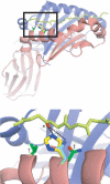
Pocket 1 of HA(306–318)–DR1. Based on modeling programs, βHis81 (yellow; 2.69Å) but not the substituted residue β81Asn (cyan; 3.75Å) of DR1 forms a short, strong hydrogen bond (dashed lines) with the carbonyl group (*) on Lys307 of HA(306–318) (light green). The residues substituted for histidine in the `rescue' mutants, β85Val (†) and β82Asn (‡), are green; α- and β-chains of DR1 are blue and red ribbons, respectively; and acidic and basic groups on the relevant side chains are red and blue, respectively. Bottom, enlargement of boxed area (pocket 1) above. Structure rendered by PyMol based on the crystal structure of HA(306–318)–DR1.
Figure 4
Figure 4
DM has a minimal effect on the dissociation of peptide from DR1βH81N. (a) Dissociation of complexes of FITC–HA(anchorless)–DR1WT, FITC–HA(308–316)–DR1βH81N and FITC–HA(anchorless)–DR1βH81N (2.5 μM), allowed to dissociate separately for 10 min as described in Figure 2a in the absence (–DM) or presence of various concentrations of DM. The fluorescence reading of complexes dissociated in the absence of DM was arbitrarily assigned a value of 1.0, and measurements are expressed as fractions of that value. Data are from one of three experiments. (b) Dissociation of complexes of DR1–H81N and FITC–HA(anchorless) or FITC–HA(308–316), allowed to dissociate as described in a in the presence (+ DM) or absence of DM (DM/DR, 1:5). The t1/2 values for all the reactions from the single-exponential curve fits are very similar: without DM, about 10 min; with DM, about 8 min; R2 = 0.959–0.997. Data are from one of at least three experiments. (c) Dissociation of complexes of HA(Y308A)–DR1WT or HA(308–316)–DR1βH81N (2.5 μM) allowed to bind to an HA(anchorless) or HA(308–316) surface, respectively, and then allowed to dissociate for 20 min in the absence of DM and for a further 20 min in the presence of 8 μM DM at a pH of 6.0. Curves were obtained by subtraction of the curve obtained with DM from the curve obtained without DM and assignment an arbitrary value of 0 for the relative units obtained at time 0. Data are from one of two experiments.
Figure 5
Figure 5
The βH81N substitution does not alter the interaction of DM with DR. (a) Tryptophan fluorescence of DR1WT or DR1βH81N (0.2 μM) in complex with HA(anchorless) or HA(308–316) incubated in citrate phosphate, pH 5.5, before and after the addition of 0.1 μM DM, monitored from 310 nm to 410 nm after excitation at 295 nm. Raw data here are from one of two experiments after subtraction of `blank' values (citrate phosphate only). (b) Average change in fluorescence (ΔTrp fluorescence) over 310–390 nm obtained from the raw data in a. *, P < 0.000001. (c,d) Fluorescence of 2 μM DR1WT (c) or DR1βH81N (d) incubated for various times at 37 °C with 60 μM FITC–HA(308–316) in citrate phosphate, pH 5.5, in the presence (+ DM) or absence (− DM) of 1 μM DM; complexes were separated from excess fluorescent peptide for fluorescence measurement. Data obtained with DM were fitted to monophasic association curves for both DR1WT and DR1βH81N. Data are from one of at least two independent experiments.
Figure 6
Figure 6
Reintroduction of an appropriate histidine residue partially restores DM-mediated dissociation. (a) Fluorescence of DR1(βH81N βV85H) and DR1(βH81N βN82H) associated with excess FITC–HA(306–318) in citrate phosphate buffer, pH 5.5. in the presence or absence of DM (DM/DR = 1:5) as described in Figure 5c,d; data from one of two experiments are fitted to biphasic or monophasic curves for groups without or with DM, respectively. (b) Dissociation of HA(308–316)–DR1(βH81N βV85H) (t1/2, 4.2 min; R2 = 0.985) and HA(308–316)–DR1(βH81N βN82H) (t1/2, 4.0 min; R2 = 0.999) complexes in the absence of DM, analyzed as described in Figure 2a. Data were fitted to a single-exponential curve and approximate t1/2 values were calculated. (c) Dissociation of FITC–HA(anchorless) in complex with DR1(βH81N βV85H) or DR1(βH81N βN82H), allowed to dissociate separately for 5 minutes as described in Figure 4a, in the absence (–DM) or presence of various concentrations of DM. Fluorescence is expressed as a fraction of complexes dissociated in the absence of DM. (d) Dissociation of HA(anchorless) from double mutants as described in Figure 2a (DM/DR = 1:5). FITC–HA(anchorless) dissociates from DR1(βH81N βN82H) with a t1/2 of about 1 min in the presence (R2 = 0.987) or absence (R2 = 0.998) of DM and dissociates from DR1(βH81N βV85H) with a t1/2 of about 4 min in the presence of DM (R2 = 0.944) and with a t1/2 of about 1 min in the absence of DM (R2 = 0.994). Data for dissociation kinetics are from one of at least four independent experiments.

Similar articles

Cited by

References

    1. Neefjes JJ, Stollorz V, Peters PJ, Geuze HJ, Ploegh HL. The biosynthetic pathway of MHC class II but not class I molecules intersects the endocytic route. Cell. 1990;61:171–183. - PubMed
    1. Peters PJ, Neefjes JJ, Oorschot V, Ploegh HL, Geuze HJ. Segregation of MHC class II molecules from MHC class I molecules in the Golgi complex for transport to lysosomal compartments. Nature. 1991;349:669–676. - PubMed
    1. Blum JS, Cresswell P. Role for intracellular proteases in the processing and transport of class II HLA antigens. Proc. Natl. Acad. Sci. USA. 1988;85:3975–3979. - PMC - PubMed
    1. Cresswell P. Assembly, transport, and function of MHC class II molecules. Annu. Rev. Immunol. 1994;12:259–293. - PubMed
    1. Sadegh-Nasseri S, McConnell HM. A kinetic intermediate in the reaction of an antigenic peptide and I-Ek. Nature. 1989;337:274–276. - PubMed

Publication types

LinkOut - more resources