. 2007 Feb;18(2):581-93.
doi: 10.1091/mbc.e06-07-0612. Epub 2006 Nov 29.

Atg27 is required for autophagy-dependent cycling of Atg9

Affiliations

Atg27 is required for autophagy-dependent cycling of Atg9

Wei-Lien Yen et al. Mol Biol Cell. 2007 Feb.

Abstract

Autophagy is a catabolic pathway for the degradation of cytosolic proteins or organelles and is conserved among all eukaryotic cells. The hallmark of autophagy is the formation of double-membrane cytosolic vesicles, termed autophagosomes, which sequester cytoplasm; however, the mechanism of vesicle formation and the membrane source remain unclear. In the yeast Saccharomyces cerevisiae, selective autophagy mediates the delivery of specific cargos to the vacuole, the analog of the mammalian lysosome. The transmembrane protein Atg9 cycles between the mitochondria and the pre-autophagosomal structure, which is the site of autophagosome biogenesis. Atg9 is thought to mediate the delivery of membrane to the forming autophagosome. Here, we characterize a second transmembrane protein Atg27 that is required for specific autophagy in yeast. Atg27 is required for Atg9 cycling and shuttles between the pre-autophagosomal structure, mitochondria, and the Golgi complex. These data support a hypothesis that multiple membrane sources supply the lipids needed for autophagosome formation.


Figures
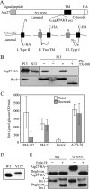
Figure 1.
Figure 1.
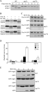
Atg27 is a type I transmembrane protein. (A) Schematic drawing of full-length Atg27. The full-length Atg27 protein contains 271 amino acids. Analysis of the primary amino acid sequence using the SignalP program indicates that Atg27 has a signal sequence (residues 1-19) and a transmembrane (TM) region (residues 199-221) according to TMHMM-prediction of helices in proteins (http://www.cbs.dtu.dk/services/TMHMM-2.0/), in a type I membrane topology. The putative PtdIns(3)phosphate-binding site (residues 188–193, KKPAKK) from the previous report (Wurmser and Emr, 2002) is indicated. Three possible membrane topologies for Atg27 are shown: I, Type II transmembrane orientation; II, two transmembrane domains; and III, Type I transmembrane protein. The asterisk marks the mutation introduced to replace glycine at position 105 with asparagine, creating a glycosylation site. (B) The C terminus of Atg27 is exposed to the cytosol. Atg27-HA (WLY1) and pep4Δ (TVY1) cells were converted into spheroplasts and then osmotically lysed. The cell lysates were centrifuged at 13,000 × g for 10 min to generate the pellet (P13) and supernatant (S13) fractions. The P13 fractions then were resuspended and subjected to treatment with 1% Triton X-100, proteinase K, both or neither on ice for 30 min. The lysates were then TCA-precipitated and analyzed by SDS-PAGE and Western blot. (C) The N-terminal 28 amino acids of Atg27 are able to direct invertase secretion. Cells expressing invertase fusion proteins (P4I-137, P4I-23, empty vector [pSEYC306], or A27I-28) were grown to early log phase. The cells were collected and subjected to an invertase activity assay as described in Materials and Methods. (D) The signal sequence of Atg27 is cleaved. Cells expressing Atg27-HA (WT) or Atg27V17P-HA (V17P) from the pAtg27–3xHA(416) or pAtg27V17P-3xHA(416) plasmids were grown to early log phase. The protein extracts were analyzed by Western blot and probed with monoclonal anti-HA antibody. (E) The lumenal region of Atg27 translocates into the ER. Cells expressing Atg27-HA (WT) and Atg27G105N-HA (G105N) were grown to early log phase. The cell lysates were subjected to endoglycosidase H treatment as described in Materials and Methods. After resolution by SDS-PAGE, the samples were analyzed by Western blot and probed with antibodies against Prc1 and HA, separately. The positions of glycosylated and deglycosylated forms of both proteins are indicated. The asterisk indicates cross-reacting bands.
Figure 2.
Figure 2.
The atg27Δ mutant is defective for autophagy-related pathways. (A) atg27Δ cells are defective in the Cvt pathway. Wild-type (WT; SEY6210), atg1Δ (WHY001), and atg27Δ (WLY2) cells were pulse-labeled for 10 min and subjected to a nonradioactive chase for 2 h. At the indicated time points cells were collected and TCA-precipitated. The cell lysates were immunoprecipitated with anti-Ape1 serum, resolved by SDS-PAGE, and then subjected to autoradiography. The positions of prApel and mApel are indicated. (B) Atg27 functions in the vesicle formation and/or completion step. Spheroplasts from the wild-type (pep4Δ vam3ts; WLY36) strain or this same strain harboring the atg1Δ (WLY74) or atg27Δ (WLY33) deletions were incubated at 37°C for 20 min, pulse-labeled with [35S]methionine/cysteine for 10 min, and then subjected to a nonradioactive chase for 27 min. The spheroplasts were osmotically lysed and separated into low-speed pellet and supernatant fractions after 5000 × g centrifugation. The pellet fractions that contained prApe1 were treated with proteinase K in the presence or absence of 0.2% Triton X-100. The resulting samples were immunoprecipitated with Ape1 antiserum and resolved by SDS-PAGE. (C) Atg27 is required for efficient pexophagy. Pex14-GFP (WT; IRA001), Pex14-GFP atg1Δ (IRA002), and Pex14-GFP atg27Δ (WLY27) strains were grown in oleic acid–containing medium to induce peroxisome proliferation and shifted to starvation medium. Protein extracts were prepared from cells at each indicated time point, resolved by SDS-PAGE, and probed with monoclonal anti-GFP antibody. The positions of Pex14-GFP and free GFP are indicated. (D) atg27Δ has an intermediate autophagy defect. atg1Δ (HAY572), wild-type (TN124), and atg27Δ (WLY3) cells expressing Pho8Δ60 were shifted from SMD to SD-N medium for 4 h. Samples at the indicated time points were collected, and protein extracts were assayed for alkaline phosphatase activity. The result represents the mean of three separate experiments, and the error bars represent the SD. (E) Wild-type (WT; SEY6210), atg1Δ (WHY001), and atg27Δ (WLY2) strains harboring a plasmid expressing GFP-Atg8 [pGFP-Aut7(414)] were grown in SMD lacking auxotrophic amino acids and shifted to SD-N. Aliquots were removed at the indicated time points. Protein extracts were prepared and resolved by SDS-PAGE. After Western blot, the membranes were probed with anti-GFP antibody.
Figure 3.
Figure 3.
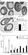
The atg27Δ mutant generated fewer autophagosomes. (A) The wild-type (pep4Δ vps4Δ; FRY143), atg1Δ (JHY28), and atg27Δ (WLY8) strains were grown to early log phase in YPD, shifted to SD-N for 5 h to induce autophagy, and examined by electron microscopy as described in Materials and Methods. Bar, 0.5 μm. (B) Quantification of the diameter of autophagic bodies (AB). To quantify the size of the accumulated ABs inside the vacuole, the diameter of ABs with a clear membrane boundary was measured. The number of ABs counted was 50 for the atg27Δ strain and 67 for wild type. (C) Quantification of autophagic body accumulation. To quantify the number of ABs accumulated, the number of autophagic bodies was counted in cells containing similar-sized vacuoles.
Figure 4.
Figure 4.
Atg27 cycles among the PAS, mitochondria, and Golgi complex. (A) Atg27 localizes to the PAS and partially to the mitochondria and Golgi. The strain expressing chromosomally tagged Atg27-GFP (WLY5) carrying either a PAS marker [pRFP-APE1(414)] or expressing chromosomally tagged Vrg4-RFP (Golgi complex marker; WLY6) were grown to early log phase or nitrogen starved for 3 h before imaging. For mitochondrial staining, the Atg27-GFP culture was incubated for 30 min in the presence of 1 μM MitoFluor Red 589 (Molecular Probes, Eugene, OR). The arrows indicate the colocalization of Atg27-GFP with mitochondria or the Golgi complex. (B) Atg27 is restricted to the PAS in atg1Δ cells. The chromosomally tagged Atg27-GFP atg1Δ strain (WLY11) carrying a PAS marker [pRFP-APE1(414)] was grown in selective medium to early log phase and then visualized by fluorescence microscopy. (C and D) The absence of Atg13, but not reduced Atg1 kinase activity, affected Atg27 localization. (C) The chromosomally tagged Atg27-GFP atg1Δ strain carrying an Atg1 kinase mutant (ATG1K54A) plasmid and (D) the Atg27-GFP atg13Δ strain (WLY18) were grown in selective SMD medium to OD600 = 0.8 and analyzed by fluorescence microscopy. Essentially identical results were obtained when cells were incubated in starvation medium. (E) Atg2 and Atg18 are required for efficient Atg27 retrograde cycling from the PAS. The chromosomally tagged Atg27-GFP atg2Δ strain (WLY78) and Atg27-GFP atg18Δ strain (WLY70) were grown in SMD medium and fixed with 1.5% formaldehyde in 50 mM potassium phosphate buffer (pH 8) for 30 min before imaging. DIC, differential interference contrast.
Figure 5.
Figure 5.
Atg27 localizes to mitochondria and the Golgi complex. The Atg27-HA (WLY1) strain was grown in YPD to OD600 = 1.0 and converted into spheroplasts. The spheroplasts were osmotically lysed and separated into pellet (P13) and supernatant (S13) fractions after a 13,000 × g centrifugation. The P13 pellet fraction was separated on a sucrose density gradient (18–54%) and centrifuged for 18 h at 176,000 × g as described in Materials and Methods. A total of 13 fractions were collected from the top to the bottom of the gradient and were subjected to immunoblot analysis with antiserum against Atg27-HA, Atg9, Pma1 (plasma membrane), Por1 (mitochondria), Mnn1 (Golgi complex), and Pho8 (vacuole).
Figure 6.
Figure 6.
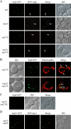
Atg27 functions before Atg1 and is required for Atg9 cycling. (A) Atg27 is required for Atg9 anterograde trafficking. The chromosomally tagged Atg9-GFP (WT; JLY44), Atg9-GFP atg1Δ (JLY45), Atg9GFP atg27Δ (JLY43), and Atg9-GFP atg1Δ atg27Δ (JLY47) strains expressing plasmid-based RFP-Atg8 were grown to OD600 = 0.8 in selective SMD medium and visualized by fluorescence microscopy. Arrows locate the PAS marker RFP-Atg8. (B) Atg9 localizes to mitochondria in the atg27Δ and atg1Δ atg27Δ mutants. The chromosomally tagged Atg9-GFP atg27Δ (JLY43) and Atg9-GFP atg1Δ atg27Δ (JLY47) strains were grown in SMD complete medium, and mitochondria were stained by incubating for 30 min in the presence of 1 μM MitoFluor Red 589. (C) The C-terminal cytosolic portion of Atg27 is not required for Atg9 cycling. Chromosomally tagged Atg9-GFP atg1Δ atg27ΔC (WLY49) cells were grown to early-log phase and collected for fluorescence microscopy. (D) Atg9 is required for Atg27 cycling from the non-PAS structures to the PAS. The chromosomally tagged Atg27-GFP atg1Δ atg9Δ strain (WLY41) expressing chromosomally tagged RFP-Ape1 was grown to early log phase in selective SMD medium before imaging. DIC, differential interference contrast.
Figure 7.
Figure 7.
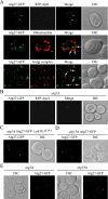
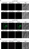
Binding to PtdIns(3)P is not required for Atg27 function. (A) Atg27 localization is not affected by Vps34. Chromosomally tagged Atg20-GFP and Atg27-GFP strains expressing plasmid-based vps34ts and RFP-Ape1 (WLY50 and WLY51, respectively) were grown in selective SMD medium to OD600 = 0.8 at 26°C or shifted to 38°C for 12 min before imaging. (B) Atg14 does not affect Atg27 PAS localization. The Atg27-GFP atg14Δ strain (WLY52) expressing RFP-Ape1 was grown in selective SMD medium and visualized by fluorescence microscopy. Bar, 5 μm.
Figure 8.
Figure 8.
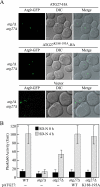
Mutation of the Atg27 putative PtdIns(3)P binding site has no effect on function. (A) The Atg27K188-193A mutant does not affect Atg9 cycling to the PAS. The chromosomally tagged Atg9-GFP atg1Δ atg27Δ strain (JLY47) expressing either plasmid-based Atg27–3xHA, Atg27K188-193A-3xHA or vector were grown in selective SMD medium and visualized by fluorescence microscopy. DIC, differential interference contrast. (B) Wild-type (TN124), atg1Δ (HAY572), and atg27Δ (WLY40) strains and the atg27Δ strain expressing plasmid-based wild-type Atg27-HA or Atg27K188-193A-HA were shifted from SMD to SD-N medium for 4 h. Samples were colleted at the indicated time points, and protein extracts were assayed for Pho8Δ60-dependent alkaline phosphatase activity. The results represent the mean of three separate experiments and the error bars represent the SD.

Similar articles

Cited by

References

    1. Abeliovich H., Zhang C., Dunn W. A., Jr, Shokat K. M., Klionsky D. J. Chemical genetic analysis of Apg1 reveals a non-kinase role in the induction of autophagy. Mol. Biol. Cell. 2003;14:477–490. - PMC - PubMed
    1. Campbell T. N., Choy F. Y. Expression of two green fluorescent protein variants in citrate-buffered media in Pichia pastoris. Anal. Biochem. 2002;311:193–195. - PubMed
    1. Cheong H., Yorimitsu T., Reggiori F., Legakis J. E., Wang C.-W., Klionsky D. J. Atg17 regulates the magnitude of the autophagic response. Mol. Biol. Cell. 2005;16:3438–3453. - PMC - PubMed
    1. Dunn W. A., Jr, Cregg J. M., Kiel J.A.K.W., van der Klei I. J., Oku M., Sakai Y., Sibirny A. A., Stasyk O. V., Veenhuis M. Peoxphagy: the selective autophagy of peroxisomes. Autophagy. 2005;1:75–83. - PubMed
    1. Gerhardt B., Kordas T. J., Thompson C. M., Patel P., Vida T. The vesicle transport protein Vps33p is an ATP-binding protein that localizes to the cytosol in an energy-dependent manner. J. Biol. Chem. 1998;273:15818–15829. - PubMed

Publication types

MeSH terms