

Androgen receptors are required for full masculinization of the ventromedial hypothalamus (VMH) in rats
- PMID: 17123532
- PMCID: PMC1828277
- DOI: 10.1016/j.yhbeh.2006.10.001
Androgen receptors are required for full masculinization of the ventromedial hypothalamus (VMH) in rats
Abstract
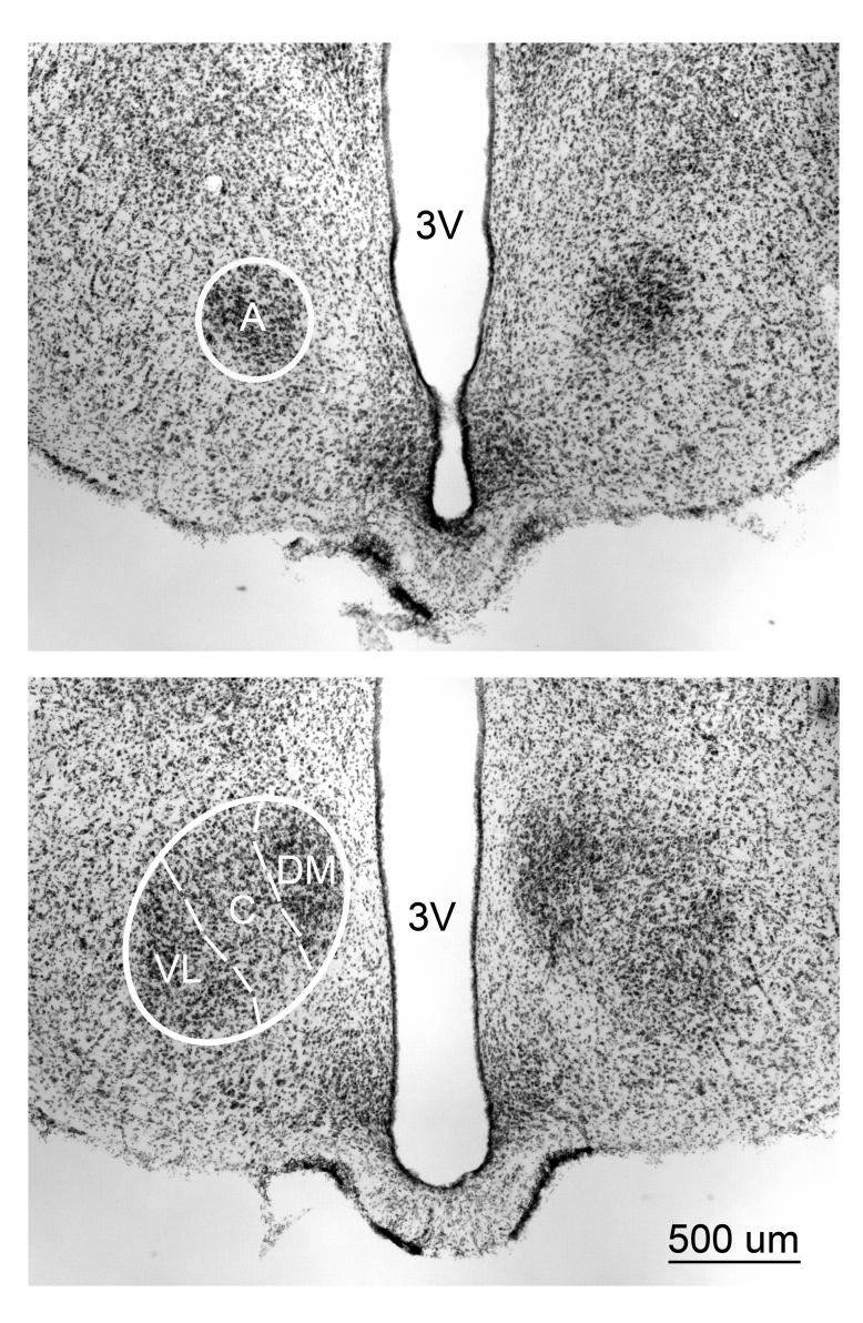
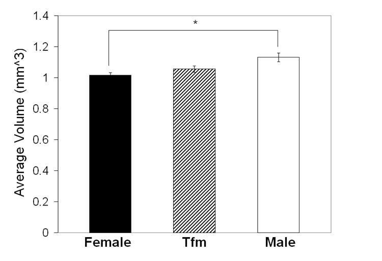
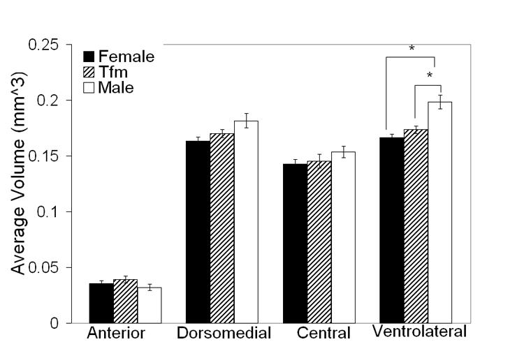
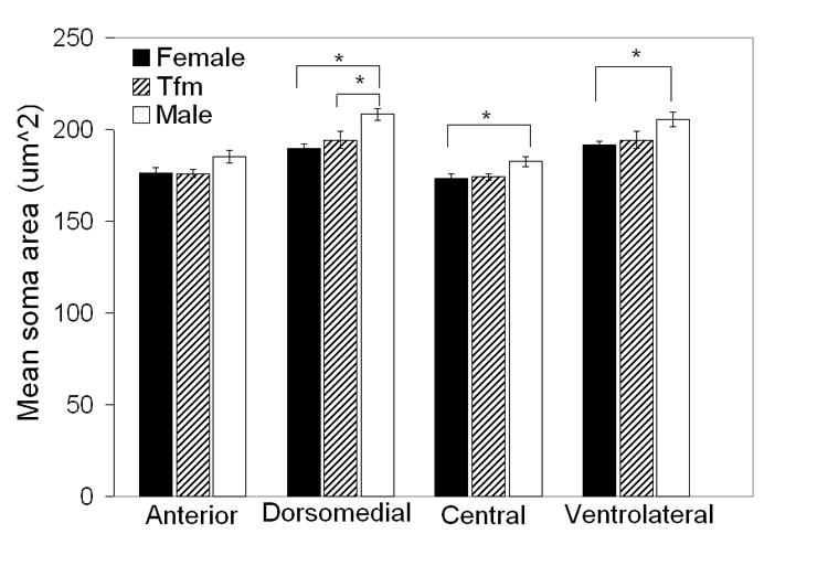
The ventromedial hypothalamus (VMH) is one of several sexually dimorphic nuclei that regulate mating behavior, and is rich in steroid hormone receptors and aromatase activity. We looked at the contribution of the androgen receptor (AR) to the volume of the VMH in rats by measuring each of the four subdivisions of the VMH in 90 day old male, female, and XY male rats carrying a mutant AR allele (tfm), which renders animals largely unresponsive to androgens. Confirming published reports, total VMH volume was greater in wild-type males than in females (P<0.01). The mean total volume of the VMH in TFM males was intermediate, but not significantly different from either females or males (Ps>0.10). The sex difference in VMH volume was primarily accounted for by the ventrolateral subdivision (VMHvl), which in both females and TFM males was significantly smaller than in wild-type males (Ps<0.005). There was no significant sex difference in the volume of the other three subdivisions of the VMH. Neuronal somata were larger in males than females in VMHvl, central VMH (VMHc) and the dorsomedial VMH (VMHdm), with TFM males having feminine neuronal somata in the VMHdm and VMHc. These data suggest that AR plays a role during sexual differentiation of the VMH, imparting its greatest effect in the VMHvl. ARs may regulate aromatase expression or activity to affect estrogen receptor activation, or may act independently of estrogen receptors to influence VMH morphology.
Figures
Similar articles
-
Selective sexual differentiation of neurone populations may contribute to sex-specific outputs of the ventromedial nucleus of the hypothalamus.J Neuroendocrinol. 2020 Jan;32(1):e12801. doi: 10.1111/jne.12801. Epub 2019 Dec 3. J Neuroendocrinol. 2020. PMID: 31605642 Free PMC article. Review.
-
Effects of the testicular feminization mutation (tfm) of the androgen receptor gene on BSTMPM volume and morphology in rats.Neurosci Lett. 2007 May 29;419(2):168-71. doi: 10.1016/j.neulet.2007.04.033. Epub 2007 Apr 21. Neurosci Lett. 2007. PMID: 17490813
-
Partial demasculinization of several brain regions in adult male (XY) rats with a dysfunctional androgen receptor gene.J Comp Neurol. 2005 Jun 27;487(2):217-26. doi: 10.1002/cne.20558. J Comp Neurol. 2005. PMID: 15880473
-
Androgen receptors are required for full masculinization of nitric oxide synthase system in rat limbic-hypothalamic region.Horm Behav. 2008 Sep;54(4):557-64. doi: 10.1016/j.yhbeh.2008.05.015. Epub 2008 Jun 6. Horm Behav. 2008. PMID: 18582470
-
Androgen action.Endocr Dev. 2014;27:28-40. doi: 10.1159/000363610. Epub 2014 Sep 9. Endocr Dev. 2014. PMID: 25247642 Review.
Cited by
-
Sex differences in glutamate transmission and plasticity in reward related regions.Front Behav Neurosci. 2024 Sep 18;18:1455478. doi: 10.3389/fnbeh.2024.1455478. eCollection 2024. Front Behav Neurosci. 2024. PMID: 39359325 Free PMC article. Review.
-
Emergence of sex differences in the development of substance use and abuse during adolescence.Pharmacol Ther. 2015 Sep;153:55-78. doi: 10.1016/j.pharmthera.2015.06.003. Epub 2015 Jun 3. Pharmacol Ther. 2015. PMID: 26049025 Free PMC article. Review.
-
Androgen receptor deficiency is associated with reduced aromatase expression in the ventromedial hypothalamus of male cichlids.Ann N Y Acad Sci. 2024 Feb;1532(1):73-82. doi: 10.1111/nyas.15096. Epub 2024 Jan 19. Ann N Y Acad Sci. 2024. PMID: 38240562 Free PMC article.
-
Selective sexual differentiation of neurone populations may contribute to sex-specific outputs of the ventromedial nucleus of the hypothalamus.J Neuroendocrinol. 2020 Jan;32(1):e12801. doi: 10.1111/jne.12801. Epub 2019 Dec 3. J Neuroendocrinol. 2020. PMID: 31605642 Free PMC article. Review.
-
Sex differences in the neural circuit that mediates female sexual receptivity.Front Neuroendocrinol. 2011 Apr;32(2):124-36. doi: 10.1016/j.yfrne.2011.02.008. Epub 2011 Feb 19. Front Neuroendocrinol. 2011. PMID: 21338620 Free PMC article. Review.
References
-
- Beach FA, Buehler MG. Male rats with inherited insensitivity to androgen show reduced sexual behavior. Endocrinology. 1977;100(1):197–200. - PubMed
-
- Christensen LW, Nance DM, Gorski RA. Effects of hypothalamic and preoptic lesions on reproductive behavior in male rats. Brain Res Bull. 1977;2(2):137–41. - PubMed
-
- Cooke BM, Breedlove SM, Jordan CL. Both estrogen receptors and androgen receptors contribute to testosterone-induced changes in the morphology of the medial amygdala and sexual arousal in male rats. Horm Behav. 2003;43(2):336–46. - PubMed
-
- Etgen AM, Morales JC. Somatosensory stimuli evoke norepinephrine release in the anterior ventromedial hypothalamus of sexually receptive female rats. J Neuroendocrinol. 2002;14(3):213–8. - PubMed
Publication types
MeSH terms
Substances
Grants and funding
LinkOut - more resources
Full Text Sources
Research Materials
