. 2006 Nov 15;20(22):3185-97.
doi: 10.1101/gad.1463206.

p63 regulates proliferation and differentiation of developmentally mature keratinocytes

Affiliations

p63 regulates proliferation and differentiation of developmentally mature keratinocytes

Amy B Truong et al. Genes Dev. .

Abstract

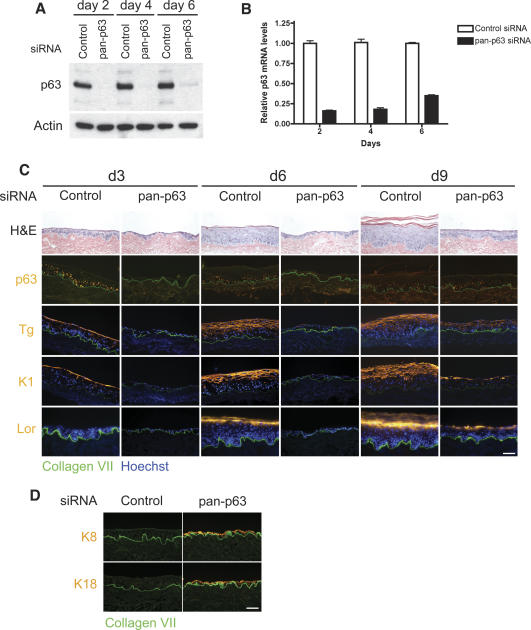
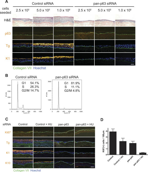
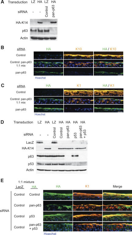
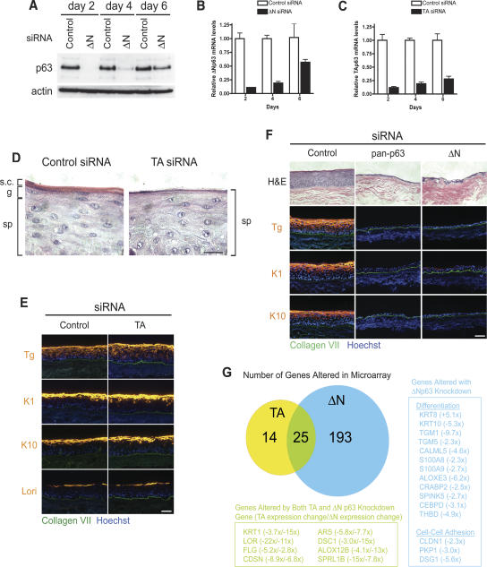
p63 is a multi-isoform p53 family member required for epidermal development. Contrasting roles for p63 in either the initial commitment to the stratified epithelial cell fate or in stem cell-based self-renewal have been proposed. To investigate p63 function in a post-developmental context, we used siRNAs directed against p63 to down-regulate p63 expression in regenerating human epidermis. Loss of p63 resulted in severe tissue hypoplasia and inhibited both stratification and differentiation in a cell-autonomous manner. Although p63-deficient cells exhibited hypoproliferation, differentiation defects were not due to tissue hypoplasia. Simultaneous p63 and p53 knockdown rescued the cell proliferation defect of p63 knockdown alone but failed to restore differentiation, suggesting that defects in epidermal proliferation and differentiation are mediated via p53-dependent and -independent mechanisms, respectively. Furthermore, DeltaNp63 isoforms are the main mediators of p63 effects, although TAp63 isoforms may contribute to late differentiation. These data indicate that p63 is required for both the proliferative and differentiation potential of developmentally mature keratinocytes.


Figures

Figure 1.
Figure 1.
p63 is required for stratification and differentiation in regenerating epidermis. (A) Western blot over a 6-d time course after introduction of siRNA oligonucleotide duplexes into primary human keratinocytes. The major band represents ΔNp63α, the predominant species in cultured keratinocytes. (Control) Control oligonucleotide; (pan-p63) oligonucleotide targeting all p63 isoforms. (B) Time course of qRT–PCR analysis of p63 mRNA levels following nucleofection with oligonucleotide duplexes targeting pan-p63 or control oligonucleotide duplexes. (C) Organotypic epidermal tissue regenerated from primary human keratinocytes after introduction of siRNA targeting all p63 isoforms (pan-p63) versus control siRNA. Tissue analyzed at days 3, 6, and 9 (d3, d6, d9, respectively) by histology and immunostaining with antibodies that recognize all p63 isoforms (p63) and the differentiation markers transglutaminase 1 (Tg), keratin 1 (K1), and loricrin (Lor), all in orange. (Green) Type VII collagen, basement membrane zone marker; (blue) Hoechst 33342 nuclear staining. Note hypoplasia and lack of differentiation marker expression in tissue lacking p63. (D) Four-day organotypic epidermal tissue generated from control or pan-p63 RNAi-treated keratinocytes was immunostained for keratins K8 and K18 (orange) and costained for type VII collagen (green); note the absence of K8/K18 in control and its appearance in p63-deficient tissue. Bar, 100 μm.
Figure 2.
Figure 2.
Effects of p63 loss on stratification and differentiation are not due to inadequate cell numbers or hypoproliferation. (A) p63 effects are independent of total cell numbers. Increasing numbers of control or p63 RNAi-treated cells were seeded onto devitalized dermis, and the tissue was harvested at 4 d. Note that even when p63 siRNA-treated cells were present in multiple layers, there was a lack in tissue polarity and organization, and differentiation marker expression was absent. Bar, 100 μm. (B) p63 knockdown results in G1 cell cycle arrest. FACS cell cycle profiles of control or pan-p63 siRNA-treated keratinocytes 60 h following transfection. Percent of cells in G1, S, or G2/M are indicated. (C) Hypoproliferation does not account for the differentiation defects observed with p63 loss. Tissue rendered hypoproliferative with 0.2 mM treatment of the DNA synthesis inhibitor HU was compared at 4 d to tissue generated from cells deficient for p63 and p53 control. Note decreased mitotic activity in HU-treated tissue but retained capacity for induction of the differentiation markers transglutaminase 1 (Tg), K1, and K10. Bar, 100 μm. (D) Quantitative mitotic index of organotypic tissue. HU indicates addition of 0.2 mM HU to the culture media.
Figure 3.
Figure 3.
Hypoproliferation induced by p63 deficiency is p53 dependent. (A) p63 knockdown increases p21 protein levels. Western blot of control or pan-p63 siRNA-transfected cells at day 2 (d2) and day 4 (d4) following siRNA transfection. (B) Simultaneous knockdown of p53 or p21 expression in combination with p63 knockdown inhibits p21 induction. Western blot of keratinocyte cell lysates; the introduced siRNA is indicated along the top, and the antibodies used for each panel are shown on the left. (C) p53 knockdown rescues the cell proliferation defect in vitro. Levels of BrdU incorporation were determined at day 2 (d2) and day 4 (d4) following transfection with the indicated siRNAs. Levels were normalized to cells receiving control siRNA, and error bars indicate the standard deviation among quadruplicate samples. (D) p53 knockdown rescues cell proliferation in organotypic epidermal tissue. Mitotic index of 4-d organotypic cultures generated from cells treated with the indicated siRNA. Cells were labeled with BrdU 12 h prior to harvesting, and tissue was stained with a BrdU antibody. (E) Rescue of cell proliferation does not rescue the differentiation defect in p63-deficient epidermal tissue. Organotypic tissues generated from the indicated siRNA-transfected cells were immunostained with antibodies against BrdU and transglutaminase 1 (Tg). Note the increase in staining with BrdU in the pan-p63/p53 double-knockdown sample but the absence of differentiation marker expression. Bar, 100 μm.
Figure 4.
Figure 4.
p63 effects on epidermal differentiation are cell autonomous. (A) Western blots of primary keratinocyte cell extracts after retroviral gene transfer and oligonucleotide nucleofection; the genes introduced are shown at the top of the panel with siRNA oligonucleotide below. Antibodies used for immunoblotting are shown at the left of each panel. LacZ (LZ)- and HA-epitope-tagged K14 were used to label cells with control and pan-p63 knockdown, respectively. (B,C) Epidermal tissue generated from LacZ-labeled control cells (top panels), HA-K14-labeled pan-p63 knockdown cells (bottom panels), or a 1:1 mixture of each (middle panels) were stained with antibodies for the differentiation markers K10 (B) and K1 (C). Tissue was harvested at 4 d. Note the complete absence of differentiation marker expression in p63 knockdown cells and the failure of immediately adjacent normal cells to rescue this. Bar, 50 μm. (D) Western blots of cell extracts following retroviral transduction and siRNA transfection. The transduced genes and siRNA oligonucleotide are indicated along the top for each sample. Antibodies detected by Western blot are indicated on the left of the panel. (E) Organotypic tissue generated from a 1:1 ratio of LacZ-expressing control cells and HA-K14-expressing cells receiving one of the following siRNAs: control, pan-p63, p53, or pan-p63 and p53. Tissue was harvested at day 4 and immunostained with antibodies recognizing HA (green) and K1 (orange). Note the presence of overlap between the HA-K14-expressing cells and K1 in the control and p53 single-knockdown mixes but the absence of overlap in tissue generated from HA-K14-expressing cells with pan-p63 knockdown alone or in combination with p53 knockdown. Bar, 50 μm.
Figure 5.
Figure 5.
ΔNp63 isoforms are required for epidermal stratification and differentiation. (A) Western blot after introduction of control oligonucleotide or oligonucleotide targeting ΔNp63 isoforms. (B,C) Time course of qRT–PCR analysis for cells treated with oligonucleotide targeting ΔNp63 isoforms (B) or TAp63 isoforms (C). (D) High-powered magnification (100×) of the tissue histology of 4-d organotypic cultures generated with control or TAp63 siRNA-transfected keratinocytes. The spinous layer (sp), granular layer (g), and stratum corneum (s.c.) are indicated. Note the absence of granules or stratum corneum from tissue generated from TAp63 knockdown cells. Bar, 20 μm. (E) Differentiation marker expression of 4-d organotypic epidermal tissue with control or TAp63 knockdown. Bar, 100 μm. (F) Control, pan, or ΔNp63 iso-specific RNAi-transfected keratinocytes were used to regenerate organotypic skin tissues. Tissue was harvested at 4 d and analyzed by histology and immunostaining for expression of the indicated differentiation proteins. Note the clear failure of stratification and differentiation with ΔNp63 isoform knockdown. Bar, 100 μm. (G) Venn diagram displaying the number of gene expression changes that accompanied TA or ΔNp63 knockdown in organotypic culture. Numbers in the Venn diagram represent genes that displayed a twofold or greater change with a p-value ≤0.05 by one-way ANOVA. Genes that met these criteria and are involved in differentiation or cell–cell adhesion are listed in the boxed regions. Numbers next to the gene name indicates the average fold change in expression, with (+) representing up-regulation and (−) representing down-regulation. For genes that changed in both TA and ΔN p63 knockdown tissue, the first number corresponds to TA knockdown samples, and the second number corresponds to ΔN knockdown samples.

Similar articles

Cited by

References

    1. Bond, J., Haughton, M., Blaydes, J., Gire, V., Wynford-Thomas, D., Wyllie, F. Evidence that transcriptional activation by p53 plays a direct role in the induction of cellular senescence. Oncogene. 1996;13:2097–2104. - PubMed
    1. Candi, E., Rufini, A., Terrinoni, A., Dinsdale, D., Ranalli, M., Paradisi, A., De Laurenzi, V., Spagnoli, L.G., Catani, M.V., Ramadan, S., et al. Differential roles of p63 isoforms in epidermal development: Selective genetic complementation in p63 null mice. Cell Death Differ. 2006;13:1037–1047. - PubMed
    1. Carroll, D.K., Carroll, J.S., Leong, C.O., Cheng, F., Brown, M., Mills, A.A., Brugge, J.S., Ellisen, L.W. p63 regulates an adhesion programme and cell survival in epithelial cells. Nat. Cell Biol. 2006;8:551–561. - PubMed
    1. Delehedde, M., Cho, S.H., Hamm, R., Brisbay, S., Ananthaswamy, H.N., Kripke, M., McDonnell, T.J. Impact of Bcl-2 and Ha-ras on keratinocytes in organotypic culture. J. Invest. Dermatol. 2001;116:366–373. - PubMed
    1. Donehower, L.A., Harvey, M., Slagle, B.L., McArthur, M.J., Montgomery, C.A., Jr., Butel, J.S., Bradley, A. Mice deficient for p53 are developmentally normal but susceptible to spontaneous tumours. Nature. 1992;356:215–221. - PubMed

Publication types

MeSH terms