. 2007 Feb;81(3):1401-11.
doi: 10.1128/JVI.01740-06. Epub 2006 Nov 15.

Retinoic acid-inducible gene I mediates early antiviral response and Toll-like receptor 3 expression in respiratory syncytial virus-infected airway epithelial cells

Affiliations

Retinoic acid-inducible gene I mediates early antiviral response and Toll-like receptor 3 expression in respiratory syncytial virus-infected airway epithelial cells

Ping Liu et al. J Virol. 2007 Feb.

Abstract

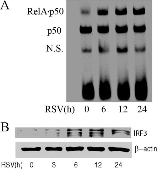
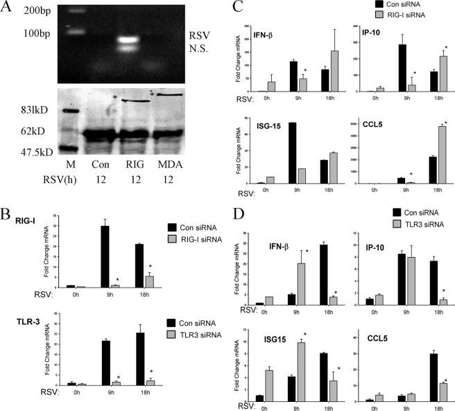
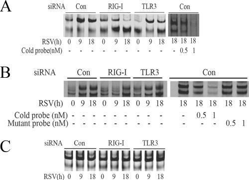
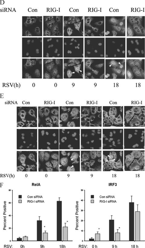
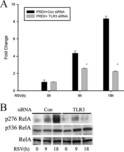
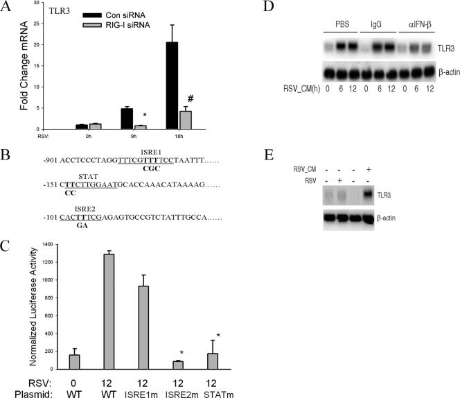
Respiratory syncytial virus (RSV) is one of the most common viral pathogens causing severe lower respiratory tract infections in infants and young children. Infected host cells detect and respond to RNA viruses using different mechanisms in a cell-type-specific manner, including retinoic acid-inducible gene I (RIG-I)-dependent and Toll-like receptor (TLR)-dependent pathways. Because the relative contributions of these two pathways in the recognition of RSV infection are unknown, we examined their roles in this study. We found that RIG-I helicase binds RSV transcripts within 12 h of infection. Short interfering RNA (siRNA)-mediated RIG-I "knockdown" significantly inhibited early nuclear factor-kappaB (NF-kappaB) and interferon response factor 3 (IRF3) activation 9 h postinfection (p.i.). Consistent with this finding, RSV-induced beta interferon (IFN-beta), interferon-inducible protein 10 (IP-10), chemokine ligand 5 (CCL-5), and IFN-stimulated gene 15 (ISG15) expression levels were decreased in RIG-I-silenced cells during the early phase of infection but not at later times (18 h p.i.). In contrast, siRNA-mediated TLR3 knockdown did not affect RSV-induced NF-kappaB binding but did inhibit IFN-beta, IP-10, CCL-5, and ISG15 expression at late times of infection. Further studies revealed that TLR3 knockdown significantly reduced NF-kappaB/RelA transcription by its ability to block the activating phosphorylation of NF-kappaB/RelA at serine residue 276. We further found that TLR3 induction following RSV infection was regulated by RIG-I-dependent IFN-beta secreted from infected airway epithelial cells and was mediated by both IFN response-stimulated element (ISRE) and signal transducer and activator of transcription (STAT) sites in its proximal promoter. Together these findings indicate distinct temporal roles of RIG-I and TLR3 in mediating RSV-induced innate immune responses, which are coupled to distinct pathways controlling NF-kappaB activation.


Figures

FIG. 1.
FIG. 1.
RSV activates NF-κB and IRF-3 in A549 cells. A549 cells were infected by the human RSV A2 strain (MOI = 1.0) for different times as indicated (in hours), and NE was prepared. (A) EMSA was performed on 5 μg NE using 0.1 nM IRDye 700-labeled DNA probe containing the κB element. The composition of the bound complexes is indicated. RelA · p50, RelA-p50 heterodimer; p50, p50 homodimer; N.S., nonspecific product. (B) Western immunoblotting was conducted on 50 μg NE. Top panel, the membrane was stained with anti-IRF-3 Ab; bottom panel, β-actin was stained as a loading control.
FIG. 2.
FIG. 2.
RIG-I mediates early innate immune response to RSV infection. (A) RNA helicase UV cross-linking and immunoprecipitation assay. A549 cells were transfected with 2 μg pT1S vector as control (Con), pT1S-FLAG-RIG-I (RIG), or pT1S-FLAG-MDA5 (MDA) for 24 h. Thereafter, cells were infected with RSV for 12 h. UV cross-linking and immunoprecipitation experiments were conducted as described in Materials and Methods. RT-PCR yielded two bands in the sample corresponding to RIG-I immunoprecipitation. The upper band was confirmed as RSV N protein RNA by sequencing (top panel); the bottom band is a nonspecific product (N.S.). Molecular sizes in base pairs (bp) are shown on the left. The bottom panel shows a Western immunoblot of the immunoprecipitates, including RIG-I and MDA5. Specific bands are indicated by asterisks. Molecular sizes are shown on the left. M, marker. (B) A549 cells were transfected with 100 nM of nonspecific siRNA as control (Con), RIG-I siRNA (RIG-I), or TLR3 siRNA (TLR3) for 48 h. Cells were infected by RSV for 0, 9, or 18 h, and total RNA was extracted. QRT-PCR was performed to determine changes in RIG-I (top panel) or TLR3 (bottom panel) expression levels as indicated. *, P is <0.01 compared to control siRNA (Student's t test). (C and D) A549 cells were transfected with control (Con), RIG-I siRNA (RIG-I), or TLR3 siRNA (TLR3) for 48 h and then RSV infected for 0, 9, or 18 h. CCL-5, IP-10, IFN-β, and ISG15 expression levels were determined by QRT-PCR; shown are the changes (n-fold) relative to unstimulated cells transfected with control siRNA (0 h). *, P is <0.05 relative to the corresponding group at the same time point (Student's t test). (C) Results of QRT-PCR of cells transfected with RIG-I siRNA and (D) cells transfected with TLR3 siRNA. Error bars indicate standard deviations.
FIG.3.
FIG.3.
RSV activates NF-κB and IRF-3 through the RIG-I pathway at the early phase of infection. (A) A549 cells were transfected with 100 nM of nonspecific siRNA as control (Con), RIG-I siRNA (RIG-I), or TLR3 siRNA (TLR3) for 48 h, followed by RSV infection for 9 or 18 h. NE from each siRNA treatment were prepared and assayed by EMSA. Shown are bound complexes on the IRDye 700-labeled κB oligonucleotides visualized by infrared scanning (left panel). A competition experiment was performed using the sample from the control siRNA-treated group that was infected with RSV for 18 h and incubated with 0, 0.5, or 1 nM unlabeled oligonucleotides (right panel). −, none. (B) IRF-3 binding at different times of RSV infection. EMSA was performed on NE using 0.1 nM IRDye 700-labeled ISRE binding site (left panel). A competition experiment with unlabeled probe and mutant probe was conducted (right panel). −, none. (C) OCT-1 binding. EMSA was performed using the same NE, binding 0.1 nM IRDye 800-labeled OCT-1 binding site. (D) A549 cells were transfected with either control (Con) or RIG-I siRNA (RIG-I) for 48 h and then RSV infected for 0, 9, or 18 h. The cells were fixed, incubated with rabbit anti-RelA Ab, and then stained with fluorescein isothiocyanate-conjugated anti-rabbit secondary Ab (top panels). The nuclei were stained with Sytox orange (middle panels). The slides were imaged using confocal microscopy, and colors were merged (bottom panels). Colocalization of RelA and nuclei is shown by light grey. White arrows indicate the cells which had RelA nuclear translocation. (E) A549 cells were treated as in (D), except that rabbit anti-IRF-3 Ab was used. Colocalization of IRF-3 and nuclei is shown by light grey and indicated by white arrows. (F) The percentages of cells with nuclei positive for RelA or IRF-3 at each time point and for each treatment were calculated based on five randomly photographed fields from two independent experiments. Asterisks indicate a significant difference between siRNA groups at the same time point of RSV infection (P < 0.05, Student's test). Error bars indicate standard deviations.
FIG.3.
FIG.3.
RSV activates NF-κB and IRF-3 through the RIG-I pathway at the early phase of infection. (A) A549 cells were transfected with 100 nM of nonspecific siRNA as control (Con), RIG-I siRNA (RIG-I), or TLR3 siRNA (TLR3) for 48 h, followed by RSV infection for 9 or 18 h. NE from each siRNA treatment were prepared and assayed by EMSA. Shown are bound complexes on the IRDye 700-labeled κB oligonucleotides visualized by infrared scanning (left panel). A competition experiment was performed using the sample from the control siRNA-treated group that was infected with RSV for 18 h and incubated with 0, 0.5, or 1 nM unlabeled oligonucleotides (right panel). −, none. (B) IRF-3 binding at different times of RSV infection. EMSA was performed on NE using 0.1 nM IRDye 700-labeled ISRE binding site (left panel). A competition experiment with unlabeled probe and mutant probe was conducted (right panel). −, none. (C) OCT-1 binding. EMSA was performed using the same NE, binding 0.1 nM IRDye 800-labeled OCT-1 binding site. (D) A549 cells were transfected with either control (Con) or RIG-I siRNA (RIG-I) for 48 h and then RSV infected for 0, 9, or 18 h. The cells were fixed, incubated with rabbit anti-RelA Ab, and then stained with fluorescein isothiocyanate-conjugated anti-rabbit secondary Ab (top panels). The nuclei were stained with Sytox orange (middle panels). The slides were imaged using confocal microscopy, and colors were merged (bottom panels). Colocalization of RelA and nuclei is shown by light grey. White arrows indicate the cells which had RelA nuclear translocation. (E) A549 cells were treated as in (D), except that rabbit anti-IRF-3 Ab was used. Colocalization of IRF-3 and nuclei is shown by light grey and indicated by white arrows. (F) The percentages of cells with nuclei positive for RelA or IRF-3 at each time point and for each treatment were calculated based on five randomly photographed fields from two independent experiments. Asterisks indicate a significant difference between siRNA groups at the same time point of RSV infection (P < 0.05, Student's test). Error bars indicate standard deviations.
FIG. 4.
FIG. 4.
The TLR3 pathway mediates the phosphorylation of NF-κB/RelA at serine 276. (A) A549 cells were transfected with control siRNA (Con) or TLR3 siRNA, and a luciferase reporter plasmid containing the PRDII domain was cotransfected for 48 h. Cells were infected with RSV for 0, 9, or 18 h before cell lysis was performed. Shown is normalized luciferase activity expressed as change (n-fold) relative to that of uninfected cells. *, P < 0.05 (Student's t test). Error bars indicate standard deviations. (B) A549 cells were transfected with control siRNA (Con) or TLR3 siRNA (TLR3) and then RSV infected for 0, 9, or 18 h. Western immunobloting was performed to detect changes in phospho-Ser276 RelA (top panel), phospho-Ser536 RelA (middle panel), and RelA (bottom panel) levels using 100 μg of whole-cell extract.
FIG. 5.
FIG. 5.
RSV-induced TLR3 expression depends on RIG-I-induced IFN-β secreted from infected cells. (A) A549 cells were transfected with control siRNA (Con) and RIG-I siRNA for 48 h and RSV infected for 0, 9, or 18 h. QRT-PCR was performed using TLR3 probe. #, P is <0.01; *, P is <0.05 relative to control siRNA at the same time point. Error bars indicate standard deviations. The results shown here are representative of two independent experiments. (B) Noncontiguous genomic sequence of hTLR3 promoter. The location relative to the major transcription start site is shown at left. Underlines, two predicted ISRE sites (ISRE1 and ISRE2) and one STAT site; bold font, site-directed mutagenesis of each individual regulatory element was performed by rolling-circle PCR. (C) A549 cells were transfected with either wild-type hTLR3/LUC reporter gene or different site mutants. Twenty-four hours later, cells were RSV infected and normalized luciferase activity was measured 12 h later. WT, wild type; *, P is <0.001 relative to wild-type hTLR3/LUC activity at 12 h. Error bars indicate standard deviations. (D) Naïve A549 cells were treated with 20% (vol/vol) UV-RSV-CM taken from RSV-infected cells for the indicated times (in hours). Prior to its addition to A549 cells, UV-RSV-CM was preincubated with PBS, rabbit IgG (IgG), or neutralizing anti-IFN-β Ab for 2 h. An autoradiogram from Northern blot hybridization is shown. Top panel, hybridization using radiolabeled TLR3 cDNA; bottom panel, hybridization with β-actin as an internal control. α, anti. (E) IFN-β-deficient Vero cells were infected with RSV for 12 h or were treated with 20% (vol/vol) UV-RSV-CM for 12 h. The conditioned medium was collected from A549 cells 24 h after RSV infection. Top panel, 20 μg total RNA was isolated and Northern blot hybridization conducted using TLR3 cDNA probe; bottom panel, β-actin hybridization. −, absent; +, present.

Similar articles

Cited by

References

    1. Aaronson, D. S., and C. M. Horvath. 2002. A road map for those who don't know JAK-STAT. Science 296:1653-1655. - PubMed
    1. Aherne, W., T. Bird, S. D. Court, P. S. Gardner, and J. McQuillin. 1970. Pathological changes in virus infections of the lower respiratory tract in children. J. Clin. Pathol. 23:7-18. - PMC - PubMed
    1. Akira, S., and K. Takeda. 2004. Toll-like receptor signalling. Nat. Rev. Immunol. 4:499-511. - PubMed
    1. Beutler, B. 2004. Inferences, questions and possibilities in Toll-like receptor signalling. Nature 430:257-263. - PubMed
    1. Brasier, A. R., J. E. Tate, and J. F. Habener. 1989. Optimized use of the firefly luciferase assay as a reporter gene in mammalian cell lines. BioTechniques 7:1116-1122. - PubMed

Publication types

MeSH terms

LinkOut - more resources