. 2007 Feb;81(3):1162-73.
doi: 10.1128/JVI.01702-06. Epub 2006 Nov 15.

Severe acute respiratory syndrome coronavirus infection of mice transgenic for the human Angiotensin-converting enzyme 2 virus receptor

Affiliations

Severe acute respiratory syndrome coronavirus infection of mice transgenic for the human Angiotensin-converting enzyme 2 virus receptor

Chien-Te K Tseng et al. J Virol. 2007 Feb.

Abstract

Animal models for severe acute respiratory syndrome (SARS) coronavirus infection of humans are needed to elucidate SARS pathogenesis and develop vaccines and antivirals. We developed transgenic mice expressing human angiotensin-converting enzyme 2, a functional receptor for the virus, under the regulation of a global promoter. A transgenic lineage, designated AC70, was among the best characterized against SARS coronavirus infection, showing weight loss and other clinical manifestations before reaching 100% mortality within 8 days after intranasal infection. High virus titers were detected in the lungs and brains of transgene-positive (Tg+) mice on days 1 and 3 after infection. Inflammatory mediators were also detected in these tissues, coinciding with high levels of virus replication. Lower virus titers were also detected in other tissues, including blood. In contrast, infected transgene-negative (Tg-) mice survived without showing any clinical illness. Pathologic examination suggests that the extensive involvement of the central nervous system likely contributed to the death of Tg+ mice, even though viral pneumonia was present. Preliminary studies with mice of a second lineage, AC63, in which the transgene expression was considerably less abundant than that in the AC70 line, revealed that virus replication was largely restricted to the lungs but not the brain. Importantly, despite significant weight loss, infected Tg+ AC63 mice eventually recovered from the illness without any mortality. The severity of the disease that developed in these transgenic mice--AC70 in particular--makes these mouse models valuable not only for evaluating the efficacy of antivirals and vaccines, but also for studying SARS coronavirus pathogenesis.


Figures

FIG. 1.
FIG. 1.
Construction and characterization of hACE2 transgene. (A) Diagram of hACE2 expression cassette. The entire open reading frame of human ACE2 (hACE2) was amplified by RT-PCR using mRNAs extracted from a human colon cancer cell line, Caco-2. The resulting cDNA of hACE2 was inserted into the expression vector pCAGGS.MCS, downstream of the CAG promoter, as described in Materials and Methods. The resulting plasmid was named pCAGGS.ACE. CMV-IE enh, CMV-IE enhancer. (B) Western blot analysis of hACE2 expression in transfected human 293 cells. Cell extracts prepared from mock-transfected (lane 1) or pCAGGS.ACE-transfected (lanes 2 and 3) human 293 cells were subjected to Western blot analysis to verify transgene expression using monoclonal antibody against hACE2. (C) Tissue expression profile of hACE2 in the transgenic mouse lineages AC70 (A) and AC63 (B). DNA-free RNAs extracted from different organs of transgenic mice at 6 to 8 weeks of age were subjected to RT-PCR analysis to evaluate the expression of hACE2 mRNA. The RT-PCR products were analyzed on 2% agarose gel. Lanes 1 to 9 represent spleen, stomach, heart, muscle, brain, kidney, lungs, intestine, and liver, respectively. The data shown are representative of two independently conducted experiments.
FIG. 2.
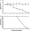
FIG. 2.
Weight loss and survival rate of SARS-CoV-infected AC70 Tg+ mice and their Tg littermates. Tg+ and Tg mice at 8 to 12 weeks of age were i.n. inoculated with 1 × 10³ TCID50 of SARS-CoV (Urbani strain) in 40 μl saline. Body weights (A) and accumulated mortality (B) of infected Tg+ (□) and Tg (○) mice were measured and recorded on a daily basis. Weight changes were expressed as the mean percent changes in infected animals (n = 10 per group, including those who died) relative to the initial weights at day 0. Error bars represent standard errors.
FIG. 3.
FIG. 3.
Kinetics of SARS-CoV replication in the lungs and brain of infected mice. Tg+ mice (□) and their Tg littermates (○) (n = 15 per group) were inoculated (i.n.) with 103 TCID50 of SARS-CoV in 40 μl saline. Three animals in each group were sacrificed daily, and virus titers in the lungs and brains were assessed by using both the standard TCID50 assay in Vero E6 cells and quantitative RT-PCR analysis, as described in Materials and Methods. The titers of infectious virus in the lungs (A) and brain (B) were calculated and expressed as log10 TCID50 virus per gram of tissue, whereas the relative copy numbers of SARS-CoV mRNA 5 (encoding M protein) of the lung (C) and brain (D) specimens as determined by Q-RT-PCR after normalization against 18S rRNA as the internal control were plotted (by the CT method). The average of mRNA 5 signals in duplicated samples of individual specimens is depicted. *, P < 0.05, and **, P < 0.01, by Student's t test, comparing Tg+ and Tg mice.
FIG. 4.
FIG. 4.
SARS-CoV replicates in the brains of mice following intraperitoneal inoculation. Tg+ (□) and Tg (○) mice (four in each group) were inoculated with 103 TCID50 of SARS-CoV. Tg+ mice started to show signs of illness at day 4. Three sick Tg+ animals, along with three apparently healthy Tg counterparts, were sacrificed at day 4 (n = 3), and the remaining mouse from each group was sacrificed at day 5 to titrate infectious virus in the brains. The infectious virus titer of individual mice is expressed as the log10 TCID50 per gram of tissue. **, P < 0.01 by Student's t test, comparing Tg+ and aged-matched Tg mice.
FIG. 5.
FIG. 5.
Histopathology and immunohistochemical analysis of SARS-CoV antigen expression in the lungs, brain, and GI tract of Tg+ mice after infection (i.n.). Paraffin-embedded lung (A to G), brain (H and I), and GI tract (J) sections of infected Tg+ mice were analyzed for the pathology and expression of the nucleocapsid protein of SARS-CoV by the methodologies described in Materials and Methods. SARS-CoV antigen (red) was readily detectable in the cytoplasm of epithelial cells of the bronchial lining (A) and pulmonary interstitium (B) at day 2. No staining was seen in the same Tg+ mouse when immunohistochemistry was performed with normal mouse ascites fluid (C). Shown are serial sections (hematoxylin and eosin in panel D and IHC in panel E) of a bronchus showing intraluminal macrophages and cellular debris in association with viral antigen. (F) Inflammatory cellular infiltrates (arrow) within smooth muscle of a pulmonary blood vessel associated with SARS-CoV antigen. (G) SARS-CoV immunostaining of a subepithelial ganglion cell in the lung at day 2. Extensive SARS-CoV antigen expression was first detected on day 3 in large numbers of morphologically intact neuronal and glial cells in the CNS (H and I). In the GI tract, the expression of SARS-CoV antigen in ganglia within the subserosal layer (arrow) was detected first at day 4 (J). Magnifications: A to F, H, and J, ×100; G, ×158; I, ×50. Staining: panels A to C and E to J, IHC with naphthol red and hematoxylin counterstaining; panel D, hematoxylin and eosin.
FIG. 6.
FIG. 6.
Expression of hACE2 in the lungs, brain, and GI tract of Tg+ mice. The paraffin-embedded sections of the lungs, brains, and GI tract were used to evaluate the expression of the hACE2 by IHC. The hACE2 antigen (red) was readily detectable primarily in the pneumocytes (A) and vascular smooth muscle in the lung (B, arrow). The hACE2 expression in the brain was also abundantly associated with choroid (C), ventricular lining (D), vascular endothelial cells (E), and patches of neuronal and glial elements (F and G). Finally, hACE2 was also found in the epithelial lining, muscularis layer, and ganglia of the GI system (H, arrow). Magnification: panels A, F, and G, ×158; panels B, D, and H, ×50; panel C, ×25; and panel E, ×100.
FIG. 7.
FIG. 7.
Expression of pulmonary cytokines and chemokines in infected mice. Lung homogenates derived from mice at indicated time intervals after infection (i.n.) were subjected to Bio-Plex analysis for assessment of the concentrations of cytokines and chemokines. Among 23 inflammatory mediators tested, the expression of IL-1β, IL-12p40, CXCL1/KC, RANTES, MCP-1, and IL-12p70 was elevated in infected Tg+, but not Tg, mice. Duplicate samples of individual specimens were assayed. The data shown are the mean ± standard error of infected animals (n = 3) at indicated time points. *, P < 0.05, and **, P < 0.01, by Student's t test, comparing Tg+ mice with aged-matched Tg controls.
FIG. 8.
FIG. 8.
Outcome in SARS-CoV-infected mice of the AC63 line. For the first experiment, Tg+ and Tg mice of the AC63 line (n = 10 each) were inoculated (i.n.) with 103 TCID50 of SARS-CoV, and the weight changes were recorded on a daily basis and expressed as the mean percent changes of infected animals (A). For the second experiment, 10 Tg+ AC63 mice were inoculated (i.n.) with 106 TCID50 of SARS-CoV. Five mice and three infected mice were sacrificed at days 5 and 8 after infection, respectively, and the titers of infectious virus in the lungs and brains were assessed and expressed as log10/gram (B). The other two infected mice were saved for observation of weight changes (C) and other clinical manifestations. **, P < 0.01 by Student's t test, comparing the virus titers between lungs and brain within Tg+ or Tg mice.

Similar articles

Cited by

References

    1. Arbour, N., G. Côté, C. Lachance, M. Tardieu, N. R. Cashman, and P. J. Talbot. 1999. Acute and persistent infection of human neural cell lines by human coronavirus OC43. J. Virol. 73:3338-3350. - PMC - PubMed
    1. Butler, N., L. Pewe, K. Trandem, and S. Perlman. 2006. Murine encephalitis caused by HCoV-OC43, a human coronavirus with broad species specificity, is partly immune-mediated. Virology 347:410-421. - PMC - PubMed
    1. Chan, K. S., J. P. Zheng, Y. W. Mok, Y. M. Li, Y. N. Liu, C. M. Chu, and M. S. Ip. 2003. SARS: prognosis, outcome and sequelae. Respirology 8:(Suppl.)S36-S40. - PMC - PubMed
    1. Coley, S. E., E. Lavi, S. G. Sawicki, L. Fu, B. Schelle, N. Karl, S. G. Siddell, and V. Thiel. 2005. Recombinant mouse hepatitis virus strain A59 from cloned, full-length cDNA replicates to high titers in vitro and is fully pathogenic in vivo. J. Virol. 79:3097-3106. - PMC - PubMed
    1. Ding, Y., L. He, Q. Zhang, Z. Huang, X. Che, J. Hou, H. Wang, H. Shen, L. Qiu, Z. Li, J. Geng, J. Cai, H. Han, X. Li, W. Kang, D. Weng, P. Liang, and S. Jiang. 2004. Organ distribution of severe acute respiratory syndrome (SARS) associated coronavirus (SARS-CoV) in SARS patients: implications for pathogenesis and virus transmission pathways. J. Pathol. 203:622-630. - PMC - PubMed

Publication types

MeSH terms

LinkOut - more resources