Review
. 2006 Nov;11(5):601-12.
doi: 10.1016/j.devcel.2006.10.010.

Catenins: keeping cells from getting their signals crossed

Affiliations
Review

Catenins: keeping cells from getting their signals crossed

Mirna Perez-Moreno et al. Dev Cell. 2006 Nov.

Abstract

Adherens junctions have been traditionally viewed as building blocks of tissue architecture. The foundations for this view began to change with the discovery that a central component of AJs, beta-catenin, can also function as a transcriptional cofactor in Wnt signaling. In recent years, conventional views have similarly been shaken about the other two major AJ catenins, alpha-catenin and p120-catenin. Catenins have emerged as molecular sensors that integrate cell-cell junctions and cytoskeletal dynamics with signaling pathways that govern morphogenesis, tissue homeostasis, and even intercellular communication between different cell types within a tissue. These findings reveal novel aspects of AJ function in normal tissues and offer insights into how changes in AJs and their associated proteins and cytoskeletal dynamics impact wound-repair and cancer.


Figures

Figure 1
Figure 1. Stages of Cadherin-Mediated Adhesion in Epithelial Cells
During the initial stages of intercellular adhesion, cells extend filopodial/lamellipodial extensions that enhance cell-cell contacts. Such dynamic membrane protrusive activity involves the actin cytoskeleton and Rho GTPases. Cadherin-catenin complexes are recruited to these nascent contacts, referred to as puncta, in a way dependent on Nectin/Afadin-based cell adhesions. cis-dimers of cadherin-catenin complexes engage in highly dynamic productive homophilic contacts, together with rearrangements of the actin cytoskeleton. These events are orchestrated by both α-catenin and p120-catenin. When more nascent contacts begin to form, the densities of AJ-associated proteins, including α-catenin, rise and promote the formation of α-catenin homodimers that may then serve as a feedback mechanism to dampen lamellipodial movements and promote the formation of radial actin cables as cell-cell junction formation progresses. At intermediate stages of intercellular adhesion, the lateral clustering of cadherins promotes the association of actin-binding and actin-polymerizing proteins. Under these conditions, membrane sealing is enhanced, and eventually radial actin cables also rearrange and get stabilized and bundled by myosin II, α-actinin, and possibly α-catenin homodimers. Establishment of mature cell contacts reorganizes the actin cytoskeleton to this more static state.
Figure 2
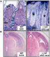
Figure 2. Conditional Ablation of α-E-Catenin Results in a Hyperproliferative State Resembling Cancer Both in Skin Epidermis and in Neuronal Progenitors in the Brain
(A) Skin. Embryonic day 18.5 skins from wild-type (WT) and K14-Cre-mediated, α-E-catenin conditionally targeted (cKO) mice were grafted onto the backs of athymic (Nude) mice, defective also in hair formation. The absence of α-E-catenin promotes epidermal hyperproliferation and the recruitment of proinflammatory infiltrates of eosinophils, macrophages, and neutrophils. By 60 days post engraftment (shown), undulating epidermal masses of α-E-catenin null cells exist, with no signs of hair follicle formation (asterisk). By 70 days, the underlying basement membrane has been invaded in discrete areas, and features of epithelial-to-mesenchymal transitions typical of squamous cell carcinoma arise (pictures kindly provided by H. Amalia Pasolli; Kobielak and Fuchs, 2006). (B) Coronal sections of developing cerebral cortex from E15.5 WT and α-E-catenin conditional mutant mice. The specific deletion of α-E-catenin in neural progenitors causes cortical hyperplasia and dysplasia when compared to controls. Note the abnormal size of the α-catenin conditional null brain, which correlates with a shortening of the cell cycle of neural progenitors, decreased apoptosis, and loss of differentiation (pictures kindly provided by V. Vasioukhin; Lien et al., 2006a).
Figure 3
Figure 3. Consequences of p120-Catenin Ablation in Skin and in the Submandibular Salivary Gland
(A) Skin sections from WT and K14-Cre mediated p120-catenin conditional mutant mice (P60). Loss of p120-catenin results in hair loss and epidermal hyperproliferation as a consequence of a chronic inflammatory skin disease. Note that the absence of p120-catenin results in the degeneration of hair follicles, enlarged blood vessels (arrow), and the recruitment of inflammatory infiltrates in the underlying dermis. Epi. epidermis; der, dermis; sf, subcutaneous fat (pictures kindly provided by H. Amalia Pasolli; Perez-Moreno et al., 2006). (B) Sections of submandibular salivary glands from P1 WT and MMTV-Cre-mediated p120-catenin conditional mutant mice. In the absence of p120-catenin, the acinar compartments in the salivary glands degenerate, and the normal ducts (white arrow) that drain into the mouth are occluded by masses of hyperproliferative cells that display features of high-grade intraepithelial neoplasia (black arrow) (pictures kindly provided by Al Reynolds; Davis and Reynolds, 2006).
Figure 4
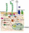
Figure 4. Role of Catenins in Mediating Signaling and Transcriptional Changes
Catenins regulate downstream signals that impact on several aspects of cell behavior. β-catenin has a well-established role in the Wnt signaling pathway, where it associates with members of the Lef/Tcf family of DNA-binding proteins to alter transcription. α-catenin orchestrates the appropriate balance between cell proliferation and differentiation by regulating a diverse number of signaling pathways including: Sonic hedgehog (Shh) and its downstream transcriptional mediator Gli; tyr kinase growth factor receptors and their downstream proliferation effectors Ras/MAPK; and NFκB, which when phosphorylated can enter the nucleus and regulate the expression of genes involved in proinflammatory responses. Loss-of-function mutations in α-catenin are associated with tissue degeneration, cancer, and marked inflammation. Loss-of-function mutations in p120-catenin have minor roles in lower eukaryotes such as Drosophila and C. elegans. However, in higher eukaryotes, alterations in p120-catenin promote the degradation of cadherins and deregulation of Kaiso. Additionally, cytosolic p120-catenin regulates the activities of Rho GTPases, inhibiting RhoA and activating Rac1 and Cdc42, thereby regulating actin cytoskeleton dynamics and cell migration. In addition, consistent with the broad roles for Rho GTPases in cell behavior, p120-catenin may also regulate several regulatory events, such as the RhoA-GTPase-mediated activation of IKK, the kinase that phosphorylates and activates NFκB. Genetic studies in mice have unambiguously demonstrated that the absence of p120-catenin is deleterious in skin, salivary gland, and brain tissues by modulating signals that promote tissue proliferation, migration, and inflammation, which may predispose the tissue to cancer.
Figure 5
Figure 5. Proposed roles of α-Catenin and p120-Catenin during Wound Healing and Cancer
Overall, the effects of α-catenin and p120-catenin are to dampen Ras/MAPK-mediated proliferation and NFκB -mediated proinflammatory responses. The links between cell-cell adhesion, proliferation, and inflammation may be key during a normal wound response. When cell-cell contacts are severed, proliferation must be triggered to repair the wound, and the immune system needs to be recruited to fight infection. As the wound is sealed and cell-cell junctions are repaired, proliferation and immune infiltration must be dampened again. In cases where α-catenin and/or p120-catenin are genetically defective, however, the cycle continues unchecked, resulting in chronic inflammation, uncontrolled proliferation, and cancer.

Similar articles

Cited by

References

    1. Adams CL, Nelson WJ, Smith SJ. Quantitative analysis of cadherin-catenin-actin reorganization during development of cell-cell adhesion. J. Cell Biol. 1996;135:1899–1911. - PMC - PubMed
    1. Adams CL, Chen YT, Smith SJ, Nelson WJ. Mechanisms of epithelial cell-cell adhesion and cell compaction revealed by high-resolution tracking of E-cadherin-green fluorescent protein. J. Cell Biol. 1998;142:1105–1119. - PMC - PubMed
    1. Akhtar N, Hotchin NA. RAC1 regulates adherens junctions through endocytosis of E-cadherin. Mol. Biol. Cell. 2001;12:847–862. - PMC - PubMed
    1. Anastasiadis PZ, Reynolds AB. The p120 catenin family: complex roles in adhesion, signaling and cancer. J. Cell Sci. 2000;113:1319–1334. - PubMed
    1. Anastasiadis PZ, Reynolds AB. Regulation of Rho GTPases by p120-catenin. Curr. Opin. Cell Biol. 2001;13:604–610. - PubMed

Publication types

LinkOut - more resources