. 2006 Nov 3;24(3):445-56.
doi: 10.1016/j.molcel.2006.09.011.

Structure and function of eukaryotic Ribonuclease P RNA

Affiliations

Structure and function of eukaryotic Ribonuclease P RNA

Steven M Marquez et al. Mol Cell. .

Abstract

Ribonuclease P (RNase P) is the ribonucleoprotein endonuclease that processes the 5' ends of precursor tRNAs. Bacterial and eukaryal RNase P RNAs had the same primordial ancestor; however, they were molded differently by evolution. RNase P RNAs of eukaryotes, in contrast to bacterial RNAs, are not catalytically active in vitro without proteins. By comparing the bacterial and eukaryal RNAs, we can begin to understand the transitions made between the RNA and protein-dominated worlds. We report, based on crosslinking studies, that eukaryal RNAs, although catalytically inactive alone, fold into functional forms and specifically bind tRNA even in the absence of proteins. Based on the crosslinking results and crystal structures of bacterial RNAs, we develop a tertiary structure model of the eukaryal RNase P RNA. The eukaryal RNA contains a core structure similar to the bacterial RNA but lacks specific features that in bacterial RNAs contribute to catalysis and global stability of tertiary structure.


Figures
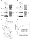
Figure 1
Figure 1
Intermolecular crosslinking analysis of eukaryal RNase P RNA-tRNA conjugates. (A) 5′-arylazido-B. subtilis mature tRNAasp crosslinks to eukaryal RNase P RNA. All reactions are identical except: (1) The reaction contained E. coli RNase P RNA but was not exposed to UV, (2) the thio-containing tRNA was not coupled to the azidophenacyl bromide, (3) no E. coli RNase P RNA, (4) RNA complementary to E. coli RNase P RNA was included instead of E. coli RNase P RNA, (5) E. coli RNase P RNA, (6) H. sapiens RNase P RNA, (7) C. elegans RNase P RNA, (8) D. melanogaster RNase P RNA, (9) S. cerevisiae RNase P RNA, (10) S. pombe RNase P RNA and (11) A. castellanii RNase P RNA was included. The S. pombe RNase P RNA-5′-arylazido-tRNA conjugates were analyzed by primer extension. Unlabeled S. pombe RNase P RNA-tRNA conjugates were prepared, purified and quantified as described in Experimental Procedures. Lane 1 and 2 contain primer extension products using oligonucleotides 150R and 250R respectively. Lanes C, U, A, and G correspond to sequencing reactions with non-crosslinked RNA template, lane N is a control primer extension without dideoxynucleotides of unmodified S. pombe RNase P RNA. The termination sites of primer extension are indicated to the right of each gel. (B) 3′-arylazido-mature RNA crosslinks to eukaryal RNase P RNA. Crosslinking and gel analysis is identical to A. (C) The secondary structure of S. pombe RNase P RNA with the crosslink sites inferred from primer extension analysis indicated by arrows. An RNase P RNA structural nomenclature is described in Marquez et al. 2005. Solid arrows indicate sites in the S. pombe RNA that crosslink to the 5′-end of tRNA. Unfilled arrows indicate sites in the S. pombe RNA that crosslink to the 3′-end of tRNA. The dashed arrow indicates the unique site crosslinked by 5′-s6G-labeled tRNA. (D) Measurement of the dissociation constant (Kd) of arylazido-mature B. subtilis tRNAasp and S. pombe RNase P RNA. Crosslinking reactions were performed in the presence of increasing amounts of S. pombe RNase P RNA (0 –2.0 X 10⁻⁵ M) incubated with 32P-labeled 5′-arylazido-mature tRNA (228 nM). (•) represents the fraction of tRNA bound to S. pombe RNase P RNA as assayed by radioactivity in the crosslinked band. The data were fit to the binding isotherm equation in Experimental Procedures.
Figure 2
Figure 2
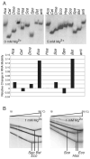
(A) Eukaryotic RNase P RNAs are affected differently by Mg2+ and are less stable than bacterial RNase P RNAs. Various eukaryotic and bacterial RNase P RNAs (Aca, A. castellanii; Cel, C. elegans; Dme, D. melanogaster; Eco, E. coli; Hsa, H. sapiens; Sce, S. cerevisiae; Spo, S. pombe; Bst, B. stearothermophilus; anti, control antisense E. coli) were folded and analyzed on 4.5% acrylamide 1X THE native gels containing 100 mM NH4OAc and various concentrations of Mg2+ (0 and 5 mM, shown left and right respectively). Minor amounts of some eukaryotic RNase P RNAs migrated in oligomeric forms as seen with bacterial RNAs and previously reported in Buck et al., 2005b (not shown). The relative change in RNA mobility between the 0 mM and 5 mM Mg2+ gels, normalized to the anti Eco control RNA, is graphed below. Relative change in mobility is defined as: (d5mM/anti d5mM)/(d0mM/anti d0mM) – 1, where d5mM is the distance the RNA traveled from the well on the 5 mM Mg2+ gel, anti d5mM is the distance the anti Eco control RNA traveled from the well on the 5 mM Mg2+ gel, d0mM is the distance the RNA traveled from the well on the 0 mM Mg2+ gel, and anti d0mM is the distance the anti Eco control RNA traveled from the well on the 0 mM Mg2+ gel. (B) The thermal stability of various eukaryotic and bacterial RNase P RNAs was analyzed by temperature gradient gel electrophoresis (TGGE). Gel conditions are as in A, except containing 1 mM Mg2+. (left) The E. coli and B. stearothermophilus RNase P RNAs melt at a higher temperature than S. pombe RNase P RNA. (right) Similarly, the E. coli RNase P RNA melts at a higher temperature than either H. sapiens or S. cerevisiae RNase P RNA. Vertical lines indicate the transition temperature, where RNA tertiary structure unfolds.
Figure 3
Figure 3
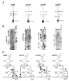
Intramolecular crosslinking analysis of circularly permuted S. pombe RNase P RNAs, cp69, cp118, cp140 and cp242. (A) Uniformly 32P–labeled photoagent-containing cpRNAs without (UV-) or with (UV+) 302 nm UV irradiation were analyzed by gel electrophoresis and autoradiography. The crosslinking reactions contained 1.0 μM photoagent containing RNAs. The cpRNA crosslink reactions resulted in crosslinked bands, designated x1, -x2, -x3 etc. (B) Primer extension mapping of crosslinked RNAs, were carried out with 5′-32P-labeled oligonucleotides complementary to S. pombe RNase P RNA (Supplemental Experimental Procedures). Primer extension mapping of some crosslinked RNAs indicated that they were circular molecules and uninformative (data not shown). Lanes C, U, A, and G correspond to sequencing reactions with non-crosslinked RNA template, lane N is a control primer extension without dideoxynucleotides of unmodified S. pombe RNase P RNA. The primer extension reactions using the crosslinked species are indicated above each lane. The termination sites of primer extension are indicated to the right of the gel. The RNA sequence of the termination sites is shown on the left. Filled circles denote the actual crosslink sites which are one nucleotide 5′ to the primer extension termination sites. (C) Crosslinking sites in the RNA secondary structures are shown. The boxed G indicates the photoagent attachment site located at the 5′ end of the cpRNA. The circled bases in the secondary structure represent the corresponding crosslinking sites. Arrowheads indicate the direction of the crosslinks.
Figure 4
Figure 4
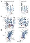
Commonalities in structure and function between the crystal structure of bacterial RNase P RNA and the modeled eukaryal RNase P RNA. (A) Coaxial stack representation of the secondary structures of B. stearothermophilus and S. pombe RNase P RNAs. The RNA that is homologous between B. stearothermophilus and S. pombe RNase P RNAs are represented as blue, non-homologous RNA is colored gray and RNA not represented in the B. stearothermophilus crystal structure is colored black in both molecules. Arrows indicate the 5′ to 3′ direction. Sites of 5′-tRNA crosslinking are represented as red spheres. B. stearothermophilus long-range tertiary interactions between helices are indicated by dashed lines, while homologous interactions are lacking in S. pombe RNA. The main site of 3′-tRNA crosslinking (P15) in B. stearothermophilus RNA is colored gold. A main site of 3′-tRNA crosslinking in S. pombe RNA is the P3 bulge-loop and, while not homologous to bacterial P15, is also colored gold. (B) Tertiary structure ribbon models of B. stearothermophilus and S. pombe RNase P RNAs, as colored in A. S. pombe RNase P RNA nucleotides 136-185 are not included in the modeling because of the inconsistency of the results between cp140 crosslink sites and the crystal structure of T. maritima RNase P RNA. The double-headed arrow indicates the long-range tertiary interaction between P5.1 and P15.1. The structure of P3 bulge-loop and P3b could not be reliably inferred from the bacterial structure and therefore has not been modeled. However, the location of 5′ and 3′-tRNA crosslinks (indicated by red spheres and regions highlighted in gold) suggest that these regions of the S. pombe RNA are in close proximity, consistent with the model. (C) Side-view of B. stearothermophilus and S. pombe RNase P RNAs. In the side-view the coaxially stack of P19, P2 and P3 is in the forefront. The general position of tRNA is indicated by a bracket.
Figure 5
Figure 5
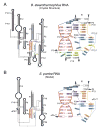
Catalytic core comparison of the bacterial RNA crystal structure and the modeled eukaryal RNase P RNAs. (A) Coaxial stack secondary structure of B. stearothermophilus RNase P RNA and a slab diagram representing base pairing and stacking interactions in the catalytic core. Joining region between P3 and P4 (J3/4) is colored in purple, P4 in red and purple, J5/15 in green, J15/15.1 in yellow, J15.2/2 in light blue, J19/4 in orange and J4/1 in dark blue. (B) Coaxial stack secondary structure of S. pombe RNase P RNA and a slab diagram representing base pairing and stacking interactions in the catalytic core. The NIHMS has received the file ‘mmc1.pdf’ as supplementary data. The file will not appear in this PDF Receipt, but it will be linked to the web version of your manuscript.

Similar articles

Cited by

References

    1. Beebe JA, Kurz JC, Fierke CA. Magnesium ions are required by Bacillus subtilis ribonuclease P RNA for both binding and cleaving precursor tRNAAsp. Biochemistry. 1996;36:10493–10505. - PubMed
    1. Brown JW. The Ribonuclease P Database. Nucleic Acids Res. 1999;27:314. - PMC - PubMed
    1. Brown JW, Nolan JM, Haas ES, Rubio MAT, Major F, Pace NR. Comparative analysis of ribonuclease P RNA using gene sequences from natural microbial populations reveals tertiary structural elements. Proc Natl Acad Sci USA. 1996;93:3001–3006. - PMC - PubMed
    1. Buck AH, Dalby AB, Poole AW, Kazantsev AV, Pace NR. Protein activation of a ribozyme: the role of bacterial RNase P protein. EMBO J. 2005;24:3360–3368. - PMC - PubMed
    1. Burgin AB, Pace NR. Mapping the active site of ribonuclease P RNA using a substrate containing a photoaffinity agent. EMBO J. 1990;9:4111–4118. - PMC - PubMed

Publication types

MeSH terms

LinkOut - more resources