. 2006 Nov 1;20(21):3022-35.
doi: 10.1101/gad.1477606.

Canonical notch signaling functions as a commitment switch in the epidermal lineage

Affiliations

Canonical notch signaling functions as a commitment switch in the epidermal lineage

Cédric Blanpain et al. Genes Dev. .

Abstract

Mammalian epidermis consists of a basal layer of proliferative progenitors that gives rise to multiple differentiating layers to provide a waterproof envelope covering the skin surface. To accomplish this, progenitor cells must detach from the basal layer, move upward, and execute a terminal differentiation program consisting of three distinct stages: spinous, granular layer, and stratum corneum. Notch signaling has been implicated in late stages of differentiation, but the commitment switch remains unknown. Here we show with loss and gain-of-function studies that active Notch intracellular domain (NICD) and its obligate canonical signaling partner RBP-J act at the basal/suprabasal juncture to induce spinous and down-regulate basal fate. Spinous layers are absent in RBP-J conditional null epidermis and expanded when Notch1 signaling is elevated transgenically in epidermis. We show that RBP-J is essential for mediating both spinous gene activation and basal gene repression. In contrast, the NICD/RBP-J target gene Hes1 is expressed in spinous layers and mediates spinous gene induction but not basal gene repression. These data uncover an early role for RBP-J and Notch in commitment of epidermal cells to terminally differentiate and reveal that spinous gene induction is mediated by a Hes1-dependent mechanism, while basal gene repression occurs independently of Hes1.


Figures
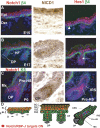
Figure 1.
Figure 1.
Notch/RBP-J-dependent signaling during epidermal development. In response to Notch signaling, NICDs are generated that associate with DNA-binding protein RBP-J to activate Notch target gene Hes1 and/or Hey1, encoding bHLH transcription factors (see also Supplementary Fig. S1). (A–C) Immunofluorescence microscopy and immunohistochemistry to monitor Notch signaling during skin embryogenesis. During the early stage of stratification (E15), Hes1 is expressed in the first suprabasal cell layer, although occasional basal cells are also positive (arrowhead). At E17, Hes1-positive cells are restricted to the spinous cell layer and in the inner core of the hair follicle (arrow). As hair follicles mature (postnatal day 0, P0), Hes1 is expressed in IRS and HS cells. Abs are color-coded according to fluorescently tagged secondary Abs. (DAPI) Blue. (D) Summary of Notch signaling patterns. (NICD1) Notch1 intracellular domain; (Hes1) Hairy Enhancer of Split 1; (Epi) epidermis; (Der) dermis; (β4) β4 integrin; (HF) hair follicle; (Mx) matrix; (pre-HS) differentiating hair shaft precursors; (ORS) outer root sheath; (IRS) inner root sheath; (SL) spinous layer; (BL) basal layer; (GL) granular layer; (SC) stratum corneum; (DP) dermal papilla, the mesenchymal component of the hair follicle.
Figure 2.
Figure 2.
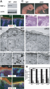
Conditional loss of RBP-J suppresses the commitment of epidermal cells to terminally differentiate. (A) Quantitative loss of expression of the Notch target Hes1 in RBP-J cKO skin. (B) Notch/RBP-J target gene Hes1 expression is greatly diminished in the absence of RBP-J. (C) K14-Cre/RBP-J cKO newborn animals display a wrinkled and translucent appearance. (D) Histology reveals a thin cKO skin epidermis. (E) Transmission electron microscopy of ultrathin sections of newborn wild-type (WT) and cKO epidermis. Boxed areas are magnified in images along bottom. Electron-dense keratohyalin granules (KG) mark the granular layer (G) cells, and are diminished in RBP-J cKO epidermis. cKO epidermis also shows an absence of spinous layer (S) cells, typified by dense keratin filament (Kf) bundles. (F–H) Immunofluorescence of spinous (F) and granular (G) markers and RT–PCR (H) revealed a marked reduction in differentiation markers in RBP-J cKO epidermis.
Figure 3.
Figure 3.
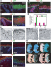
Conditional loss of RBP-J does not disrupt basal gene expression but does decrease proliferation. (A–D) Basal markers Keratin 5, Keratin 14, p63, and integrins are not altered in RBP-J KO epidermis. Immunostaining (A,B), FACS analysis (C), and RT–PCR (D) for basal markers shows that these markers are maintained if not slightly increased in the absence of RBP-J KO. (E,F) Staining and quantification of BrdU incorporation (E) and phospho-Histone H3 (F) demonstrates diminished proliferation in RBP-J KO epidermis. (G) Comparable immunofluorescence between wild-type (WT) and RBP-J cKO newborn skin stained with Abs against Keratin 6 (K6), a marker of hyperproliferative disorders in the epidermis.
Figure 4.
Figure 4.
Increased Notch signaling through NICD promotes activation of the target gene Hes1 and the first step of lineage commitment in the epidermis. (A) Skin-specific expression of the NICD-ires-GFP bicistronic transgene during embryogenesis, as measured by GFP epifluorescence. (B) NICD transgene expression results in NICD/RBP-J target gene Hes1 induction in the epidermis. (C) NICD transgenic animals display expansion of spinous layers as marked by Keratin 1 (K1) staining. (D)RT–PCR for spinous (K1) and granular markers (Filaggrin and Loricrin) shows altered differentiation in NICD1-expressing epidermis. (E) Ultrastructural analyses show that keratohyalin granules (G) are reduced and dense keratin bundles typical of spinous layers (S) are increased in NICD1 epidermis. (Kf) Keratin filaments. (F,G) Immunofluorescence microscopy reveals that both Loricrin and Filaggrin, the principal components of granular cells, are decreased in NICD transgenic epidermis. (H) Barrier assay, as determined by penetration of blue dye. The barrier is normally acquired and dye excluded by E17.5, but is defective in NICD transgenic animals, even after birth.
Figure 5.
Figure 5.
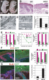
Increased Notch signaling results in inhibition of basal character and blistering disorder. (A) NICD1 transgenic animals display skin blistering at birth. (B) Histology reveals skin blistering at birth in NICD1 animals. (C) While the BL remains intact, there is a marked decrease in hemidesmosomes in NICD1 epidermis as quantified in D. (E) Temporal reduction in surface integrins and their mRNAs correlates with NICD induction in transgenic basal cells, analyzed by FACS (dispase and trypsin-treated epidermis, GFP+ gated cells). (F) Immunofluorescence microscopy and RT–PCR to monitor NICD-mediated changes in expression of basal markers. (G) Presence of atypical suprabasal S-phase epidermal cells in E18 transgenic mice pulsed 4 h with BrdU. (Graph) Quantification of suprabasal mitotic cells is determined by FACS analysis of BrdU-positive cells in α6-negative cells.
Figure 6.
Figure 6.
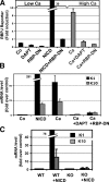
Calcium induces spinous differentiation through canonical Notch/RBP-J signaling. (A–C) Calcium induces RBP-J reporter gene activity (A), and endogenous K1/K10 gene expression (B) by a Notch/RBP-J-dependent mechanism, as this effect is blocked by expression of a dominant-negative RBP-J. (C) Stimulation of spinous markers by NICD1 is completely blocked in the absence of RBP-J. RBP-J reporter assays and quantifications of K1 and K10 were performed on FACS-isolated MK cultured in low-calcium (differentiation-restricted) or high-calcium (differentiation-promoting) medium, as indicated. Where noted, cells were also transduced with IRES-GFP retroviral expression vectors encoding NICD, dominant-negative RBP-J (RBP-DN), or empty vector (Co, control). Where indicated, cells were also treated with DAPT (N-[N-{3,5-difluorophenacetyl}-lalanyl]-S-phenylglycine t-butyl ester) to inhibit Notch processing and NICD production (Geling et al. 2002).
Figure 7.
Figure 7.
The Notch target gene Hes1 induces spinous gene expression. (A) Endogenous Hes1 and Hey1 can both be induced by either calcium or NICD1 expression. (B) Hes1 but not Hey1 can induce expression of spinous markers K1 and K10. MK were cultured in low calcium and infected with IRES-GFP retroviral expression vectors encoding NICD, Hes1, Hey1, or empty vector (Co, control). Proper expression was confirmed by immunoblot (see Supplementary Fig. S5B).
Figure 8.
Figure 8.
Basal markers are reduced by active Notch signaling. (A) Coculturing K14-GFP MK with cells expressing the Notch ligand Jagged1 (Jag1) results in a reduction in surface α6 integrin levels. MK were cocultured and then subjected to FACS for quantification. (B) MK were infected as in Figure 7 and then assayed for basal layer genes as monitored by RT–PCR of GFP-FACS-isolated keratinocyte mRNAs. (C) The absence of down-regulation of α6 integrin expression by NICD1 in RBP-J cKO cells demonstrated that Notch repression of basal integrin is RBP-J dependent. (D,E) MK were infected with NICD, Hes1, and Hey1, but assayed by FACS (D) and RT–PCR (E) for basal marker expression, and neither Hes1 nor Hey1 repressed basal markers.
Figure 9.
Figure 9.
A model depicting the roles of Notch signaling pathways in governing the transition from proliferation to differentiation in epidermal progenitors. Upon ligand engagement, Notch is activated, releasing NICD. NICD plays two roles in driving the transition from basal to spinous cells. NICD interacts with RBP-J to drive expression of Hes1, a canonical target gene. This leads to the downstream induction of spinous layer genes encoding differentiation-specific proteins. NICD represses basal genes including integrins, allowing basal cells to detach from the BL during stratification by a mechanism independent of Hes1.

Similar articles

Cited by

References

    1. Apelqvist, A., Li, H., Sommer, L., Beatus, P., Anderson, D.J., Honjo, T., Hrabe de Angelis, M., Lendahl, U., Edlund, H. Notch signalling controls pancreatic cell differentiation. Nature. 1999;400:877–881. - PubMed
    1. Artavanis-Tsakonas, S., Rand, M.D., Lake, R.J. Notch signaling: Cell fate control and signal integration in development. Science. 1999;284:770–776. - PubMed
    1. Bardin, A.J., Le Borgne, R., Schweisguth, F. Asymmetric localization and function of cell-fate determinants: A fly's view. Curr. Opin. Neurobiol. 2004;14:6–14. - PubMed
    1. Barolo, S., Walker, R.G., Polyanovsky, A.D., Freschi, G., Keil, T., Posakony, J.W. A notch-independent activity of suppressor of hairless is required for normal mechanoreceptor physiology. Cell. 2000;103:957–969. - PubMed
    1. Beres, T.M., Masui, T., Swift, G.H., Shi, L., Henke, R.M., MacDonald, R.J. PTF1 is an organ-specific and Notch-independent basic helix–loop–helix complex containing the mammalian Suppressor of Hairless (RBP-J) or its paralogue, RBP-L. Mol. Cell. Biol. 2006;26:117–130. - PMC - PubMed

Publication types

MeSH terms