. 2006 Nov 7;103(45):16704-9.
doi: 10.1073/pnas.0608358103. Epub 2006 Oct 30.

Coupling of STIM1 to store-operated Ca2+ entry through its constitutive and inducible movement in the endoplasmic reticulum

Affiliations

Coupling of STIM1 to store-operated Ca2+ entry through its constitutive and inducible movement in the endoplasmic reticulum

Yoshihiro Baba et al. Proc Natl Acad Sci U S A. .

Abstract

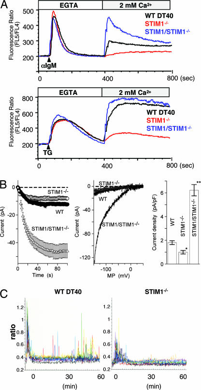
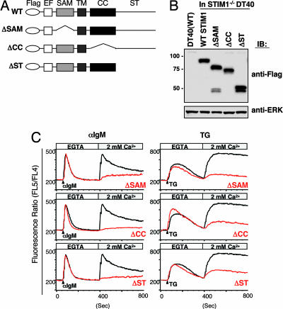
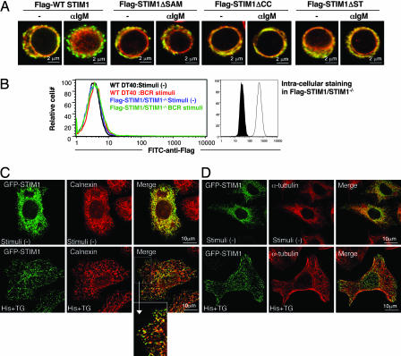
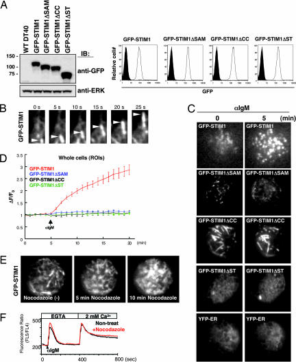
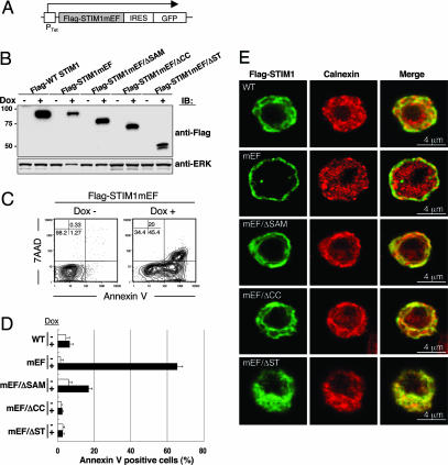
Depletion of intracellular calcium (Ca(2+)) stores induces store-operated Ca(2+) (SOC) entry across the plasma membrane (PM). STIM1, a putative Ca(2+) sensor in the endoplasmic reticulum (ER), has been recently shown to be necessary for SOC channel activation. Here we show that STIM1 dynamically moves in tubulovesicular shape on the ER and its subcompartment in resting living cells, whereas, upon Ca(2+) store depletion, it is rapidly redistributed into discrete puncta that are located underneath, but not inserted into the PM. Normal constitutive movement of STIM1 is mediated through the coiled-coil and Ser/Thr-rich C-terminal domains in the cytoplasmic region of STIM1, whereas subsequent inducible puncta formation further requires the sterile alpha motif domain protruding into the ER lumen. Each of these three domains (coiled-coil, Ser/Thr-rich, and sterile alpha motif) was essential for activating SOC channels. Hence, our findings based on structure-function experiments suggest that constitutive dynamic movement of STIM1 in the ER and its subcompartment is obligatory for subsequent depletion-dependent redistribution of STIM1 into puncta underneath the PM and activation of SOC channels.


Conflict of interest statement

Conflict of interest statement: A.W. is on the advisory board of the RIKEN Institute and encouraged T.K. to pursue this work.

Figures

Fig. 1.
Fig. 1.
Suppression of SOC influx, Icrac, and Ca2+ oscillation in STIM1-deficient DT40 B cells. (A) The Ca2+ mobilization profiles were monitored by Ca2+ add-back using Indo-1 imaging. Ca2+ release was first evoked by the stimulation of BCR [anti-IgM mAb (M4)] (Upper) or TG (Lower) in Ca2+-free conditions (0.5 mM EGTA), and Ca2+ influx was induced by restoring the extracellular Ca2+ concentration to 2 mM in WT (black lines), STIM1-deficient (STIM1−/−; red lines), or Flag-tagged STIM1 transduced STIM1-deficient (STIM1/STIM1−/−; blue lines) DT40 B cells. FL4, 500–520 nm; FL5, 400–420 nm. (B) Average time courses of ionic currents evoked by 10 μM IP3 at −130 mV in WT DT40 cells (•; n = 10), STIM1−/− cells (○; n = 24), and STIM1/STIM1−/− cells (△; n = 14). Cytosolic calcium was clamped to near zero with 10 mM EGTA. Data are means ± SE. The whole-cell configuration of the patch-clamp recording was established at time 0 (Left). Representative leak-subtracted current–voltage relationships of WT DT40 (black), STIM1−/− (gray), and STIM1/STIM1−/− (black) are shown (Center). A comparison of current density was also done at −130 mV. Columns are the means ± SE (Right). (C) Ca2+ oscillation in STIM1-deficient DT40 B cells after BCR stimulation. Ca2+ responses in single cells upon BCR stimulation with anti-IgM mAb (M4) (1 μg/ml) were monitored in Fura-2 loaded WT DT40 (Left) and STIM1−/− (Right) cells.
Fig. 2.
Fig. 2.
STIM1 utilizes its multiple functional domains for Ca2+ influx. (A) Schematic representation of STIM1 mutants and the functional domains, including Flag-tagged, SAM, coiled-coil (CC), and Ser/Thr-rich C-terminal (ST) domains. Flag-tagged STIM1 cDNAs encoding deletion mutations were transfected into STIM1-deficient DT40 B cells. EF, EF-hand motif; TM, a single transmembrane. (B) Expression of Flag-STIM1 in various mutant DT40 cells. Whole-cell lysates were fractionated by SDS/PAGE and immunoblotted with anti-Flag mAb (Upper) or anti-ERK Ab (Lower). (C) STIM1 utilizes the multiple functional domains for Ca2+ influx. Intracellular Ca2+ release and influx in response to BCR with anti-IgM mAb (Left) and TG stimulation (Right) in STIM1-deficient DT40 cells expressing WT Flag-STIM1 (black lines) or mutants (red lines) were measured by Ca2+ add-back methods.
Fig. 3.
Fig. 3.
Requirement of multiple functional domains for redistribution of STIM1 to punctate subcellular compartments. (A) Flag-STIM1 (green) and calnexin (red) immunofluorescence staining of DT40 mutants before and after BCR stimulation (10 min) in the absence of extracellular Ca2+. All images were taken with confocal microscopy. (B) Extracellular and intracellular staining of Flag-STIM1. Flow cytometry using FITC-anti-Flag mAb was conducted in nonpermeabilized WT DT40 B cells before (black line) and after (red line) BCR stimulation or STIM1-deficient DT40 B cells expressing Flag-STIM1 before (blue line) and after (green line) BCR stimulation (Left). Permeabilized cells were stained with FITC-anti-Flag mAb. Filled and open histograms represent WT DT40 B cells and STIM1-deficient DT40 B cells expressing Flag-STIM1, respectively (Right). (C and D) Subcellular distribution of STIM1 in HeLa cells. HeLa cells expressing GFP-STIM1 were stimulated with 100 μM histamine (His) and 2 μM TG for 10 min and then fixed and processed for immunofluorescence with anti-calnexin Ab (C) or anti-α-tubulin mAb (D). An enlargement of the boxed region in the merge images from C is also shown. All images were taken with confocal microscopy.
Fig. 4.
Fig. 4.
Microtubule-dependent dynamic behavior of GFP-STIM1 in living B cells. (A) cDNAs of GFP-tagged STIM1, STIM1 lacking SAM, CC, or ST domains were transduced in STIM1-deficient DT40 B cells, and these proteins were detected by immunoblotting with anti-GFP Ab (Left Upper). ERK protein was detected as a loading control (Left Lower). GFP expression in various DT40 mutants was detected by flow cytometry (Right). Filled and open histograms represent WT DT40 B cells and GFP-STIM1-transfected cells, respectively. (B) GFP signals of WT STIM1 near the PM were monitored by total internal reflection fluorescence microscopy at steady state. The movement of GFP-STIM1 was detected as tubular granules (arrowheads). Stills from the associated time-lapse Movies 1 and 2, taken from 410 s, are shown. (C) The movement of WT GFP-STIM1 and mutants were monitored before and after BCR stimulation in the absence of extracellular Ca2+. Stills from the associated time-lapse total internal reflection fluorescence movies are shown (Movies 1 and 2 for WT GFP-STIM1, Movies 3 and 4 for GFP-STIM1ΔSAM, Movies 5 and 6 for GFP-STIM1ΔCC, and Movies 7 and 8 for GFP-STIM1ΔST). The dynamics of YFP-ER are also shown (Movie 9). (D) Kinetic analysis of the average region of interest intensities of GFP-WT STIM1 (red line; n = 14), GFP-STIM1ΔSAM (blue line; n = 12), GFP-STIM1ΔCC (black line; n = 6), and GFP-STIM1ΔST (green line; n = 8). Cells were stimulated with anti-IgM mAb after 5 min in the absence of extracellular Ca2+. ΔF/Fo, the ratios of average intensities of regions of interest at indicated times after stimulation (F) and at steady state (Fo). Regions of interest are for whole cells. Error bars represent standard deviations of mean. (E) Dynamic behavior of GFP-STIM1 in DT40 cells treated with nocodazole (10 μM). Stills from the associated time-lapse Movie 10 are shown. (F) After 1 h of incubation of nocodazole, intracellular Ca2+ release and influx in response to BCR stimulation in DT40 B cells expressing Flag-STIM1 were monitored by Ca2+ add-back methods.
Fig. 5.
Fig. 5.
Expression of STIM1 EF-hand mutants, but not double mutants of EF hand and its functional domains, induces cell death. (A) Schematic representation of the doxycycline-inducible construct of STIM1 EF-hand mutants. cDNA of Flag-tagged STIM1 EF-hand mutants (mEF; D76A/D78A/N80A/E87A) and double mutation of mEF in addition with ΔSAM (mEF/ΔSAM), ΔCC (mEF/ΔCC), and ΔST (mEF/ΔST) linked by an internal ribosome entry site (IRES) to a cDNA encoding EGFP were transfected into STIM1-deficient DT40 B cells. Ptet, doxycycline responsible promoter. (B) Expression of STIM1 mEF mutants in DT40 cells after doxycycline induction for 24 h was detected by immunoblotting with anti-Flag mAb (Upper). ERK protein was detected as a loading control (Lower). (C) The expression of STIM1 EF-hand mutants induced cell death. STIM1-deficient DT40 cells expressing Flag-STIM1mEF were stained with phycoerythrin-annexin V and 7-amino-actinomycin D before and after 24 h doxycycline (Dox) induction and were analyzed by flow cytometry. The induction of STIM1 mutants was monitored by GFP (data not shown), and doxycycline-induced cell death was detected in the GFP-positive gate. (D) Cells were stained with phycoerythrin-annexin V before and after doxycycline induction for 24 h and were analyzed by flow cytometry. Bar graphs show the percentage of annexin V-positive cells among total (Dox−) and GFP+ (Dox+) cells. These results are representative of three independent experiments (means ± SE). (E) Localization of various STIM1 EF-hand mutants. Various Flag-STIM1 (green) and calnexin (red) immunofluorescence staining was shown after 24 h of doxycycline induction.

Similar articles

Cited by

References

    1. Berridge MJ, Lipp P, Bootman MD. Nat Rev Mol Cell Biol. 2000;1:11–21. - PubMed
    1. Lewis RS. Annu Rev Immunol. 2001;19:497–521. - PubMed
    1. Gallo EM, Cante-Barrett K, Crabtree GR. Nat Immunol. 2006;7:25–32. - PubMed
    1. Parekh AB, Putney JW., Jr Physiol Rev. 2005;85:757–810. - PubMed
    1. Venkatachalam K, van Rossum DB, Patterson RL, Ma HT, Gill DL. Nat Cell Biol. 2002;4:E263–E272. - PubMed

Publication types

MeSH terms

LinkOut - more resources