. 2006 Oct;2(10):e116.
doi: 10.1371/journal.ppat.0020116.

EC5S ubiquitin complex is recruited by KSHV latent antigen LANA for degradation of the VHL and p53 tumor suppressors

Affiliations

EC5S ubiquitin complex is recruited by KSHV latent antigen LANA for degradation of the VHL and p53 tumor suppressors

Qi-Liang Cai et al. PLoS Pathog. 2006 Oct.

Abstract

Cellular protein degradation pathways can be utilized by viruses to establish an environment that favors their propagation. Here we report that the Kaposi's sarcoma-associated herpesvirus (KSHV)-encoded latency-associated nuclear antigen (LANA) directly functions as a component of the EC5S ubiquitin complex targeting the tumor suppressors von Hippel-Lindau (VHL) and p53 for degradation. We have characterized a suppressor of cytokine signaling box-like motif within LANA composed of an Elongin B and C box and a Cullin box, which is spatially located at its amino and carboxyl termini. This motif is necessary for LANA interaction with the Cul5-Elongin BC complex, to promote polyubiquitylation of cellular substrates VHL and p53 in vitro via its amino- and carboxyl-terminal binding domain, respectively. In transfected cells as well as KSHV-infected B lymphoma cells, LANA expression stimulates degradation of VHL and p53. Additionally, specific RNA interference-mediated LANA knockdown stabilized VHL and p53 in primary effusion lymphoma cells. Thus, manipulation of tumor suppressors by LANA potentially provides a favorable environment for progression of KSHV-infected tumor cells.


Conflict of interest statement

Competing interests. The authors have declared that no competing interests exist.

Figures

Figure 1
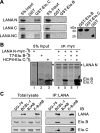
Figure 1. LANA Associates with the Elongin BC Complex
(A) The amino-terminal domain of LANA directly interacts with Elongin C but not Elongin B in vitro. The cDNA encoding N (1–340 aa), C (762-1162 aa), or NC (1–327̂929-1162 aa) truncation of LANA with myc tagged were expressed in a coupled transcription/translation system in the presence of [35S]methionine. Aliquots (20 μl) of the translation products were pulled down by GST, GST–Elongin C, or GST–Elongin B, respectively. The pulldown complex as well as a 1-μl aliquot of the transcription/translation reaction (Input) were fractionated by SDS-PAGE, and detected by a PhosphorImager. The proteins of GST and GST fused with Elongin B or Elongin C were shown by Coomassie staining in the right panel. (B) Elongin C is critical for LANA binding to Elongin B. In vitro produced and [35S]-labeled proteins LANA N (1–340 aa), Elongin C, and Elongin B were incubated with anti-myc antibodies. The interacting complexes were resolved by SDS-PAGE and examined by a PhosphorImager. (C) LANA associates with Elongin BC complex in KSHV-infected cells. Cell lysates of BCBL-1, DG-75, BJAB, or BJAB with KSHV infection (K-BJAB) were subjected to IP against LANA or Elongin C (Elo. C) followed by IB against LANA, Elongin C, and Elongin B (Elo. B).
Figure 2
Figure 2. The LANA–Elongin BC Complex Assemble with the Cul5/Rbx1 Module
(A) LANA protein associates with the Cul5/Rbx1 module but not Cul2/Rbx1 module, and (B) Elongin BC complex increases interaction of LANA with the Cul5/Roc1 module. Saos-2 cells were transfected with expression vector encoding the indicated proteins in the figure. The lysates underwent IP using anti-myc antibodies. Crude extracts (left panels) and immune complexes (right panels) were separated by SDS-PAGE and IB with the indicated antibodies.
Figure 3
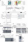
Figure 3. LANA Associates with the Elongin BC Complex and Cul5 through the SOCS-Box–Like Motif
(A) KSHV LANA contains a putative SOCS-box motif which includes a consensus of BC-box and Cul-box motifs. Upper panels show hydrophobic sequence alignment of the BC-box and Cul-box motifs from viral LANA and cellular SOCS-box–containing proteins that bind Elongin BC and Cullin proteins individually. Conserved residues with similarity are shaded in the upper panel. Lower panels show ORF73 proteins from other gammaherpesvirus contain an SOCS-box–like motif which is similar to KSHV LANA. HVS, herpesvirus saimiri; RRV, rhesus monkey rhadinovirus. The site mutations of LANA in leucine (L) and/or cystine (C) are indicated with underlining. (B) LANA associates with the Elongin BC complex through the BC-box motif. Saos-2 cells were transfected with expression vector encoding the indicated proteins. The WT LANA and its mutant with BC-box (ΔBC) deletion and point mutation (LPCF, L213G or C217G) within BC box were compared. Since LANA lost its normally top bands (approximately 230 kDa) in the L213G mutant, another two lower bands of LANA (between 190 kDa and 230 kDa) were specifically present. The lysates underwent IP and IB assays as described previously. RD, relative density. (C) LANA associates with Cullin 5 through the Cul-box motif. Saos-2 cells were transfected with expression vector encoding the indicated proteins. The WT LANA with Cul-box (ΔCul) or SOCS-box (ΔSCOS) deletion was compared. The lysates underwent IP and IB assays as described previously. Crude extracts (left panels) and immune complexes (right panels) are shown. (D) LANA assembles with Elongin BC complex and Cul5/Rbx1 module to reconstitute a multiprotein complex with E3 ubiquitin ligase activity. Left panel shows the LANA–Elongin BC–Cul5–Rbx1 complex had ubiquitin ligase activity. The cell lysates of WT LANA (Figure 3C, lane 4) or SOCS-box–like motif deletion mutant (Figure 3C, lane 3) as control was subjected to anti-myc immunoaffinity purification. The purified LANA immune complex (IC) was incubated with various combinations of Uba1 (E1), Ubc5a (E2), and GST-ubiquitin (GST-Ub) in the absence or presence of ATP, to assess its ability to stimulate ubiquitylation in vitro. The proteins were separated by SDS-PAGE and IB using anti-GST antibodies. Right panel shows SOCS-box–like motif deletion reduced the capacity of LANA to induce polyubiquitylation. Saos-2 cells were transfected with WT LANA or its SOCS mutant. After 24-h transfection, cells were treated with MG132 (0.5 μM) for 2 h. Lysates were subjected to IB against Ub or myc antibodies.
Figure 4
Figure 4. Binding to the Elongin BC Complex Stabilizes LANA
(A) The stabilities of WT LANA and its SOCS motif mutant. Five million Saos-2 cells were transfected with 5 μg of LANA-WT (left panel) or LANA-ΔSOCS (right panel) expression vector in the presence of 5 μg of Elongin B and 5 μg of Elongin C expression vectors, as indicated. Twenty-four hours after transfection, cells were treated with 100 μg/ml cyclohexamide (CHX) for different times (0, 5, 10, and 24 h). Aliquots (40 μg) of each whole-cell extract underwent IB with the indicated antibodies. (B) The relative levels of WT LANA and its SOCS mutant coexpression with Elongin BC complex. Protein density was quantitated by densitometry of immunoblots using Odyssey Image v1.2 from three separate experiments.
Figure 5
Figure 5. The Tumor Suppressors p53 and VHL Are Inhibited in the KSHV Latently Infected and LANA-Expressing Cells
(A) IB analysis of KSHV-positive cell lines (BC-3, BCBL-1, JSC-1, and BC-1) and KSHV-negative cell lines (BJAB, DG-75, and Louckes) against p53, VHL, and β-actin. Ten million of each cell line were lysed and coimmunoprecipitated using anti-VHL antibodies followed by detection with anti-VHL. Cell lysate (5%) was used in IB assays with anti-p53 and anti–β-actin. The data show that p53 and VHL in KSHV-positive cells are more strongly inhibited than in KSHV-negative cells. (B) The levels of p53 and VHL protein in B-lymphoma cells with KSHV latent infection. Ten million BJAB- or KSHV-infected BJAB (k-BJAB) cells were lysed and coimmunoprecipitated using anti-VHL antibodies followed by detection with anti-VHL. Cell lysate (5%) was used in IB assays against LANA, p53, and β-actin. The data showed that the tumor suppressor proteins p53 and VHL were inhibited during KSHV infection. (C) p53 and VHL are inhibited in the LANA stable expressing cells. Ten million 293/LANA or 293/Vector stable cell lysates were analyzed by IB against LANA, p53, and VHL as described previously. The data showed that LANA decreases tumor suppressor p53 and VHL levels. (D) p53 and VHL turnover in the LANA knockdown BCBL-1 cells. Cell lysates from stable BCBL-1 cells with LANA or control firefly luciferase knockdown (RNAi) were subjected to IB assays (left panel) against LANA, p53, VHL, and β-actin, or total RNA underwent RT-PCR analysis (right panel). RD, relative density.
Figure 6
Figure 6. Distinct Domains of LANA Interact with Tumor Suppressors p53 and VHL
(A) The amino-terminal domain of LANA binding VHL and carboxyl-terminal domain binding p53 in vitro. The cDNA encoding N (1–340 aa), C (762-1162 aa), and NC (1–327̂929-1162 aa) LANA with myc tag, HA-VHL, and p53 were translated in a coupled transcription/translation system in the presence of [35S]methionine. Fifteen-microliter aliquots of the translation products were immunoprecipitated with anti-myc antibodies. Immunoprecipitated proteins (IP) and a 1-μl aliquot of the transcription/translation reaction (Input) were fractionated by SDS-PAGE and detected by autoradiography. RD, relative density. (B) LANA is associated with p53 and VHL in cells. Saos-2 cells were cotransfected with expression vector encoding the indicated proteins. The lysates underwent IP using anti-myc antibodies. Crude extracts (left panels) and immune complexes (right panels) were separated by SDS-PAGE and IB with antibodies indicated. Asterisk indicates IgG heavy chain.
Figure 7
Figure 7. LANA Promotes Polyubiquitylation of Tumor Suppressors p53 and VHL
(A) LANA induces p53 and VHL polyubiquitylation. Saos-2 (left panel) or 786-O (right panel) cells were cotransfected with different combinations of myc-LANA (WT and ΔSOCS), myc-p53 (or HA-VHL), and HA-Ub as indicated. At 48 h posttransfection, transfected cells were harvested, lysed, and protein normalized. Total protein (40 μg) was subjected to resolve and IB assays against myc (LANA), p53 (or VHL), and β-actin. The data showed LANA can induce p53 and VHL ubiquitylation in vivo. (B) The LANA–Elongin BC–Cul5–Rbx1 complex induces p53 and VHL polyubiquitylation in vitro. The cell lysates of WT LANA (Figure 3C, lane 4) or SOCS-box–like motif deletion mutant (Figure 3C, lane 3) as control was subjected to anti-myc immunoaffinity purification. The purified LANA immune complex (IC) was incubated with various combinations of Uba1 (E1), Ubc5a (E2), His-Ub, or GST-VHL (or GST-p53) in the presence of ATP in vitro. The proteins were separated by SDS-PAGE and IB against GST antibody. Left panel, GST-p53; right panel, GST-VHL.
Figure 8
Figure 8. A Model for KSHV LANA Assembles EC5S Ubiquitin Complex to Target Downstream Substrates for Degradation
LANA is predicted to form a complex with Cul5/Rbx1 that interacts with Elongin BC but not LANA ΔSOCS (Cul box and BC box) mutant. LANA acts as adapter to link substrates which bind at its amino (1) or carboxyl (2) -terminal domain (like VHL and p53) to EC5S ubiquitin complex and induces the pathway of ubiquitin E1 activation, E2 conjugation, and substrate polyubiquitylation as well as 26S proteasome–mediated degradation.

Similar articles

Cited by

References

    1. Chang Y, Cesarman E, Pessin MS, Lee F, Culpepper J, et al. Identification of herpesvirus-like DNA sequences in AIDS-associated Kaposi's sarcoma. Science. 1994;266:1865–1869. - PubMed
    1. Boshoff C, Schulz TF, Kennedy MM, Graham AK, Fisher C, et al. Kaposi's sarcoma-associated herpesvirus infects endothelial and spindle cells. Nat Med. 1995;1:1274–1278. - PubMed
    1. Arvanitakis L, Mesri EA, Nador RG, Said JW, Asch AS, et al. Establishment and characterization of a primary effusion (body cavity-based) lymphoma cell line (BC-3) harboring Kaposi's sarcoma-associated herpesvirus (KSHV/HHV-8) in the absence of Epstein-Barr virus. Blood. 1996;88:2648–2654. - PubMed
    1. Hayward GS. Human herpesvirus 8 latent-state gene expression and apoptosis in Kaposi's sarcoma lesions. J Natl Cancer Inst. 1999;91:1705–1707. - PubMed
    1. Watanabe T, Sugaya M, Atkins AM, Aquilino EA, Yang A, et al. Kaposi's sarcoma-associated herpesvirus latency-associated nuclear antigen prolongs the life span of primary human umbilical vein endothelial cells. J Virol. 2003;77:6188–6196. - PMC - PubMed

Publication types

MeSH terms