Modulation of glutaredoxin in the lung and sputum of cigarette smokers and chronic obstructive pulmonary disease
- PMID: 17064412
- PMCID: PMC1633737
- DOI: 10.1186/1465-9921-7-133
Modulation of glutaredoxin in the lung and sputum of cigarette smokers and chronic obstructive pulmonary disease
Abstract
Background: One typical feature in chronic obstructive pulmonary disease (COPD) is the disturbance of the oxidant/antioxidant balance. Glutaredoxins (Grx) are thiol disulfide oxido-reductases with antioxidant capacity and catalytic functions closely associated with glutathione, the major small molecular weight antioxidant of human lung. However, the role of Grxs in smoking related diseases is unclear.
Methods: Immunohistochemical and Western blot analyses were conducted with lung specimens (n = 45 and n = 32, respectively) and induced sputum (n = 50) of healthy non-smokers and smokers without COPD and at different stages of COPD.
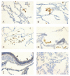
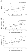
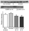
Results: Grx1 was expressed mainly in alveolar macrophages. The percentage of Grx1 positive macrophages was significantly lower in GOLD stage IV COPD than in healthy smokers (p = 0.021) and the level of Grx1 in total lung homogenate decreased both in stage I-II (p = 0.045) and stage IV COPD (p = 0.022). The percentage of Grx1 positive macrophages correlated with the lung function parameters (FEV1, r = 0.45, p = 0.008; FEV1%, r = 0.46, p = 0.007, FEV/FVC%, r = 0.55, p = 0.001). Grx1 could also be detected in sputum supernatants, the levels being increased in the supernatants from acute exacerbations of COPD compared to non-smokers (p = 0.013) and smokers (p = 0.051).
Conclusion: The present cross-sectional study showed that Grx1 was expressed mainly in alveolar macrophages, the levels being decreased in COPD patients. In addition, the results also demonstrated the presence of Grx1 in extracellular fluids including sputum supernatants. Overall, the present study suggests that Grx1 is a potential redox modulatory protein regulating the intracellular as well as extracellular homeostasis of glutathionylated proteins and GSH in human lung.
Figures


Similar articles
-
Expression of glutaredoxin is highly cell specific in human lung and is decreased by transforming growth factor-beta in vitro and in interstitial lung diseases in vivo.Hum Pathol. 2004 Aug;35(8):1000-7. doi: 10.1016/j.humpath.2004.04.009. Hum Pathol. 2004. PMID: 15297967
-
Glutathione S-transferase omega in the lung and sputum supernatants of COPD patients.Respir Res. 2007 Jul 6;8(1):48. doi: 10.1186/1465-9921-8-48. Respir Res. 2007. PMID: 17617905 Free PMC article.
-
[The change of concentration of endothelin derived from alveolar macrophages and in induced sputum in patients with chronic bronchitis].Zhonghua Jie He He Hu Xi Za Zhi. 2001 Jun;24(6):351-4. Zhonghua Jie He He Hu Xi Za Zhi. 2001. PMID: 11802988 Chinese.
-
Interleukin-18 in induced sputum: association with lung function in chronic obstructive pulmonary disease.Respir Med. 2009 Jul;103(7):1056-62. doi: 10.1016/j.rmed.2009.01.011. Epub 2009 Feb 8. Respir Med. 2009. PMID: 19208460
-
Ageing and long-term smoking affects KL-6 levels in the lung, induced sputum and plasma.BMC Pulm Med. 2011 May 11;11:22. doi: 10.1186/1471-2466-11-22. BMC Pulm Med. 2011. PMID: 21569324 Free PMC article.
Cited by
-
Thioredoxins, glutaredoxins, and peroxiredoxins--molecular mechanisms and health significance: from cofactors to antioxidants to redox signaling.Antioxid Redox Signal. 2013 Nov 1;19(13):1539-605. doi: 10.1089/ars.2012.4599. Epub 2013 Mar 28. Antioxid Redox Signal. 2013. PMID: 23397885 Free PMC article. Review.
-
Glutathione-S-transferases in lung and sputum specimens, effects of smoking and COPD severity.Respir Res. 2008 Dec 13;9(1):80. doi: 10.1186/1465-9921-9-80. Respir Res. 2008. PMID: 19077292 Free PMC article.
-
Mechanistic and kinetic details of catalysis of thiol-disulfide exchange by glutaredoxins and potential mechanisms of regulation.Antioxid Redox Signal. 2009 May;11(5):1059-81. doi: 10.1089/ars.2008.2291. Antioxid Redox Signal. 2009. PMID: 19119916 Free PMC article. Review.
-
Oxidative and anti-oxidative status in muscle of young rats in response to six protein diets.Sci Rep. 2017 Oct 13;7(1):13184. doi: 10.1038/s41598-017-11834-5. Sci Rep. 2017. PMID: 29030561 Free PMC article.
-
Redox Regulator GLRX Is Associated With Tumor Immunity in Glioma.Front Immunol. 2020 Nov 30;11:580934. doi: 10.3389/fimmu.2020.580934. eCollection 2020. Front Immunol. 2020. PMID: 33329553 Free PMC article.
References
-
- Rahman I, MacNee W. Lung glutathione and oxidative stress: implications in cigarette smoke-induced airway disease. Am J Physiol. 1999;277:L1067–88. - PubMed
-
- Cantin AM, North SL, Hubbard RC, Crystal RG. Normal alveolar epithelial lining fluid contains high levels of glutathione. J Appl Physiol. 1987;63:152–157. - PubMed
Publication types
MeSH terms
Substances
LinkOut - more resources
Full Text Sources
Medical

