. 2006 Oct 23;175(2):305-13.
doi: 10.1083/jcb.200608009.

Phosphorylation and activity of the tumor suppressor Merlin and the ERM protein Moesin are coordinately regulated by the Slik kinase

Affiliations

Phosphorylation and activity of the tumor suppressor Merlin and the ERM protein Moesin are coordinately regulated by the Slik kinase

Sarah C Hughes et al. J Cell Biol. .

Abstract

Merlin and Moesin are closely related members of the 4.1 Ezrin/Radixin/Moesin domain superfamily implicated in regulating proliferation and epithelial integrity, respectively. The activity of both proteins is regulated by head to tail folding that is controlled, in part, by phosphorylation. Few upstream regulators of these phosphorylation events are known. In this study, we demonstrate that in Drosophila melanogaster, Slik, a Ste20 kinase, controls subcellular localization and phosphorylation of Merlin, resulting in the coordinate but opposite regulation of Merlin and Moesin. These results suggest the existence of a novel mechanism for coordinate regulation of cell proliferation and epithelial integrity in developing tissues.


Figures
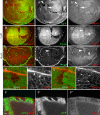
Figure 1.
Figure 1.
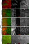
Slik activity affects Merlin protein localization in a dose-dependent manner. Mitotic clones of slik1 mutant cells are marked by the lack of a GFP marker (A′, arrowhead; and D, E, and F′) or the lack of phospho-Moesin (B′ and C′, arrowheads). (A–A′′) Sections taken below the apical surface show a marked increase in Merlin staining within homozygous slik1 clones (A′′, arrowhead), with a concomitant decrease in Merlin staining within the wild-type sister clone (A′′, arrow) marked by the increased expression of GFP (A′, arrow). (B–C′′) Optical sections taken either 1 (B–B′′) or 6 μm (C–C′′) beneath the apical surface demonstrate that in slik1 clones, Merlin is mislocalized away from the apical surface. An apical section (B′′, arrowheads) shows decreased Merlin staining in the slik1 clone, whereas more basal Merlin staining is increased in slik1 cells (C′′, arrowheads). (D–E′) Optical cross sections of clones showing the reduction of Merlin apically (D′, arrows) and an increase in punctate Merlin staining basally (E′, arrows). (F–F′′) A similar effect on Merlin protein staining and localization are seen in the follicle cell epithelium surrounding the developing oocyte. Bars, 10 μm.
Figure 2.
Figure 2.
Slik activity alters the subcellular localization and trafficking of Merlin protein in S2 cells. (A–D) Different subcellular localizations of Merlin are observed after transient expression using a short pulse of heat shock–driven expression of GFP-tagged Merlin (hs-Mer+GFP). (A) High levels of Merlin protein localized completely at the membrane. These levels are increased from what is seen with a pulse of Merlin expression alone. (B) Moderate levels of mostly membranous Merlin localization with a small number of cytoplasmic punctate structures. (C) Merlin localized to numerous small cytoplasmic vesicles throughout the cell. (D) Merlin localized to fewer, larger cytoplasmic vesicles. (E–K) Histograms of cells displaying various phenotypes (in A–D) at different time points (1, 3, and 6 h) after Merlin expression. The y axis corresponds to the percentage of each phenotype observed in the field of cells counted. Error bars indicate SD of at least three replicates. (E) Trafficking of wild-type Merlin. A progression from the plasma membrane to large endocytic vesicles is observed over time as previously described (McCartney and Fehon, 1996; LaJeunesse et al., 1998). (F) Merlin trafficking in cells that express both Merlin and Slik. There is a slower progression of Merlin moving from the plasma membrane into cytoplasmic vesicles. In addition, there is an increase in the number of cells exhibiting high levels of Merlin that remain at the membrane (A). (G) Cells in which both Merlin and Slikkd are expressed. Merlin localization in these cells is very similar to that observed in cells expressing Merlin alone (E). (H) A phosphomimetic form of Merlin, MerT616D, is retained at the plasma membrane for an extended time compared with Mer+. (I) Nonphosphorylatable Merlin (MerT616A) traffics away from the plasma membrane faster than Mer+. (J) Merlin coexpressed with wild-type Moesin trafficking similarly to Merlin expression alone (E). (K) Merlin trafficking in cells that express Merlin, Moesin, and Slik. Merlin localization patterns are similar to the expression of Merlin alone (E). Bar, 2 μm.
Figure 3.
Figure 3.
Slik activity can alter Merlin phosphorylation. (A) Merlin protein was immunoprecipitated from third instar imaginal disc cell lysates. Merlin protein migrates as two prominent bands and one or more minor bands in wild-type (WT) lysates. Numbers below the lanes represent the ratio of the top (phosphorylated) bands to the bottom (unphosphorylated) band in each lane (top bracket vs. the bottom bracket). For wild type and UAS-slik, n = 6, and for UAS-slikkd, n = 4. In cell lysates from imaginal discs in which Slik is overexpressed (UAS-slik), the more hyperphosphorylated (slower migrating) bands are relatively more abundant, as evidenced by the increased ratio of top to bottom bands. Expression of the kinase-inactive Slik (UAS-slik kd) has a similar phosphorylation pattern to wild type. All samples in this blot are from the same experiment but have been rearranged for order. (B) To confirm that the shift observed in migration of the Merlin bands is caused by phosphorylation, samples were treated with λ phosphatase. All Merlin staining is reduced to a single species after this treatment. The samples in this blot are representative examples taken from a separate experiment than that shown in A. In this case, the UAS-slik kd sample was under loaded. (C) The phosphorylation patterns of MerT616D and MerT616A in the presence or absence of coexpressed Slik kinase in S2 cells. The slowest migrating form of MerT616D is enhanced relative to MerT616A. Neither pattern is altered by the coexpression of Slik. (D) In vitro GST pull-down assay showing a direct interaction between the S35-labeled Slik protein (arrow) and both GST-Merlin and GST-Moesin but not with GST alone. This blot is taken from a single experiment. A background band above Slik is also present in the GST control. FT, flow through.
Figure 4.
Figure 4.
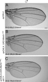
Slik activity inhibits Merlin function genetically. The phenotypic modification of an activated Merlin protein (Mer1-600) by reduction in Slik function was analyzed. (A) A representative example of a male wild-type wing from flies carrying the apGAL4 driver, which is expressed in the dorsal surface of the wing. (B) A representative example of a wing in which activated Merlin (Mer1-600) is expressed in the dorsal surface of the wing under the apGAL4 driver. There is a mean 15% decrease in wing area from the wild type. (C) Removal of one copy of slik1 in the wings expressing Mer1-600 produces a further reduction in wing area by a mean of 18% from the wild type. Thus, reduction of Slik function enhances the activated Merlin phenotype. Measurements in each panel represent the mean area of the wing (millimeters squared) for each genotype. For each combination, at least 10 wings were measured. Bar, 200 μm.
Figure 5.
Figure 5.
Slik affects Merlin independently of Moesin. The effect of Slik on Merlin was analyzed in the presence or absence of Moesin. (A–A′′) An activated Moesin (MoeT559D) transgene was expressed in slik1 clones using the MARCM technique. Mutant clones are positively marked with GFP. Within slik1 clones, the expression of a constitutively active Moesin does not rescue the mislocalization of Merlin protein away from the apical membrane. (B–B′′) A Moesin RNAi transgene expressed under the control of the enGAL4 driver in the posterior compartment produces a subtle increase in Merlin staining. (C–C′′) Coexpression of a Moesin RNAi transgene to knock down Moesin expression together with UAS-slik in posterior cells results in a clear increase in Merlin protein staining in the apical domain. Note that the boundary between the anterior and posterior compartments is clearly defined in C′′. (D–D′′) Coexpression of a Moesin RNAi transgene with UAS-slikkd does not alter Merlin staining or localization, indicating that the kinase activity of Slik is required. (E–E′′) Expression of UAS-slik using enGAL4 in the presence of normal levels of Moesin has no detectable effect on Merlin protein staining in the posterior compartment of a third imaginal wing disc. Arrows mark the anterior-posterior boundary, with posterior to the right. Bar, 10 μm.
Figure 6.
Figure 6.
Schematic diagram of functional relationships between Slik, Merlin, Moesin, and the regulation of tissue integrity and proliferation in developing epithelia. As demonstrated in this study, Slik activity simultaneously promotes Moesin function and inhibits Merlin. Our previous results have shown that Moesin functions to negatively regulate Rho activity and promote epithelial integrity (Speck et al., 2003). Merlin functions to restrict proliferation in the same epithelia. Thus, the net result of Slik activity is to drive proliferation and simultaneously stabilize epithelial integrity.

Similar articles

Cited by

References

    1. Bilder, D. 2004. Epithelial polarity and proliferation control: links from the Drosophila neoplastic tumor suppressors. Genes Dev. 18:1909–1925. - PubMed
    1. Bilder, D., M. Li, and N. Perrimon. 2000. Cooperative regulation of cell polarity and growth by Drosophila tumor suppressors. Science. 289:113–116. - PubMed
    1. Brand, A.H., and N. Perrimon. 1993. Targeted gene expression as a means of altering cell fates and generating dominant phenotypes. Development. 118:401–415. - PubMed
    1. Bretscher, A., K. Edwards, and R.G. Fehon. 2002. ERM proteins and Merlin: integrators at the cell cortex. Nat. Rev. Mol. Cell Biol. 3:586–599. - PubMed
    1. Bryant, P.J., and P. Simpson. 1984. Intrinsic and extrinsic control of growth in developing organs. Q. Rev. Biol. 59:387–415. - PubMed

Publication types

MeSH terms