. 2006 Nov 1;25(21):5083-93.
doi: 10.1038/sj.emboj.7601383. Epub 2006 Oct 12.

An extended consensus motif enhances the specificity of substrate modification by SUMO

Affiliations

An extended consensus motif enhances the specificity of substrate modification by SUMO

Shen-Hsi Yang et al. EMBO J. .

Abstract

Protein modification by SUMO conjugation is an important regulatory event. Sumoylation usually takes place on a lysine residue embedded in the core consensus motif psiKxE. However, this motif confers limited specificity on the sumoylation process. Here, we have probed the roles of clusters of acidic residues located downstream from the core SUMO modification sites in proteins such as the transcription factor Elk-1. We demonstrate that these are functionally important in SUMO-dependent transcriptional repression of Elk-1 transcriptional activity. Mechanistically, the acidic residues are important in enhancing the efficiency of Elk-1 sumoylation by Ubc9. Similar mechanisms operate in other transcription factors and phosphorylation sites can functionally substitute for acidic residues. Thus, an extended sumoylation motif, termed the NDSM (negatively charged amino acid-dependent sumoylation motif), helps define functional SUMO targets. We demonstrate that this extended motif can be used to correctly predict novel targets for SUMO modification.


Figures
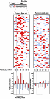
Figure 1
Figure 1
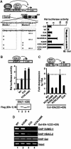
The region downstream from the core SUMO motif is rich in acidic residues. The left panel shows an alignment of the 10-amino-acid tail region located downstream from known characterised SUMO sites found in human transcriptional regulatory proteins (see Supplementary Table S1 for more details). Only acidic (shaded red) and basic (shaded blue) residues are depicted. The graph shown below indicates the percentage enrichment of acidic (red) and basic (blue) residues at each position, relative to the average occurrence of each type of amino acid in the downstream region of a control data set derived from shuffled ψKxE motifs. The panel on the right portrays the same information, but is a random selection from the total core consensus ψKxE data set (see Supplementary Table S2 for details).
Figure 2
Figure 2
Functional importance of acidic groups located downstream from the core SUMO site. (A) Reporter gene analyses of the activities of the indicated mutant versions of Gal-Elk-1(233–428) fusion proteins in 293 cells. The structure of the LexA-Gal4-driven luciferase reporter used and the domain structure of the Elk-1 moiety is diagrammed and the sequence of the R-motif shown. The SUMO acceptor lysines are shaded grey and downstream acidic residues underlined. The boxes represent 14-amino-acid modules consisting of the core SUMO consensus site ψKXE followed by 10 downstream amino acids (tail). Assays were performed on the reporter in the presence of co-transfected LexA-VP16. The identity of the mutations in Elk-1 are indicated, with E3A representing the simultaneous mutation of E255, E256 and E258 to alanine. Western blots (IB) of Gal-Elk-1 protein levels are shown on the right. Data are shown relative to the activity of wild-type (WT) GAL-Elk-1(223–428) (taken as 1). Luciferase assays are representative of at least two independent experiments (s.e.'s are shown; n=2). (B) Reporter gene analyses of the activities of the indicated WT and mutant versions of full-length Elk-1 proteins in 293 cells. Assays were performed on an egr-1 promoter-driven luciferase reporter (shown schematically). A Western blotting showing the relative expression levels of the Flag-tagged Elk-1 proteins is show below the graph. Data are representative of at least two independent experiments (s.e.'s are shown; n=2). (C) Reporter gene analyses of the activities of the indicated WT and mutant versions of Gal-Elk-1(223–428) fusion proteins in 293 cells in the presence of TSA. Assays were performed on a Gal4-driven luciferase reporter (shown schematically). Data are shown as fold derepression by TSA relative to the activity of each construct in the absence of TSA. Luciferase assays are representative of at least two independent experiments (s.e.'s are shown; n=2). (D) ChIP analysis in the presence of the indicated WT and mutant versions of Gal-Elk-1(223–428) fusion proteins in 293 cells on the promoter of a Gal4-driven E1b luciferase reporter. ChIPs were performed with the indicated antibodies.
Figure 3
Figure 3
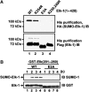
Role of acidic residues in Elk-1 sumoylation. (A) Sumoylation of Elk-1 in vivo. The indicated mutant versions of His-Flag-tagged Elk-1(1–428) were coexpressed with HA-tagged SUMO-1 and His-tagged Elk-1 was precipitated from cell lysates. Sumoylated Elk-1 was detected by immunoblotting (IB) with an anti-HA antibody and total Elk-1 with an anti-Flag antibody. (B) Elk-1 sumoylation in vitro. Purified wild-type (WT) and E3A mutant GST-Elk-1(201–260) (2 μg) were sumoylated in vitro with 500 ng Ubc9 for the indicated times. Sumoylated Elk-1 was detected by IB with an anti-SUMO-1 antibody and total GST-Elk-1 with an anti-GST antibody.
Figure 4
Figure 4
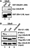
The acidic residues are important in Ubc9–Elk-1 interactions. (A) GST pulldown assay of myc-tagged Ubc9 by the indicated GST-Elk-1 fusion proteins. (B) Co-immunoprecipitation analysis of Ubc9 with Elk-1 from 293 cells co-transfected with expression vectors encoding myc-tagged Ubc9 and the indicated Flag-tagged Elk-1 proteins. Immunoprecipitation (IP) was performed with an anti-Elk-1 antibody, and precipitated Elk-1 and Ubc9 were detected by immunoblotting (IB) with the indicated antibodies.
Figure 5
Figure 5
Role of acidic residues in sumoylation of MEF2A. (A) Schematic illustration of the Gal4-driven luciferase reporter and the Gal4-MEF2A(266–413) fusion protein. The sequence of the module containing the core SUMO consensus site ψKXE followed by 10 downstream amino acids is shown. The SUMO acceptor lysines are shaded grey and downstream acidic residues underlined. The residues altered in each mutant protein are indicated. (B) Reporter gene analyses of the activities of the indicated mutant versions of GAL-MEF2A(266–413) fusion proteins in 293 cells. Data are presented relative to the activity of the wild-type (WT) protein (taken as 1). (C) Western blots (IB) of Gal-MEF2A protein levels. (D) Sumoylation of MEF2A in vivo. WT or mutant versions (S400A/D404A or S400A/R403E) of His-Flag-tagged MEF2A(1–486) were coexpressed in 293 cells with myc-tagged SUMO-1 and His-tagged MEF2A was precipitated from cell lysates. Sumoylated MEF2A was detected by immunoblotting (IB) with an anti-myc antibody and total MEF2A with an anti-Flag antibody. The numbers under each lane represent the average binding in three independent experiments, compared to WT MEF2A (taken as 100%).
Figure 6
Figure 6
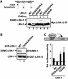
Role of acidic residues in sumoylation of LRH-1. (A) Sumoylation of LRH-1 in vivo. The sequence of the module in LRH-1 containing the core SUMO consensus site ψKXE followed by 10 downstream amino acids is shown. Annotation is as in Figure 5A. The indicated wild-type and mutant myc-tagged LRH-1 proteins were coexpressed in 293 cells with HA-tagged SUMO-1 and PIASxα where indicated. LRH-1- and SUMO-conjugated species were detected by immunoblotting (IB) with an anti-myc antibody. (B) LRH-1 sumoylation in vitro. Purified wild-type (WT) and D229A/E336A mutant GST-LRH-1(134–257) (2 μg) were sumoylated in vitro with 20 ng Ubc9 for 1 h. Sumoylated LRH-1 was detected by immunoblotting. (C) Reporter gene analyses of the activities of the indicated WT and mutant versions of full-length LRH-1 proteins in His-SUMO-HeLa cells. Assays were performed on an SHP promoter-driven luciferase reporter (shown schematically). Data are representative of at least two independent experiments (s.e.'s are shown; n=2).
Figure 7
Figure 7
Identification of a basic patch on Ubc9 required for efficient sumoylation of an NDSM-containing substrate. (A) In vitro sumoylation assay of GST-Elk-1(201–260) (top panel) and GST-RanGAP-1(418–587) (third panel) (2 μg) using the indicated Ubc9 mutants (500 ng). Inputs of the GST fusion proteins (second and fourth panels; anti-GST immunoblot (IB)) and Ubc9 mutants (bottom panel; Coomassie stained) are shown. Sumoylated substrates are detected using anti-SUMO-1 IB. The sequences immediately downstream from the major SUMO motifs found in Elk-1 and RanGAP-1 are shown at the top. Downstream acidic residues are highlighted in red. (B) In vitro time-course sumoylation assay of GST-Elk-1(201–260) (left) and GST-RanGAP-1(418–587) (right) using the indicated Ubc9 mutants for the indicated times. Proteins levels and detection were as in (A). (C) In vitro sumoylation assay of wild-type and E3A mutant GST-Elk-1(201–260) (top panel) (2 μg) using the indicated wild-type and mutant Ubc9 (500 ng). Inputs of the GST fusion proteins (bottom panel; anti-GST IB) are shown. Sumoylated substrates are detected using anti-SUMO-1 IB. (D, E) In vitro sumoylation assay of wild-type and E3A mutant GST-LRH-1(134–257) (2 μg) (top panel) using either low (10 ng) or high (50 ng) amounts of the indicated wild-type and mutant Ubc9. Sumoylation assays were carried out in the absence (D) or presence (E) of PIASxα (62.5 ng). Inputs of the GST fusion proteins (bottom panel; anti-GST IB) are shown. Sumoylated substrates are detected using anti-SUMO-1 IB. (F) GST pulldown assay of Ubc9 (3 μg) by GST-Elk-1(201–260) (top panel) and GST-RanGAP-1(418–587) (third panel) (3 μg each). Ubc9 and inputs of the GST fusion proteins (second and fourth panels) were detected using IB with anti-Ubc9 or anti-GST antibodies, respectively. (G) Ubc9 structural models showing the charged surface area (left-hand panel, created using WebLab ViewerPro 4.0) and a space filling model (right-hand panel, created using Cn3D version 4.1). The left-hand model shows basic areas as blue and acidic areas as red. The locations of the basic patch created by K59 and R61 (shown as blue on the right-hand model), K74 (yellow) and the active site (yellow) are indicated. (H) Schematic model depicting the interactions between Ubc9 and the NDSM. The preponderance of acidic residues within positions 3–6 is denoted by (E/D)4 within the tail. The ψKxE core engages with the active site, whereas the tail region binds to the basic patch.
Figure 8
Figure 8
Identification of novel SUMO substrates using an extended SUMO consensus motif. (A) Sequences of the SUMO sites within known and novel substrates predicted using NDSM motif (see Supplementary Table S3 for complete data set). (B) Gene ontologies showing high levels of over-representation in the NDSM-predicted data set (see Supplementary Table S4 for complete data set). Data are represented as fold increase over a control data set generated with a shuffled ψKxE motif with downstream acidic residues. P-values are generated using a χ2 test. (C) Identification of SUMO-conjugated proteins. Proteins conjugated to His-tagged SUMO were precipitated from normal HeLa or His-SUMO-HeLa cells and identified by Western analysis (IB) with the indicated antibodies (right-hand panels). The input proteins are shown on the left. Bands corresponding to sumoylated proteins are indicated by the suffix ‘S-'. A colour version of this figure is available at The EMBO Journal Online.

Similar articles

Cited by

References

    1. Anckar J, Hietakangas V, Denessiouk K, Thiele DJ, Johnson MS, Sistonen L (2006) Inhibition of DNA binding by differential sumoylation of heat shock factors. Mol Cell Biol 26: 955–964 - PMC - PubMed
    1. Bernier-Villamor V, Sampson DA, Matunis MJ, Lima CD (2002) Structural basis for E2-mediated SUMO conjugation revealed by a complex between ubiquitin-conjugating enzyme Ubc9 and RanGAP1. Cell 108: 345–356 - PubMed
    1. Binda O, Roy JS, Branton PE (2006) RBP1 family proteins exhibit SUMOylation-dependent transcriptional repression and induce cell growth inhibition reminiscent of senescence. Mol Cell Biol 26: 1917–1931 - PMC - PubMed
    1. Chalkiadaki A, Talianidis I (2005) SUMO-dependent compartmentalization in promyelocytic leukemia protein nuclear bodies prevents the access of LRH-1 to chromatin. Mol Cell Biol 25: 5095–5105 - PMC - PubMed
    1. Gill G (2005) Something about SUMO inhibits transcription. Curr Opin Genet Dev 15: 536–541 - PubMed

Publication types

MeSH terms