Bidirectional control of CNS capillary diameter by pericytes
- PMID: 17036005
- PMCID: PMC1761848
- DOI: 10.1038/nature05193
Bidirectional control of CNS capillary diameter by pericytes
Abstract
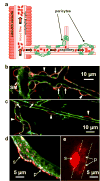
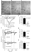
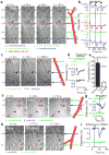
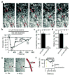
Neural activity increases local blood flow in the central nervous system (CNS), which is the basis of BOLD (blood oxygen level dependent) and PET (positron emission tomography) functional imaging techniques. Blood flow is assumed to be regulated by precapillary arterioles, because capillaries lack smooth muscle. However, most (65%) noradrenergic innervation of CNS blood vessels terminates near capillaries rather than arterioles, and in muscle and brain a dilatory signal propagates from vessels near metabolically active cells to precapillary arterioles, suggesting that blood flow control is initiated in capillaries. Pericytes, which are apposed to CNS capillaries and contain contractile proteins, could initiate such signalling. Here we show that pericytes can control capillary diameter in whole retina and cerebellar slices. Electrical stimulation of retinal pericytes evoked a localized capillary constriction, which propagated at approximately 2 microm s(-1) to constrict distant pericytes. Superfused ATP in retina or noradrenaline in cerebellum resulted in constriction of capillaries by pericytes, and glutamate reversed the constriction produced by noradrenaline. Electrical stimulation or puffing GABA (gamma-amino butyric acid) receptor blockers in the inner retina also evoked pericyte constriction. In simulated ischaemia, some pericytes constricted capillaries. Pericytes are probably modulators of blood flow in response to changes in neural activity, which may contribute to functional imaging signals and to CNS vascular disease.
Figures


Comment in
-
Neuroscience: controlled capillaries.Nature. 2006 Oct 12;443(7112):642-3. doi: 10.1038/443642a. Nature. 2006. PMID: 17035989 No abstract available.
Similar articles
-
Neuroscience: controlled capillaries.Nature. 2006 Oct 12;443(7112):642-3. doi: 10.1038/443642a. Nature. 2006. PMID: 17035989 No abstract available.
-
Capillary pericytes regulate cerebral blood flow in health and disease.Nature. 2014 Apr 3;508(7494):55-60. doi: 10.1038/nature13165. Epub 2014 Mar 26. Nature. 2014. PMID: 24670647 Free PMC article.
-
Active role of capillary pericytes during stimulation-induced activity and spreading depolarization.Brain. 2018 Jul 1;141(7):2032-2046. doi: 10.1093/brain/awy143. Brain. 2018. PMID: 30053174 Free PMC article.
-
A tight squeeze: how do we make sense of small changes in microvascular diameter?J Physiol. 2023 Jun;601(12):2263-2272. doi: 10.1113/JP284207. Epub 2023 May 9. J Physiol. 2023. PMID: 37036208 Free PMC article. Review.
-
Calcium signalling in pericytes.J Vasc Res. 2014;51(3):190-9. doi: 10.1159/000362687. Epub 2014 Jun 4. J Vasc Res. 2014. PMID: 24903335 Review.
Cited by
-
Integrative analysis of the human brain mural cell transcriptome.J Cereb Blood Flow Metab. 2021 Nov;41(11):3052-3068. doi: 10.1177/0271678X211013700. Epub 2021 May 22. J Cereb Blood Flow Metab. 2021. PMID: 34027687 Free PMC article.
-
Role of endothelium-pericyte signaling in capillary blood flow response to neuronal activity.J Cereb Blood Flow Metab. 2021 Aug;41(8):1873-1885. doi: 10.1177/0271678X211007957. Epub 2021 Apr 14. J Cereb Blood Flow Metab. 2021. PMID: 33853406 Free PMC article.
-
Emerging Role of Pericytes and Their Secretome in the Heart.Cells. 2021 Mar 4;10(3):548. doi: 10.3390/cells10030548. Cells. 2021. PMID: 33806335 Free PMC article. Review.
-
Pericytes in the eye.Pflugers Arch. 2013 Jun;465(6):789-96. doi: 10.1007/s00424-013-1272-6. Epub 2013 Apr 9. Pflugers Arch. 2013. PMID: 23568370 Review.
-
Brain pericytes from stress-susceptible pigs increase blood-brain barrier permeability in vitro.Fluids Barriers CNS. 2012 Jun 29;9(1):11. doi: 10.1186/2045-8118-9-11. Fluids Barriers CNS. 2012. PMID: 22569151 Free PMC article.
References
-
- Attwell D, Iadecola C. The neural basis of functional brain imaging signals. Trends Neurosci. 2002;25:621–625. - PubMed
-
- Cohen Z, Molinatti G, Hamel E. Astroglial and vascular interactions of noradrenaline terminals in the rat cerebral cortex. J Cereb Blood Flow Metab. 1997;17:894–904. - PubMed
-
- Berg BR, Cohen KD, Sarelius IH. Direct coupling between blood flow and metabolism at the capillary level in striated muscle. Am J Physiol. 1997;272:H2693–2700. - PubMed
Publication types
MeSH terms
Substances
Grants and funding
LinkOut - more resources
Full Text Sources
Other Literature Sources
Miscellaneous

