. 2006 Oct 9;175(1):55-66.
doi: 10.1083/jcb.200604009.

Promyelocytic leukemia nuclear bodies behave as DNA damage sensors whose response to DNA double-strand breaks is regulated by NBS1 and the kinases ATM, Chk2, and ATR

Affiliations

Promyelocytic leukemia nuclear bodies behave as DNA damage sensors whose response to DNA double-strand breaks is regulated by NBS1 and the kinases ATM, Chk2, and ATR

Graham Dellaire et al. J Cell Biol. .

Abstract

The promyelocytic leukemia (PML) nuclear body (NB) is a dynamic subnuclear compartment that is implicated in tumor suppression, as well as in the transcription, replication, and repair of DNA. PML NB number can change during the cell cycle, increasing in S phase and in response to cellular stress, including DNA damage. Although topological changes in chromatin after DNA damage may affect the integrity of PML NBs, the molecular or structural basis for an increase in PML NB number has not been elucidated. We demonstrate that after DNA double-strand break induction, the increase in PML NB number is based on a biophysical process, as well as ongoing cell cycle progression and DNA repair. PML NBs increase in number by a supramolecular fission mechanism similar to that observed in S-phase cells, and which is delayed or inhibited by the loss of function of NBS1, ATM, Chk2, and ATR kinase. Therefore, an increase in PML NB number is an intrinsic element of the cellular response to DNA damage.


Figures
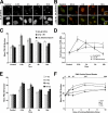
Figure 1.
Figure 1.
PML NB number increases in response to DSBs in NHDFs. NHDF cells (GM05757) were treated with varying doses of IR, etoposide (20 μM VP16), or 1.5 μM doxorubicin for 30 min to induce DSBs. (A) IF analysis of PML NB number in maximum-intensity Z projections of NHDFs after etoposide; time after treatment is indicated in hours. (B) IF analysis of the distribution of PML NBs in relation to DSBs in NHDFs after 2 Gy IR or VP16. γ-H2AX is used as a marker for chromatin containing DSBs, and asterisks mark the time points in which the maximum fluorescence intensity of γ-H2AX was first detected. Arrowheads indicate juxtaposition of γ-H2AX and PML NBs at 18 h after DNA damage (inset). Images represent a single focal plane. (C) Comparison of mean PML NB number over time after IR, VP16, and doxorubicin treatment. (D) Comparison of fold increase in PML NB number over time after IR, VP16, and doxorubicin treatment. (E and F) Response of PML NBs to graded doses of IR expressed as a function of time (E) or at each time point as a function of dose (F). Bars, 5 μm.
Figure 2.
Figure 2.
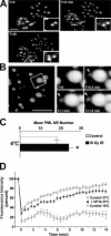
PML protein and PML NB dynamics after DSB induction. (A) PML microbody formation occurs rapidly after treatment with etoposide. Two U-2 OS human osteosarcoma cells stably expressing GFP-PML IV protein were imaged by fluorescence microscopy before (T = 0) and after addition of etoposide (20 μM VP16; T = 5 min and T = 2 h). Enlarged region of the cell marked by white asterisk is shown at each time point. White arrowheads indicate newly formed microbodies after VP16 treatment. (B) Formation of PML microbodies in response to DNA DSBs occurs by supramolecular fission from preexisting parental PML NBs. A U-2 OS cell expressing GFP-PML IV was visualized before (T = 0) and during treatment with 20 μM VP16 over several minutes (T = 0.5, 1.0, and 1.5 min are shown). Arrowhead indicates fission of a PML microbody from a larger parental PML NB. (C) PML NBs increase in number in cells irradiated on ice. NHDFs (GM05757s) were incubated on ice for 20 min and either fixed (Control) or irradiated on ice (10 Gy IR) before fixation. Mean PML NB number increases significantly between control (17 ± 1; n = 30) and cells irradiated with 10 Gy IR on ice (24 ± 2; n = 30; *, P = 0.0008). (D) Dynamics of the PML protein within PML NBs is affected by DNA damage and reduced temperature. Asynchronous U-2 OS GFP-PML IV cells were subjected to treatment with etoposide (20 μM VP16 for 30 min) before mobility of PML protein within PML NBs was analyzed by FRAP at 37°C (n = 20). Mobility of the PML protein at PML NBs in DNA damaged cells is compared with control untreated cells (n = 20) at 37°C and at 15°C (n = 7). Data are presented as the mean fluorescence recovery plotted as percent of initial fluorescence intensity of the PML NB over 14 min. Error bars represent the standard error. Bars, 5 μm.
Figure 3.
Figure 3.
PML NBs lose positional stability when chromatin is damaged in their vicinity. (A) UV laser–induced DSBs alter the positional stability of PML NBs. A single U-2 OS cell expressing GFP-PML IV is shown, in which DSBs were created in a laser track along a defined ROI (∼0.5 × 10 μm; rectangular box) by photoinduction; PML NB movement was tracked over time. PML NBs (arrowheads) along the laser path (rectangular box) move toward and aggregate with one large PML NB (arrow) adjacent to the laser track. PML NB number (NB#) is shown before laser induction of DSBs and 22 min after induction. (B) Confirmation of laser-induced DSBs by IF detection of γ-H2AX. The same cell shown in A was fixed at 60 min after laser induction of DSBs and processed for immunodetection of PML and γ-H2AX. PML NB number at this time point is indicated (NB#). Bars, 5 μm.
Figure 4.
Figure 4.
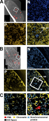
Ultrastructural analysis of PML NBs in NHDFs by correlative LM/ESI before and after etoposide-induced DNA damage. Regions of interest containing a PML NB, which are shown at higher magnification in subsequent images, are delineated by white boxes. (A) LM/ESI of a single NHDF (GM05757) cell, fluorescently labeled for PML protein. Elemental maps of nitrogen (N) and phosphorus (P), and the merged maps of a PML NB and its surrounding nucleoplasm reveal protein-based (cyan) and nucleic acid-based (yellow) components. Chromatin appears yellow in the merged image because of high N and P content. A single PML NB is shown at higher magnification (cyan, as indicated by the arrow) making many contacts to the surrounding chromatin (yellow), and has radial symmetry typical of PML NBs in unstressed cells. (B) LM/ESI of a single NHDF (GM05757) treated with 20 μM etoposide (VP16) for 30 min, fluorescently labeled for PML protein. After treatment with VP16, the protein core of PML NB is disrupted in response to DSB induction; few contacts with chromatin remain, and radial symmetry is lost. (C) PML NB in B, at higher magnification (left), and a cartoon representation of the same EM micrograph (right), where PML protein–containing protein structures (red), chromatin (yellow), and other nonchromosomal protein (blue) are shown. Redistribution of PML microbodies along chromatin fibers (asterisks) is observed, and larger interchromatin spaces (black areas) are apparent. PML protein localization was determined by immunogold detection of PML (white dots). Bars, 500 nm.
Figure 5.
Figure 5.
The increase in PML NB number in response to DSBs is independent of new protein translation and p53. NHDF cells (GM05757) in the presence or absence of 150 μM cycloheximide (CHX), Saos-2 human osteosarcoma cells, and isogenic HCT116 human colon carcinoma cells (+ or − p53) were treated with etoposide (20 μM VP16) for 30 min (*, P < 0.0001). (A) Western blot analysis of PML protein levels after etoposide treatment in the presence or absence of cycloheximide. NHDFs were treated with etoposide (20 μM VP16 for 30 min) and harvested at the indicated times for SDS-PAGE and Western blot analysis. Ratio of PML protein levels in the control lane to PML protein at the indicated time points after etoposide treatment are shown normalized against actin. (B) Comparison of mean PML NB number after VP16 treatment in NHDFs, NHDFs treated with cycloheximide (+CHX), and Saos-2 cells. (C) Comparison of mean PML NB number after VP16 treatment in isogenic HCT116 and HCT116 p53-null cells. (D) Comparison of DNA synthesis activity of NHDFs, NHDFs treated with cycloheximide (+CHX), and Saos-2 cells at 18 h after VP16 treatment. 18 h after VP16treatment, cells were incubated with BrdU, fixed, and processed for immunodetection of BrdU, and DNA was counterstained with DAPI. Asterisks represent BrdU-positive cells. Bars, 5 μm.
Figure 6.
Figure 6.
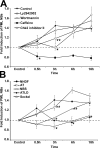
The increase in PML NB number in response to DSBs is delayed or inhibited in the presence of PI3 kinase inhibitors and in DNA repair–deficient cell lines. (A) Comparison of effects of DNA repair kinase inhibitors on the increase in PML NB number in response to DSBs. NHDF cell line GM05757 (control) was pretreated with 10 μM Chk2 kinase inhibitor (Chk2 inhibitor II) or various PI3 kinase inhibitors (5 mM caffeine, 20 μM wortmannin, or 50 μM LY2942002) for 30 min before treatment with etoposide (20 μM VP16 for 30 min; *, P < 0.0001; **, P < 0.001). (B) Comparison of the fold increase in PML NB number after etoposide treatment (20 μM VP16 for 30 min) in NHDF cells and DNA repair–deficient human fibroblast cell lines. AT, ataxia telangiectasia; NBS, Nijmegan breakage syndrome; ATLD, AT-like disorder; Seckel, Seckel syndrome. *, P < 0.0001; **, P < 0.02.
Figure 7.
Figure 7.
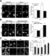
PML NB induction in response to DSBs requires NBS1, Chk2, and ATR-function. Cells were treated with etoposide (20 μM VP16 for 30 min), left to recover for 3 h, and processed for IF detection of PML. DNA was counterstained with DAPI. PML NB number is indicated in maximum-intensity Z projections of IF images of control and etoposide-treated cells (left) and a comparison of mean PML NB number (right) is shown. Error bars represent the SEM. Bars, 5 μm. (A) Comparison of the PML NB number between etoposide-treated Chk2-null (Chk2 −/−) and wild-type Chk2 (Chk2 WT) MEFs (*, P < 0.001). (B) Comparison of PML NB number between Tert-immortalized human NBS fibroblasts infected with an empty retroviral vector (NBST pBabe) or a retroviral vector carrying wild-type human NBS1 (*, P < 0.02). (C) Disruption of ATR kinase function inhibits the increase in PML NB number in response to DSBs. U-2 OS cells expressing a doxycycline-inducible, kinase-inactive, dominant-negative ATR kinase (ATR-DN) were treated with (+ Dox) or without doxycycline for 24 h before etoposide treatment. Besides PML, IF detection of ATR-DN (Fl-ATR-DN) is also shown. Cells with high ATR-DN expression (arrow) contain fewer PML NBs after etoposide treatment than cells with low expression (arrowhead). White asterisks indicate two cells with similar PML NB number in cells not expressing ATR-DN. DNA was counterstained with DAPI. *, P < 0.0001; **, P < 0.001.
Figure 8.
Figure 8.
Summary of the biophysical and molecular mechanisms responsible for the increase in PML NB number in response to DSBs. (A) Model of the biophysical effect on PML NBs by changes in chromatin structure or tensegrity. Chromatin is constrained and under tension (double-headed arrows) by tethering to subnuclear compartments such as the nucleolus, nuclear lamina, and, possibly, PML NBs. DSB-induced changes in chromatin structure or tensegrity alter the balance of forces constraining chromatin within the nucleus; this biophysical phenomenon destabilizes PML NBs that are tethered to chromatin, resulting in microbody formation by fission from preexisting NBs. (B) Model for PML NB number increase in response to DSBs. Initially, PML NB number increases because of biophysical changes in chromatin after DSBs, as in A. The second phase of the PML NB response to DNA damage requires ongoing DNA repair processes, which can be inhibited by low temperatures (4°C) by inhibition (caffeine) or loss of ATR kinase function (ΔATR) and, to a lesser extent, by loss of function of NBS1 (ΔNBS1) or Chk2 (ΔChk2), whose activation is affected by inhibition of ATM (wortmannin). (C) Summary of DNA repair kinase pathways implicated in phosphorylation of the PML protein in response to DNA DSBs. It is currently unknown if ATM can directly phosphorylate PML.

Similar articles

Cited by

References

    1. Arienti, K.L., A. Brunmark, F.U. Axe, K. McClure, A. Lee, J. Blevitt, D.K. Neff, L. Huang, S. Crawford, C.R. Pandit, et al. 2005. Checkpoint kinase inhibitors: SAR and radioprotective properties of a series of 2-arylbenzimidazoles. J. Med. Chem. 48:1873–1885. - PubMed
    1. Bernardi, R., P.P. Scaglioni, S. Bergmann, H.F. Horn, K.H. Vousden, and P.P. Pandolfi. 2004. PML regulates p53 stability by sequestering Mdm2 to the nucleolus. Nat. Cell Biol. 6:665–672. - PubMed
    1. Bradshaw, P.S., D.J. Stavropoulos, and M.S. Meyn. 2005. Human telomeric protein TRF2 associates with genomic double-strand breaks as an early response to DNA damage. Nat. Genet. 37:193–197. - PubMed
    1. Bristow, R.G., and R.P. Hill. 2005. Molecular and cellular basis of radiotherapy. In The Basic Science of Oncology, 4th edition. I.F. Tannock, R.P. Hill, R.G. Bristow, and L. Harrington, editors. McGraw-Hill, New York. 261–288.
    1. Buscemi, G., C. Savio, L. Zannini, F. Micciche, D. Masnada, M. Nakanishi, H. Tauchi, K. Komatsu, S. Mizutani, K. Khanna, et al. 2001. Chk2 activation dependence on Nbs1 after DNA damage. Mol. Cell. Biol. 21:5214–5222. - PMC - PubMed

Publication types

MeSH terms