. 2007 Jan;27(1):170-81.
doi: 10.1128/MCB.01456-06. Epub 2006 Oct 9.

The mitogen-activated protein kinase (MAPK)-activated protein kinases MK2 and MK3 cooperate in stimulation of tumor necrosis factor biosynthesis and stabilization of p38 MAPK

Affiliations

The mitogen-activated protein kinase (MAPK)-activated protein kinases MK2 and MK3 cooperate in stimulation of tumor necrosis factor biosynthesis and stabilization of p38 MAPK

N Ronkina et al. Mol Cell Biol. 2007 Jan.

Abstract

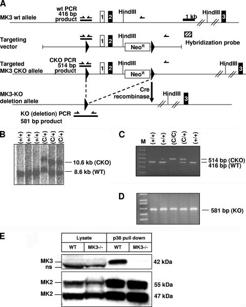
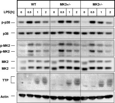
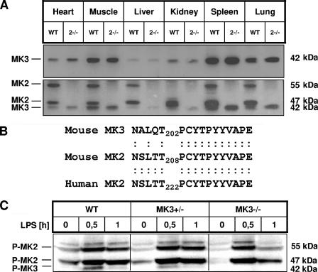
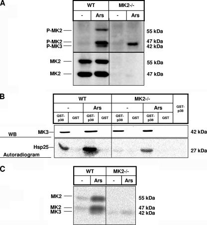
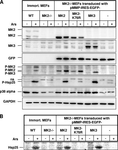
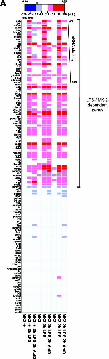
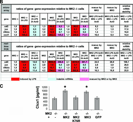
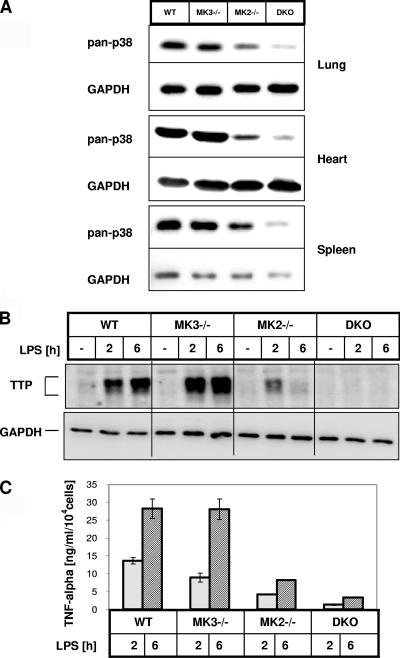
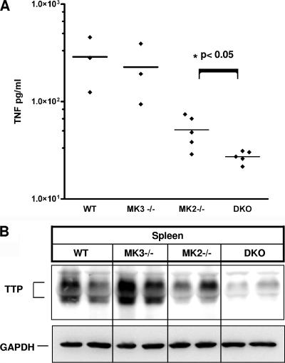
MK2 and MK3 represent protein kinases downstream of p38 mitogen-activated protein kinase (MAPK). Deletion of the MK2 gene in mice resulted in an impaired inflammatory response although MK3, which displays extensive structural similarities and identical functional properties in vitro, is still present. Here, we analyze tumor necrosis factor (TNF) production and expression of p38 MAPK and tristetraprolin (TTP) in MK3-deficient mice and demonstrate that there are no significant differences with wild-type animals. We show that in vivo MK2 and MK3 are expressed and activated in parallel. However, the level of activity of MK2 is always significantly higher than that of MK3. Accordingly, we hypothesized that MK3 could have significant effects only in an MK2-free background and generated MK2/MK3 double-knockout mice. Unexpectedly, these mice are viable and show no obvious defects due to loss of compensation between MK2 and MK3. However, there is a further reduction of TNF production and expression of p38 and TTP in double-knockout mice compared to MK2-deficient mice. This finding, together with the observation that ectopically expressed MK3 can rescue MK2 deficiency similarly to MK2, indicates that both kinases share the same physiological function in vivo but are expressed to different levels.


Figures

FIG. 1.
FIG. 1.
Gene targeting strategy of MK3 deletion and absence of MK3 protein in MK3−/− tissues. (A) The MK3-CKO construct is composed of a 7-kb fragment of the MK3 gene with a 3-kb short arm and a 4-kb long arm flanking exons 1 and 2 and the neomycin resistance selectable marker gene (Neor). Exons 1 and 2 and Neor are flanked with LoxP sites as indicated by solid triangles. Positions of the Hind III restriction sites are indicated. Also indicated are positions of PCR primers, amplicons, and product sizes used for genotyping. (B) Southern blot analysis showing wild-type (+/+) and targeted (+/C) ES cell clones. PCR genotyping results from different sets of tail biopsies distinguishing WT (+) and conditional (C/C and C/+) alleles. (D) PCR results identifying KO (−) alleles after the MK3 KO allele was converted via germ line Cre-mediated deletion by crossing protamine-Cre transgenic mice (129SvEvBrd) with MK3-CKO mice. (E) Endogenous MK3 and MK2 were precipitated from 1 mg of spleen lysates by means of recombinant GST-p38 bound to glutathione-Sepharose 4B. Beads and 0.2 mg of whole lysates were subjected to SDS-PAGE and Western blotting using anti-MK3 antiserum (upper blot) and anti-MK2 antibodies (lower blot). There is a specific 42-kDa MK3 band precipitated from WT tissues which completely disappears in MK3−/− tissues. Endogenous MK3 is not detectable in whole cell lysates.ns, nonspecific band.
FIG. 2.
FIG. 2.
Analysis of p38/MK2 signaling in MK3-deficient macrophages. There were no significant changes in p38 MAPK, MK2, and TTP expression and phosphorylation levels in MK3-deficient cells. Peritoneal macrophages from WT, MK3+/−, and MK3−/− mice were serum starved for several hours and then stimulated with 10 ng/ml LPS for the times indicated. Lysates were then immunoblotted for total and phosphorylated p38 MAPK and MK2 and with an antiserum that recognizes both the phosphorylated and nonphosphorylated forms of TTP.
FIG. 3.
FIG. 3.
Analysis of MK2 and MK3 expression and phosphorylation. MK3 expression in different tissues. GST-p38 was bound to glutathione-Sepharose 4B (Amersham Pharmacia Biotech). The beads were incubated with the lysates from different WT and MK2−/− tissues. (A) The blot was developed against MK3 using anti-MK3-specific antiserum. The same blot was redeveloped against MK2 using anti-MK2 antibodies. (B) Comparison of the phosphorylation sites of MK2 and MK3, recognized by the phospho-specific antiserum against human phospho-T222 MK2 (MK2 pT222). (C) Detection of phospho-MK2 and phospho-MK3 in LPS-stimulated macrophages by Western blotting. Peritoneal macrophages from WT, MK3+/−, and MK3−/− mice were serum starved for several hours and then stimulated with 10 ng/ml LPS for the times indicated. Lysates were then immunoblotted using antibodies against phosphorylated MK2 (pT222) that recognize the phosphorylated MK3 as well (see panel B).
FIG. 4.
FIG. 4.
In vivo phosphorylation and activation of MK3 by arsenite. Primary WT and MK2−/− MEF cells were starved overnight and then stimulated with 200 μM arsenite (Ars) for 30 min. The protein lysates were prepared. (A) The lysates were immunoblotted for phosphorylated MK2/MK3 and for total MK2. (B) MK2/MK3 kinase activity against recombinant Hsp25. Endogenous MK2 and MK3 were precipitated indirectly by using catalytically dead mutant GST-p38 (TGY/AGF) coupled to glutathione-Sepharose beads. The beads were used in a kinase reaction with recombinant Hsp25 as a substrate. The reaction mixture was resolved by SDS-PAGE immunoblotted for MK3 (WB), and then Hsp25 phosphorylating activity was detected by phosphorimaging (autoradiogram). (C) In-gel Hsp25 kinase assay of precipitates from GST-p38 (TGY/AGF) pull-down in WT and MK2−/− MEFs.
FIG. 5.
FIG. 5.
Rescue of Hsp25 phosphorylation and p38 level in MK2-deficient MEFs by overexpression of MK3. MK2−/− immortalized MEF cells were transduced with pMMP-IRES-EGFP vector expressing EGFP in parallel with wild-type MK2, a catalytically dead mutant of MK2-K76R, or MK3 and as a negative control with empty vector expressing EGFP alone. Cells were starved overnight and then were left unstimulated (Ars−) or were stimulated for 40 min with 200 μM arsenite (Ars+). (A) In vivo rescue of Hsp25 phosphorylation and p38-alpha protein levels by overexpressed MK3. The lysates were immunoblotted for phosphorylated and total Hsp25 and p38-alpha and for phosphorylated MK2/3. As an expression control the lysates were immunoblotted for MK2, MK3, and GFP. (B) Cell lysates were incubated in vitro with recombinant Hsp25 and [γ-33P]ATP, and Hsp25 phosphorylating activity was detected by phosphorimaging.
FIG.6.
FIG.6.
Rescue of mRNA stability and stimulation of CXCL1 production in MK2-deficient MEFs by MK2 and MK3. (A) DNA microarray analysis of mRNA stability. cRNAs derived from the indicated samples were labeled with Cy3 and hybridized to 12 microarrays containing probes for 155 inflammatory genes including 19 housekeeping genes. A total of 129 genes with fluorescence intensity values of >100 in MK2-reconstituted cells stimulated with LPS were regarded as significantly expressed and selected for further analysis. Expression values of each indicated sample were divided by those of the MK2 KO control samples. The resulting ratios were log 2 transformed and are grouped according to dependence on MK2 and LPS (ratio of MK2 treated for 2 h with LPS/MK2−/− of >2.0) followed by their sensitivity to ActD treatment in the absence of MK2 as an indirect indicator of mRNA stability (ratio of MK2 −/− treated for 2 h with LPS/MK2 −/− treated for 2 h with LPS and for 2 h with ActD). Transcripts whose mRNA levels decreased by 50% (socs3) down to 4% (csf2) are indicated. (B) Shown are the relative mRNA expression values for the four most unstable mRNAs as determined by microarray experiments or by real-time PCR (C) CXCL1 production of LPS-stimulated MK2-deficient MEFs transduced with constructs coding for catalytically active MK2 and MK3, for MK2-K76R, and as control for GFP alone or nontransduced (−). Asterisks indicate statistically significant difference (single-sided t test, P < 0.05).
FIG.6.
FIG.6.
Rescue of mRNA stability and stimulation of CXCL1 production in MK2-deficient MEFs by MK2 and MK3. (A) DNA microarray analysis of mRNA stability. cRNAs derived from the indicated samples were labeled with Cy3 and hybridized to 12 microarrays containing probes for 155 inflammatory genes including 19 housekeeping genes. A total of 129 genes with fluorescence intensity values of >100 in MK2-reconstituted cells stimulated with LPS were regarded as significantly expressed and selected for further analysis. Expression values of each indicated sample were divided by those of the MK2 KO control samples. The resulting ratios were log 2 transformed and are grouped according to dependence on MK2 and LPS (ratio of MK2 treated for 2 h with LPS/MK2−/− of >2.0) followed by their sensitivity to ActD treatment in the absence of MK2 as an indirect indicator of mRNA stability (ratio of MK2 −/− treated for 2 h with LPS/MK2 −/− treated for 2 h with LPS and for 2 h with ActD). Transcripts whose mRNA levels decreased by 50% (socs3) down to 4% (csf2) are indicated. (B) Shown are the relative mRNA expression values for the four most unstable mRNAs as determined by microarray experiments or by real-time PCR (C) CXCL1 production of LPS-stimulated MK2-deficient MEFs transduced with constructs coding for catalytically active MK2 and MK3, for MK2-K76R, and as control for GFP alone or nontransduced (−). Asterisks indicate statistically significant difference (single-sided t test, P < 0.05).
FIG. 7.
FIG. 7.
Analysis of MK2/MK3 DKO mice. (A) Western blot detection of p38 MAPK levels in total lysates from MK2/MK3 DKO and, as controls, in MK2- and MK3-single knockout and WT mouse tissues. (B) Peritoneal-derived macrophages were stimulated with 1 μg/ml LPS for the times indicated. The TTP level in total lysates from WT, MK2−/−, MK3−/−, and MK2/MK3 DKO cells was analyzed by Western blotting. (C) TNF ELISA of macrophage culture supernatants. BMDMs were cultivated for 7 days, harvested, and counted, and equal numbers of cells were transferred to a 96-well plate. Cells were induced with 1 μg/ml LPS or left untreated as control for 2 and 6 h. TNF levels in the supernatants were measured by ELISA. For each genotype, three independent measurements were made; average and standard deviations are shown. The experiment is representative of two similar experiments.
FIG. 8.
FIG. 8.
Analysis of MK2/MK3 DKO mice in a septic shock in vivo model. Five animals of each genotype were injected intraperitoneally with LPS (5 mg per kg of body weight) diluted in PBS. Ninety minutes after injection mice were sacrificed, and serum and spleen were immediately isolated. (A) TNF level in serum was quantified by ELISA. A statistically significant difference between MK2−/− and DKO is indicated (single-sided t test, P = 0.03). (B) Spleen cell lysates were prepared using kinase assay lysis buffer and analyzed by Western blotting against TTP.

Similar articles

Cited by

References

    1. Adams, R. H., A. Porras, G. Alonso, M. Jones, K. Vintersten, S. Panelli, A. Valladares, L. Perez, R. Klein, and A. R. Nebreda. 2000. Essential role of p38α MAP kinase in placental but not embryonic cardiovascular development. Mol. Cell 6:109-116. - PubMed
    1. Allen, M., L. Svensson, M. Roach, J. Hambor, J. McNeish, and C. A. Gabel. 2000. Deficiency of the stress kinase p38α results in embryonic lethality: characterization of the kinase dependence of stress responses of enzyme-deficient embryonic stem cells. J. Exp. Med. 191:859-870. - PMC - PubMed
    1. Beardmore, V. A., H. J. Hinton, C. Eftychi, M. Apostolaki, M. Armaka, J. Darragh, J. McIlrath, J. M. Carr, L. J. Armit, C. Clacher, L. Malone, G. Kollias, and J. S. Arthur. 2005. Generation and characterization of p38β (MAPK11) gene-targeted mice. Mol. Cell. Biol. 25:10454-10464. - PMC - PubMed
    1. Ben-Levy, R., S. Hooper, R. Wilson, H. F. Paterson, and C. J. Marshall. 1998. Nuclear export of the stress-activated protein kinase p38 mediated by its substrate MAPKAP kinase-2. Curr. Biol. 8:1049-1057. - PubMed
    1. Brook, M., C. R. Tchen, T. Santalucia, J. McIlrath, J. S. Arthur, J. Saklatvala, and A. R. Clark. 2006. Posttranslational regulation of tristetraprolin subcellular localization and protein stability by p38 mitogen-activated protein kinase and extracellular signal-regulated kinase pathways. Mol. Cell. Biol. 26:2408-2418. - PMC - PubMed

Publication types

MeSH terms

Substances