. 2006 Nov;99(4):1207-23.
doi: 10.1111/j.1471-4159.2006.04185.x. Epub 2006 Oct 5.

Manipulating Kv4.2 identifies a specific component of hippocampal pyramidal neuron A-current that depends upon Kv4.2 expression

Affiliations

Manipulating Kv4.2 identifies a specific component of hippocampal pyramidal neuron A-current that depends upon Kv4.2 expression

Aaron Lauver et al. J Neurochem. 2006 Nov.

Abstract

The somatodendritic A-current, I(SA), in hippocampal CA1 pyramidal neurons regulates the processing of synaptic inputs and the amplitude of back propagating action potentials into the dendritic tree, as well as the action potential firing properties at the soma. In this study, we have used RNA interference and over-expression to show that expression of the Kv4.2 gene specifically regulates the I(SA) component of A-current in these neurons. In dissociated hippocampal pyramidal neuron cultures, or organotypic cultured CA1 pyramidal neurons, the expression level of Kv4.2 is such that the I(SA) channels are maintained in the population at a peak conductance of approximately 950 pS/pF. Suppression of Kv4.2 transcripts in hippocampal pyramidal neurons using an RNA interference vector suppresses I(SA) current by 60% in 2 days, similar to the effect of expressing dominant-negative Kv4 channel constructs. Increasing the expression of Kv4.2 in these neurons increases the level of I(SA) to 170% of the normal set point without altering the biophysical properties. Our results establish a specific role for native Kv4.2 transcripts in forming and maintaining I(SA) current at characteristic levels in hippocampal pyramidal neurons.


Figures

Fig. 1
Fig. 1
Differential inactivation properties allow the separation of ISA from other voltage-gated potassium currents. (a) Inactivation of native Kv channels in response to pre-pulse potentials applied for 1 s from −100 to +20 mV in 10-mV steps. Currents are elicited by a test pulse to +50 mV. Current traces bunch together in a middle voltage range from −50 to −30 mV (b) Native Kv currents separate into two clear components of inactivating current, ISA and IAD, and a sustained component, ISS. Voltage pre-pulses between −30 and −40 mV selectively suppress ISA without inactivating IAD. (c) Difference currents measured between recordings from holding potentials of −100 mV and −30 mV isolate the ISA current.
Fig. 2
Fig. 2
Activation and inactivation properties of characterized voltage-gated potassium currents. (a) Isolated ISA current traces in response to 500-ms depolarizations from −70 to +50 mV. ISA isolated as a difference current between recordings obtained with a 500-ms pre-pulse to −100 mV versus a pre-pulse to −30 mV. Inactivation of isolated ISA is very rapid and nearly complete in the first 50 ms. Peak activation and steady-state inactivation curves for isolated ISA current. (b) Isolated IAD current traces in response to 500-ms depolarizations from −60 to +80 mV. IAD was isolated from ISS by subtraction of traces obtained with a 500-ms pre-pulse to +50 mV from traces recorded from a holding potential of −30 mV. The accuracy of the subtraction was improved by stepping the +50-mV traces back to −30 mV for 15 ms before the test pulse was applied to provide enough time for most of the ISS channels to close, but not allow the IAD channels to recover from inactivation. Following isolation, it is clear that IAD inactivation is slower than ISA, as evident by comparison with traces in (a). Peak activation and steady-state inactivation curves for isolated IAD current. (c) Isolated ISS current traces in response to 500-ms depolarization from −60 to +80 mV. ISS isolated by pulsing to +50 mV for 500 ms, to inactivate IAD, then shutting the ISS channels with a 10-ms pulse to −30 mV before re-opening by a test pulse. Peak activation curve for isolated ISS current.
Fig. 3
Fig. 3
HpTx3 selectively suppresses ISA in a voltage-dependent manner. (a) Total outward current in cultured hippocampal pyramidal neuron before and after application of 100 nm HpTx3. Selective suppression of the rapidly inactivating ISA current is evident by the restricted effect of the toxin during the first 50 ms of the depolarization before ISA inactivates, see Fig. 2(a and b). Fractional loss of ISA is highly voltage dependent (n = 3).
Fig. 4
Fig. 4
pSuperRed-Kv4.2i selectively suppresses the expression of Kv4.2. (a) pSuperRed expressed DsRed2 from a CMV promoter and shRNAs from the H1 Pol III promoter. ShRNA sequences are directionally inserted into the BglII-HindIII sites. (b) pSuperRed clearly marks transfected pyramidal neurons in culture which are expressing interfering RNAs. (c) pSuperRed-Kv4.2i strongly suppresses the expression of Kv4.2 in COS7 cells. (d) Summary data shows that pSuperRed-Kv4.2i selectively suppresses Kv4.2 and not other potential A-current subunits. Control pSuperRed has no effect on expression of any subunit (each condition is an average of nine separate transfections).
Fig. 5
Fig. 5
Kv4.2 RNA interference selectively suppresses ISA. (a) Representative ISA current traces from neuron expressing pSuperRed (Control) or pSuperRed-Kv4.2i (Kv4.2-RNAi). ISA component of current was recorded as described in Fig. 1(c). (b) Summary data shows Kv4.2-RNAi selectively suppresses the ISA component of voltage-gated potassium current and has no significant effect on IAD or ISS (untransfected, n = 9; pSuperRed, n = 12; pSuperRed-Kv4.2i, n = 7). (c) ISA currents in response to a pulse to +50 mV are scaled by normalized to peak current during the pulse and overlapped from control and Kv4.2-RNAi expressing neurons. The shape of the ISA current during the pulse to +50 mV is similar in both conditions, showing that the residual ISA current is kinetically similar to the total ISA current. The variable baseline level in the pre-pulse current is a subtraction artifact, as described in Materials and methods, that does not affect the test or tail pulse currents. (d) ISA current activation and inactivation curves for recordings from control and Kv4.2-RNAi expressing neurons. (e) Voltage-dependence for inactivation time constant for the ISA current in control and Kv4.2-RNAi expressing neurons (pSuperRed, n = 9; pSuperRed-Kv4.2i, n = 4). Both show a similar range of time constants and slowing of inactivation with depolarization.
Fig. 6
Fig. 6
Increasing the expression of Kv4.2 selectively increases ISA. (a) Representative ISA current traces from neuron expressing empty pSuperRed (Control) or pCMV-Kv4.2. ISA component of current was recorded as described in Fig. 1(c). (b) Summary data shows Kv4.2 expression selectively increases the ISA component of voltage-gated potassium current and has no significant effect on IAD or ISS. (un-transfected, n = 9; pSuperRed, n = 12; pCMV-Kv4.2, n = 8). (c) ISA currents in response to a pulse to +50 mV are normalized to peak current during the pulse and overlapped from control and Kv4.2-expressing neurons. The shape of the ISA current during the pulse to +50 mV is similar in both conditions, showing that over-expressed ISA current is kinetically similar to the native ISA current. The variable baseline level in the pre-pulse current is a subtraction artifact, as described in Materials and methods, that does not affect the test or tail pulse currents. (d) ISA current activation and inactivation curves for recordings from control and Kv4.2-expressing neurons. (e) Voltage-dependence for inactivation time constant for the ISA current in control and Kv4.2-expressing neurons (pSuperRed, n = 9; pCMV-Kv4.2, n = 4). Both show a similar range of time constants and slowing of inactivation with depolarization.
Fig. 7
Fig. 7
Modulating ISA amplitude selectively modulates the amplitude of the fast component for recovery from inactivation. (a) Representative traces during recovery from inactivation for neurons transfected with Kv4.2, pSuperRed (control) or pSuperRed-Kv4.2i measured using two pulse protocols with a variable interpulse time at −90 mV. Currents are scaled to the same size. (b) Recovery from inactivation shows a two-exponential time course for all three types of neurons [average values of τf = 34.1 ± 2.4 ms and τs = 542 ± 123 ms, with no significant differences in taus among the different groups (n = 16)]. The relative size of the fast component to the slow component is increased with Kv4.2 expression and decreased with Kv4.2-RNAi, as expected if this component is because of ISA (pSuperRed-Kv4.2i, n = 7; pSuperRed, n = 7; pCMV-Kv4.2, n = 4). (c) Comparison of the fraction of total A-current that is because of ISA: Fraction ISA = (ISA/ (ISA + IAD) (see Figs 5b and 6b) to the fraction of the total recovery from inactivation that occurs with a fast kinetic: Fraction Fast Recovery = (Af/(Af + As) (see Fig. 7b). The data vary along the identity line as expected if the fast recovery component is because of ISA and the slow recovery component is as a result of IAD.
Fig. 8
Fig. 8
Expression of a dominant-negative Kv4 construct suppresses ISA to a similar extent as Kv4.2 RNA interference. (a) Summary graph comparing the suppression of ISA by Kv4.2-RNAi (n = 7) with the suppression by Kv4DN-EGFP (n = 8). Both approaches suppress current to the same extent. (b) Inactivation recovery traces for control neurons transfected with EGFP compared with Kv4DN-EGFP, recorded in outside-out patches. The currents are significantly smaller with Kv4DN-EGFP and recover more slowly. (c) Fits to normalized recovery data show the two exponential components of total A-current recovery. Amplitude of the fast component decreases with Kv4DN-EGFP expression. (d) Summary data comparing the change in the amplitude of the fast recovery time constant following Kv4.2-RNAi (n = 7) or Kv4DN-EGFP (n = 3) expression. Both suppress the fast component of recovery to a similar extent.
Fig. 9
Fig. 9
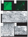
Sindbis vectors express Kv4.2 and Kv4 dominant-negative constructs in CA1 pyramidal neurons in organotypic culture. (a) High-powered view of CA1 pyramidal neurons infected with Sindbis-Kv4.2 that co-expresses EGFP. Entire cell fills evenly with green fluorescence. Scale bar = 5 μm. (b) Single CA1 pyramidal neuron infected with Sindbis-Kv4DN-EGFP. In these cells there is an uneven distribution of green fluorescence that is restricted to the somatodendritic compartments. Labeling hot spots are indicated. Scale bar = 10 μm. (c) Electron micrograph from the hippocampal CA1 region showing a neuronal perikaryon from a pyramidal neuron infected with Sindbis-Kv4DN-EGFP. Immunoperoxidase labeling for Kv4DN-EGFP (open arrows) is seen as a black precipitate distributed mainly along the ER membrane. The labeled soma is receiving a synaptic input from an unlabeled terminal (UT). G, Golgi stacks; M, mitochondri; Nu, nucleus. Scale bar = 1 μm. (d) Electron micrograph from CA1 pyramidal neuron proximal dendrite of neuron infected with Kv4DN-EGFP. Immunoperoxidase labeling (open arrows) is diffusely distributed within the cytoplasm and appears to be most prevalent along membranes of the ER as well as on elements of the cytoskeleton. Immunoperoxidase labeling is also evident within a multivesicular body (MVB). The dendrite receives multiple asymmetric (curved arrow) synapses from unlabeled terminals (UT2-4) that show no immunoperoxidase label. M, mitochondrion. Scale bar = 1 μm. (e) Subcellular distribution for Kv4DN-EGFP in dendrites quantitated by immunogold labeling. Gold particles are found primarily in the cytoplasm and are most prevalent along the membranes of the smooth endoplasmic reticulum (SER; arrows). Note a coated vesicle (CV) near the SER. Scale bars = 0.2 μm.
Fig. 10
Fig. 10
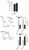
Regulation of ISA expression is similar in dissociated hippocampal pyramidal neurons and CA1 pyramidal neurons in organotypic culture. (a) Peak conductance density for ISA in dissociated hippocampal pyramidal neurons (n = 9) and organotypic CA1 pyramidal neurons (n = 7) are not significantly different. (b) Sindbis-Kv4DN-EGFP suppresses ISA current amplitude in CA1 pyramidal neurons in organotypic culture. Representative ISA currents obtained from these studies are shown as different currents obtained by subtracting a depolarization to 0 mV with a pre-pulse to −40 mV from a matched recording with a pre-pulse to −80 mV. (c) Summary data show that suppression of ISA by Kv4DN-EGFP is similar in organotypic dissociated culture (n = 9) to suppression seen in dissociated culture (n = 8). (d) Increasing expression of Kv4.2 in CA1 pyramidal neurons by Sindbis-Kv4.2 increases the amplitude of ISA. ISA was isolated as described in (b). (e) Summary data shows that the increase in current is similar for pCMV-Kv4.2 expression in dissociated pyramidal neurons (n = 8) and Sindbis-Kv4.2 infection of CA1 pyramidal neurons in organotypic culture (n = 5).

Similar articles

Cited by

References

    1. An WF, Bowlby MR, Betty M, et al. Modulation of A-type potassium channels by a family of calcium sensors. Nature. 2000;403:553–556. - PubMed
    1. Bahring R, Dannenberg J, Peters HC, Leicher T, Pongs O, Isbrandt D. Conserved Kv4 N-terminal domain critical for effects of Kv channel-interacting protein 2.2 on channel expression and gating. J. Biol. Chem. 2001;276:23 888–23 894. - PubMed
    1. Baldwin TJ, Tsaur ML, Lopez GA, Jan YN, Jan LY. Characterization of a mammalian cDNA for an inactivating voltage-sensitive K+ channel. Neuron. 1991;7:471–483. - PubMed
    1. Barry DM, Xu H, Schuessler RB, Nerbonne JM. Functional knockout of the transient outward current, long-QT syndrome, and cardiac remodeling in mice expressing a dominant-negative Kv4α subunit. Circ. Res. 1998;83:560–567. - PubMed
    1. Bernard C, Legros C, Ferrat G, Bischoff U, Marquardt A, Pongs O, Darbon H. Solution structure of hpTX2, a toxin from Heteropoda venatoria spider that blocks Kv4.2 potassium channel. Protein Sci. 2000;9:2059–2067. - PMC - PubMed

Publication types

MeSH terms

Substances