. 2006 Oct;116(10):2777-90.
doi: 10.1172/JCI28828.

Tumors induce a subset of inflammatory monocytes with immunosuppressive activity on CD8+ T cells

Affiliations

Tumors induce a subset of inflammatory monocytes with immunosuppressive activity on CD8+ T cells

Giovanna Gallina et al. J Clin Invest. 2006 Oct.

Abstract

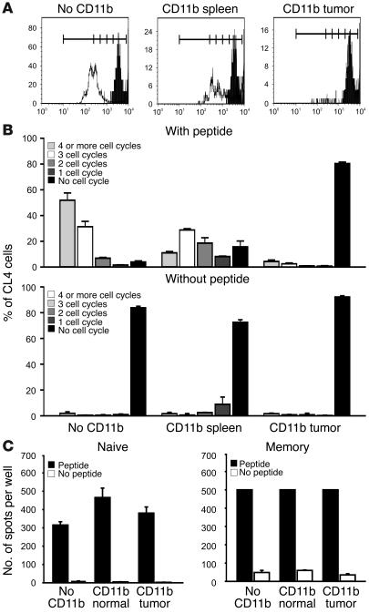
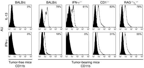
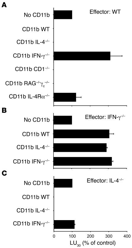
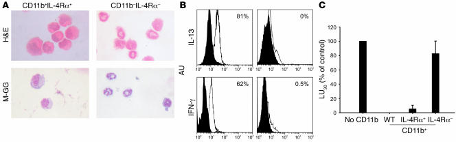
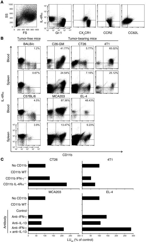
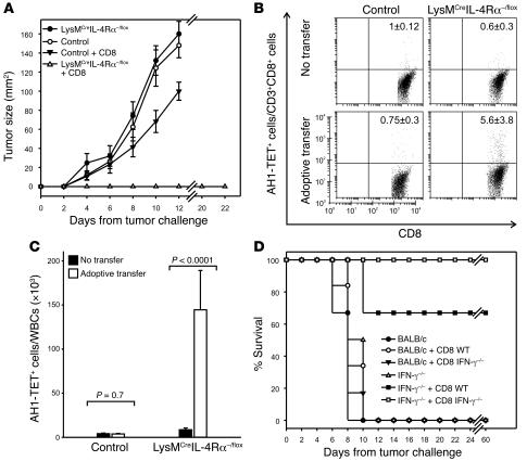
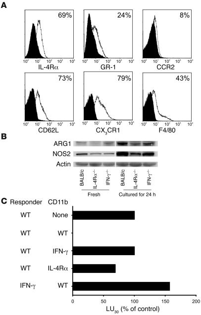
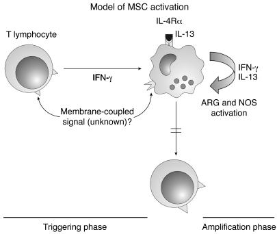
Active suppression of tumor-specific T lymphocytes can limit the efficacy of immune surveillance and immunotherapy. While tumor-recruited CD11b+ myeloid cells are known mediators of tumor-associated immune dysfunction, the true nature of these suppressive cells and the fine biochemical pathways governing their immunosuppressive activity remain elusive. Here we describe a population of circulating CD11b+IL-4 receptor alpha+ (CD11b+IL-4Ralpha+), inflammatory-type monocytes that is elicited by growing tumors and activated by IFN-gamma released from T lymphocytes. CD11b+IL-4Ralpha+ cells produced IL-13 and IFN-gamma and integrated the downstream signals of these cytokines to trigger the molecular pathways suppressing antigen-activated CD8+ T lymphocytes. Analogous immunosuppressive circuits were active in CD11b+ cells present within the tumor microenvironment. These suppressor cells challenge the current idea that tumor-conditioned immunosuppressive monocytes/macrophages are alternatively activated. Moreover, our data show how the inflammatory response elicited by tumors had detrimental effects on the adaptive immune system and suggest novel approaches for the treatment of tumor-induced immune dysfunctions.


Figures

Figure 1
Figure 1. MSCs inhibit antigen-induced T lymphocyte proliferation but not IFN-γ release.
(A) CFSE-labeled splenocytes derived from mice transferred with HA-specific CD8+ (CL4) T cells and primed with HA-encoding vaccinia virus were stimulated with the HA peptide in the absence (No CD11b) or in the presence of CD11b+ cells magnetically purified from either the spleen or the tumor infiltrate of tumor-bearing mice and admixed at a 1:10 ratio. The cultures were stimulated for 60 hours with the relevant peptide, and the clonotypic proliferation was evaluated as CFSE dilution by flow cytometry. (B) Data are presented as the mean percentage of CL4 cells in each cycle from triplicate wells (± SEM); 1 of 2 representative experiments is reported. (C) MSCs do not suppress IFN-γ production from naive or antigen-experienced (memory) CD8+ T cells. MSCs were isolated from the spleens of normal or tumor-bearing mice, and 6 × 10⁵ cells were admixed with 3 × 10⁶ splenocytes containing naive (2.4 × 10⁵) or in vivo antigen-experienced (2.1 × 10⁵) HA-specific CD8+ T cells stimulated with the relevant peptide in an ELISPOT assay. Data (mean ± SEM) are derived from triplicate wells of 1 representative experiment.
Figure 2
Figure 2. CD11b+ splenocytes from tumor-bearing mice produce both IFN-γ and IL-13.
CD11b+ cells isolated from the spleens of normal BALB/c and different tumor-bearing mice (WT BALB/c, IFN-γ–/–, CD1–/–, and RAG–/–γc–/– KO mice) were analyzed by flow cytometry for intracellular presence of IL-13 (top panels) and IFN-γ (bottom panels). Open histograms indicate staining with IL-13 or IFN-γ antibody; filled histograms indicate staining with isotype-matched antibody controls. Data are representative of at least 3 determinations in separate experiments. MFI, mean fluorescence intensity (a log scale of 100–104 was used).
Figure 3
Figure 3. Evaluation of cytokines and cytokine receptors required by tumor-induced CD11b+ cells to inhibit generation of alloreactive and tumor-specific CTLs.
CD11b+ sorted cells from tumor-bearing WT or different KO mice were added at a final concentration of 3% to a mixed leukocyte culture set up with BALB/c effectors stimulated with an equal number of C57BL/6 splenocytes. After 5 days, cytotoxic activity was tested in a 5-hour 51Cr-release assay against either a syngeneic control target (CT26, H-2d) or an allogeneic target (MBL-2, H-2b). Effector lymphocytes were taken from WT (A), IFN-γ KO (B), or IL-4 KO (C) mice. LU30, defined as the number of lymphocytes necessary to achieve 30% specific lysis of 2 × 10³ target cells in a 5-hour assay, was calculated on the basis of the total number of viable cells recovered from the cultures. Data are expressed as the ratio between the LU30 measured in cultures containing the third-party cells and in control cultures set up in the absence of third-party cells. Data are mean ± SEM from 3 experiments.
Figure 4
Figure 4. Morphological and functional characterization of tumor-induced CD11b+IL-4Rα+ and CD11b+IL-4Rα cells.
Splenocytes were sorted by microbeads to select CD11b+ cells, and this cellular population was subsequently sorted by flow cytometry to select IL-4Rα+ (left panels) and IL-4Rα (right panels) cells. (A) The sorted cells were stained with H&E and May-Grünwald-Giemsa stain (M-GG). Pictures were taken under microscopy at ×40 magnification. CD11b+IL-4Rα+ cells showed a monocytic morphology, while CD11b+IL-4Rα cells comprised granulocytes at various differentiation stages, including immature cells with band morphology. (B) Both populations were analyzed by flow cytometry for their expression of IL-13 (top panels) and IFN-γ (bottom panels). Open histograms indicate staining with IL-13 or IFN-γ antibody; filled histograms indicate isotype-matched antibody control. Data are representative of at least 3 determinations in separate experiments. (C) The 2 populations were added at a final concentration of 3% to alloantigen-stimulated cultures (see above). CD11b+IL-4Rα+ but not CD11b+IL-4Rα cells suppressed the generation of alloreactive CTLs (P < 0.01 and P = 0.48, respectively). Results are shown as the fraction of LU30 measured in the control allo-cultures lacking third-party CD11b+ cells. Data are mean ± SEM from 3 experiments.
Figure 5
Figure 5. Phenotypic characterization of tumor-induced CD11b+IL-4Rα+ cells.
(A) CD11b+ cells from spleens of mice inoculated 9 days earlier with C26-GM tumor cell lines were enriched through immunomagnetic microbeads and stained with the indicated antibody. FS, forward scatter; SS, side scatter. (B) Blood cells (top rows) and spleen cells (bottom rows) from mice of various strains inoculated with different tumors were stained for CD11b and IL-4Rα. The following tumor cell lines were used: the BALB/c-derived colon carcinoma CT26 (closely related to the C26 tumor used in our previous experiments) and mammary carcinoma 4T1; and the C57BL/6–derived EL-4 lymphoma and MCA203 fibrosarcoma. Data are from a single experiment representative of 3. (C) CD11b+ cells were sorted from the spleens of tumor-bearing WT or KO BALB/c mice and added at a final concentration of 3% to a mixed leukocyte culture set up as previously described (top panels). CD11b+ cells sorted from the spleens of tumor-bearing C57BL/6 mice were added at a final concentration of 3% to a mixed leukocyte culture set up with C57BL/6 effectors stimulated with an equal number of γ-irradiated BALB/c splenocytes, in the presence or absence of anti–IFN-γ and/or anti–IL-13 mAbs added at the beginning and at the third day of culture (bottom panels).
Figure 6
Figure 6. Adoptive cell transfer is effective only in mice with a myeloid-restricted inactivation of IL-4Rα.
(A) LysMCreIL-4R–/flox mice and control littermates were challenged s.c. on day 0 with C26-GM cells and were given, 24 hours later, CD8+ T cells purified from the spleens and lymph nodes of mice vaccinated 14 days before with γ-irradiated C26-GM cells. Tumor size is indicated as the product of the 2 main perpendicular diameters measured with a caliper. Mice with tumors had to be euthanized on day 12, since they were showing severe signs of distress. (B) The CD3+CD8+ cells positive for the Ld tetramer loaded with AH1 peptide were determined in the blood of tumor-bearing mice 8 days after tumor inoculation. One dot plot for each group is reported. Numbers indicate the percentages of AH1-TET+ cells among gated CD3+CD8+ T lymphocytes (mean ± SEM, n = 5–6). (C) The absolute numbers of CD3+CD8+AH1-TET+ cells were calculated for the total number of circulating white blood cells (WBCs) and reported as mean ± SEM. The background staining given by control β-gal876–884/Ld TET was subtracted for each determination. (D) IFN-γ–/– and WT BALB/c mice were challenged s.c. on day 0 with C26-GM cells and given, 24 hours later, CD8+ T cells purified from the spleens and lymph nodes of IFN-γ–/– and WT BALB/c mice, vaccinated 14 days before with γ-irradiated C26-GM cells. Results, expressed as survival rates after tumor challenge, are from 1 representative experiment with 6 mice per group.
Figure 7
Figure 7. IFN-γ and IL-13 cooperate in the activation of immunosuppressive pathways.
(A) Kinetics of IL-4Rα expression at the cell surface of tumor-induced CD11b+ splenocytes. CD11b+ splenocytes from tumor-free (normal) or tumor-bearing mice (tumor) were incubated for 72 hours alone, with IFN-γ, with a neutralizing antibody against mouse IL-13, or with both. Isotype-matched antibodies were included as negative controls. IL-4Rα protein expression on the cell surface was analyzed at various time points by flow cytofluorimetry and reported as the mean percentage of positive cells ± SEM of 3 separate determinations. Differences between diverging kinetics values were statistically significant (P < 0.05). (BD) Effect of CD11b+ cell exposure to IL-13 and IFN-γ. CD11b+ cells were sorted from the spleens of tumor-free and tumor-bearing mice. These last cells were analyzed immediately (Fresh) or cultured for 48 hours in medium alone (No cytokine) or in the presence of IFN-γ, IL-13, or both. Some samples were treated either with IFN-γ for 24 hours followed by addition of IL-13 for a further 24 hours (IFN-γ → IL-13) or with the inverse cytokine combination (IL-13 → IFN-γ). Cells were then analyzed for expression of Arg1 and Nos2 mRNA by real-time PCR (B), for Arg1 and Nos2 protein levels by Western blotting (C), and for relative enzyme activity (D). Log2FC is the log-transformed gene expression value of the target gene, normalized to an endogenous reference and relative to a calibrator sample. ND, not done. Data for real-time-PCR, Western blot, and enzyme activity are from the same experiment representative of 3.
Figure 8
Figure 8. Phenotypic and functional characterization of CD11b+ cells infiltrating the tumor.
(A) CD11b+ cells were separated from the disaggregated nodules of C26-GM tumor and stained with the indicated mAbs. (B) ARG1 and NOS2 protein expression level in CD11b+ cells isolated from tumors of WT, IL-4Rα–/–, and IFN-γ–/– BALB/c mice, analyzed immediately (Fresh) and after 24 hours of culture in standard medium. (C) Cytotoxicity of WT and IFN-γ–/– BALB/c splenocytes (Responder), stimulated with an equal number of γ-irradiated C57BL/6 splenocytes and cocultured without (None) or with CD11b+ cells sorted from tumors of WT, IL-4Rα–/–, and IFN-γ–/– BALB/c mice. Data are the mean of 2 separate experiments.
Figure 9
Figure 9. Mechanisms of MSC-dependent suppression.
In the “triggering phase,” T lymphocytes activated by exposure to the antigen release IFN-γ that, possibly in conjunction with a not yet identified membrane signal, activates inflammatory monocyte precursors. Whereas in resting condition this would result in induction of classically activated macrophages, monocytes conditioned by tumors show a different program because of the expression of IL-4Rα and the ability to release both IL-13 and IFN-γ. The IFN-γ production, initially sustained by activated T lymphocytes, is subsequently amplified by the cytokine released from activated monocytes. IFN-γ allows the prolonged expression and signaling of IL-4Rα after engagement by IL-13 released via an autocrine circuit. During the “amplification phase,” the cytokines are thus able to maintain a prolonged activation of the enzymes NOS and ARG, which ultimately originate the immunosuppressive mediators acting on the CD8+ T cells. This model supports the view that a prolonged stimulation of MSCs is necessary to mediate a full inhibition of CD8+ T cells and explains why T cells can initially proliferate in the presence of spleen-derived MSCs. On the other hand, ARG and NOS are constitutively activated in tumor-infiltrating MSCs, which account for the prompt immunosuppression provided by these cells (Figure 1).

Comment in

Similar articles

Cited by

References

    1. Bronte V., et al. Apoptotic death of CD8+ T lymphocytes after immunization: induction of a suppressive population of Mac-1+/Gr-1+ cells. J. Immunol. 1998;161:5313–5320. - PMC - PubMed
    1. Bronte V., et al. Unopposed production of granulocyte-macrophage colony-stimulating factor by tumors inhibits CD8+ T cell responses by dysregulating antigen-presenting cell maturation. J. Immunol. 1999;162:5728–5737. - PMC - PubMed
    1. Kusmartsev S., Nefedova Y., Yoder D., Gabrilovich D.I. Antigen-specific inhibition of CD8+ T cell response by immature myeloid cells in cancer is mediated by reactive oxygen species. J. Immunol. 2004;172:989–999. - PubMed
    1. Almand B., et al. Increased production of immature myeloid cells in cancer patients: a mechanism of immunosuppression in cancer. J. Immunol. 2001;166:678–689. - PubMed
    1. Bronte V., et al. Identification of a CD11b+/Gr-1+/CD31+ myeloid progenitor capable of activating or suppressing CD8+ T cells. Blood. 2000;96:3838–3846. - PMC - PubMed

Publication types

MeSH terms