Review
. 2006 Oct 5;52(1):77-92.
doi: 10.1016/j.neuron.2006.09.021.

Mechanisms of neuropathic pain

Affiliations
Review

Mechanisms of neuropathic pain

James N Campbell et al. Neuron. .

Abstract

Neuropathic pain refers to pain that originates from pathology of the nervous system. Diabetes, infection (herpes zoster), nerve compression, nerve trauma, "channelopathies," and autoimmune disease are examples of diseases that may cause neuropathic pain. The development of both animal models and newer pharmacological strategies has led to an explosion of interest in the underlying mechanisms. Neuropathic pain reflects both peripheral and central sensitization mechanisms. Abnormal signals arise not only from injured axons but also from the intact nociceptors that share the innervation territory of the injured nerve. This review focuses on how both human studies and animal models are helping to elucidate the mechanisms underlying these surprisingly common disorders. The rapid gain in knowledge about abnormal signaling promises breakthroughs in the treatment of these often debilitating disorders.


Figures

Figure 1
Figure 1. Animal Models of Neuropathic Pain
(A) Four different nerve injury models are shown. In the spinal nerve ligation (SNL) model, one or more spinal nerves going to the foot are ligated and cut (Kim and Chung, 1992). In the partial sciatic ligation (PSL) model, a portion of the sciatic nerve is tightly ligated (Seltzer et al., 1990). The chronic constriction injury (CCI) model involves placement of four loose chromic-gut ligatures on the sciatic nerve. An immune response to the sutures leads to nerve swelling and nerve constriction. In the spared nerve injury (SNI) model, the common peroneal and tibial nerves are cut, sparing the sural nerve (Decosterd and Woolf, 2000). In each model, only a portion of the afferents going to the foot are lesioned. (B and C) Each of these nerve injury models leads to hyperalgesia, which is manifest by enhanced responses to mechanical, heat, and/or cooling stimuli. (B) To test for mechanical hyperalgesia, Von Frey monofilaments with different bending forces are applied to the plantar surface of the foot. The threshold force for paw withdrawal decreases dramatically after the nerve injury (adapted with permission [Li et al., 2000]) (C) To test for heat hyperalgesia, a radiant heat source is focused onto the plantar surface of the foot, and the reaction time for paw withdrawal is measured. The difference in reaction time between the ipsilateral and contralateral foot is calculated. After the SNL, the withdrawal of the ipsilateral foot is faster than the contralateral foot (negative latency difference), indicating the presence of heat hyperalgesia (adapted from Kim and Chung [1992] reprinted from Pain, pp. 355–363, copyright 1992, with permission from the International Association for the Study of Pain). Data are presented as mean ± SEM.
Figure 2
Figure 2. A Spinal Nerve Injury Leads to Alterations at Many Sites along the Neural Axis for Pain
Eight different sites of pathophysiological changes are shown. (1) Spontaneous neural activity and ectopic sensitivity to mechanical stimuli develops at the site of nerve injury. (2) The expression of different molecules in the dorsal root ganglion of the injured nerve is up- or downregulated, reflecting the loss of trophic support from the periphery. Spontaneous neural activity develops in the dorsal root ganglia. (3) The distal part of the injured nerve undergoes Wallerian degeneration, exposing the surviving nerve fibers from uninjured portions of the nerve to a milieu of cytokines and growth factors. (4) Partial denervation of the peripheral tissues leads to an excess of trophic factors from the partly denervated tissue that can lead to sensitization of primary afferent nociceptors. (5) The expression of different molecules in the dorsal root ganglion of the uninjured nerve is up- or downregulated, reflecting the enhanced trophic support from the periphery. (6) Sensitization of the postsynaptic dorsal horn cell develops, leading to an augmentation of the response to cutaneous stimuli. (7) Activated microglial cells contribute to the development of this dorsal horn sensitization. (8) Changes in descending modulation of dorsal horn neurons also may contribute to the enhanced responsiveness of dorsal horn neurons.
Figure 3
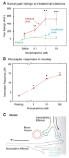
Figure 3. A Model for Sympathetically Maintained Pain
(A) After acutely relieving pain by performing a sympathetic block, norepinephrine in physiological concentrations was injected intra-dermally in a blinded fashion into the previously hyperalgesic area. The norepinephrine injections into the affected, but not the unaffected, extremity produced pain. Norepinephrine does not induce pain in control subjects. These data suggest that the nociceptors are sensitized to catechols in patients with SMP (adapted with permission [Ali et al., 2000]). ACUC, area under curve. (B) In primates, normal nociceptors do not respond to catechol administration. However, in a monkey-spinal nerve ligation model, nociceptors developed a response to the α 1 adrenergic agonist phenylephrine administered topically to their receptive field (adapted and used with permission [Ali et al., 1999]). AP, action potentials. (C) A model to account for SMP. After a partial nerve lesion, some afferent fibers still remain in the skin. Factors released in the skin induce the sympathetic efferents to sprout into more superficial areas of the skin (Yen et al., 2006). These factors also lead the nociceptors to express α 1 adrenergic receptors. Now, the release of norepinephrine from the sympathetic terminals leads to activation of the nociceptive terminals, which accounts for the coupling of sympathetic activity with nociceptor responses. Data are presented as mean ± SEM.
Figure 4
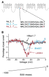
Figure 4. A Genetic Basis for Erythromelalgia
(A) Schematic of the Nav 1.7 voltage-gated sodium channel that is found exclusively in sensory and sympathetic neurons. A number of point mutations have been identified in this sodium channel in families of patients with erythromelalgia (Waxman and Dib-Hajj, 2005). The two mutations shown here produced single amino acid substitutions at the sites indicated. (B) Whole-cell-patch-clamp recordings in transfected HEK293 cells revealed that these point mutations lead to an augmentation of the response of the channel to a slow depolarizing ramp (100 mV in 500 ms). These mutations would be expected to increase the excitability of the peripheral sensory neuron. (Adapted with permission [Cummins et al., 2004]. Copyright 2004 by the Society for Neuroscience.).
Figure 5
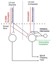
Figure 5. Central Sensitization Mechanisms Involved in the Spinal Nerve Ligation Model
Spontaneous activity from the injured afferents (L5) and intact nociceptors (L4) may sensitize central pain-signaling neurons. The spontaneous activity in the L5 fibers is restricted to myelinated afferents. Nociceptive C fibers from L4 spontaneously discharge and may themselves be sensitized. The enhanced discharge of the primary afferents leads to augmented response of dorsal horn cells to nociceptor input and increased synaptic efficacy of inputs from mechanoreceptors (mechanism for allodynia). Alterations in descending modulation and inhibitory interneuron function also likely play a role.
Figure 6
Figure 6
Microglial Cells in the CNS Are Activated following Peripheral Nerve Injury and Release Cytokines that Alter the Responses of Dorsal Horn Cells

Similar articles

Cited by

References

    1. Abbadie C, Lindia JA, Cumiskey AM, Peterson LB, Mudgett JS, Bayne EK, DeMartino JA, MacIntyre DE, Forrest MJ. Impaired neuropathic pain responses in mice lacking the chemokine receptor CCR2. Proc Natl Acad Sci USA. 2003;100:7947–7952. - PMC - PubMed
    1. Ahlgren SC, Levine JD. Mechanical hyperalgesia in streptozotocin-diabetic rats. Neuroscience. 1993;52:1049–1055. - PubMed
    1. Alessandri-Haber N, Dina OA, Yeh JJ, Parada CA, Reichling DB, Levine JD. Transient receptor potential vanilloid 4 is essential in chemotherapy-induced neuropathic pain in the rat. J Neurosci. 2004;24:4444–4452. - PMC - PubMed
    1. Ali Z, Ringkamp M, Hartke TV, Chien HF, Flavahan NA, Campbell JN, Meyer RA. Uninjured C-fiber nociceptors develop spontaneous activity and alpha adrenergic sensitivity following L6 spinal nerve ligation in the monkey. J Neurophysiol. 1999;81:455–466. - PubMed
    1. Ali Z, Raja SN, Wesselmann U, Fuchs PN, Meyer RA, Campbell JN. Intradermal injection of norepinephrine evokes pain in patients with sympathetically maintained pain. Pain. 2000;88:161–168. - PubMed

Publication types

LinkOut - more resources