. 2006 Oct 18;25(20):4752-62.
doi: 10.1038/sj.emboj.7601351. Epub 2006 Sep 28.

Mdm2 is involved in the ubiquitination and degradation of G-protein-coupled receptor kinase 2

Affiliations

Mdm2 is involved in the ubiquitination and degradation of G-protein-coupled receptor kinase 2

Alicia Salcedo et al. EMBO J. .

Abstract

G-protein-coupled receptor kinase 2 (GRK2) is a central regulator of G-protein-coupled receptor signaling. We report that Mdm2, an E3-ubiquitin ligase involved in the control of cell growth and apoptosis, plays a key role in GRK2 degradation. Mdm2 and GRK2 association is enhanced by beta(2)-adrenergic receptor stimulation and beta-arrestin. Increased Mdm2 expression accelerates GRK2 proteolysis and promotes kinase ubiquitination at defined residues, whereas GRK2 turnover is markedly impaired in Mdm2-deficient cells. Moreover, we find that activation of the PI3K/Akt pathway by insulin-like growth factor-1 alters Mdm2-mediated GRK2 degradation, leading to enhanced GRK2 stability and increased kinase levels. These data put forward a novel mechanism for controlling GRK2 expression in physiological and pathological conditions.


Figures
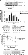
Figure 1
Figure 1
(A) Association of endogenous Mdm2 and GRK2. HeLa cells growing in 150 mm dishes were serum-starved for 2 h and incubated with an inverse β-agonist (betaxolol, 10 μM) for 15 min to lessen basal Mdm2/GRK2 association before challenging with 10 μM isoproterenol for the indicated times. Cells were lysed in buffer A as detailed in Materials and methods and lysates subjected to immunoprecipitation with anti-Mdm2 (SMP14) antibody. Immunoprecipitates were resolved by SDS–PAGE and GRK2 associated to the Mdm2 immunocomplex was detected by Western blot analysis with a specific antibody. 1% of the total lysate used for immunoprecipitation was loaded as input signal. (B) The Mdm2/GRK2 association is enhanced by β2AR stimulation and β-arrestin expression. HEK-293 cells were transiently transfected with plasmids encoding GRK2, β2AR, Mdm2 and β-arrestin1 when indicated. Cells were challenged with 10 μM isoproterenol for different times and GRK2/Mdm2 co-immunoprecipitation assessed as detailed in Materials and methods. GRK2 presence in the non-stimulated Mdm2 immunocomplex was taken as the control condition. Data were normalized by total Mdm2 and depicted as mean±s.e.m. of four independent experiments. A representative gel is shown in the bottom panel. (C) Normal decay of GRK2 protein is impaired upon of β-arrestin1 and β-arrestin2 knockdown. GRK2 protein levels were examined by immunoblot analysis in both wild-type and β-arrestin1/2-deficient MEFs upon treatment with cycloheximide (CHX) for the indicated times. The amount of GRK2 at 0 h was defined as 100%, and data normalized by actin protein levels. In both panels, data are the mean±s.e.m. of four independent experiments (*P<0.05). Representative gels are shown.
Figure 2
Figure 2
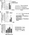
(A) Mdm2 increases degradation of GRK2 and of a slow-turnover GRK2 mutant. HEK-293 cells were transiently transfected with wild-type GRK2 or the GRK2-K220R mutant in the presence or absence of Mdm2, and kinase turnover assessed by pulse–chase experiments as described in Materials and methods. 35S-labeled proteins immunoprecipitated with the anti-GRK2 antibody AbFP2 were resolved by SDS–PAGE followed by fluorography and densitometry. 35S-labeled GRK2 band densities were then normalized to total GRK2 present in the immunoprecipitates, as determined by Western blot analysis. Data are mean±s.e.m. of 3–4 independent experiments performed in duplicate, *P<0.05, **P<0.01. A representative gel autoradiography is shown. (B, C) Effect of LMB treatment on GRK2 turnover. HEK-293 cells transfected with GRK2 and Mdm2 or an empty vector were treated with LMB or vehicle before and during pulse–chase experiments as detailed in Materials and methods, and GRK2 turnover determined as in previous figures. Data are the mean±s.e.m. of three independent experiments performed in duplicate, *P<0.05, **P<0.01. Representative fluorographs are shown. In panel C, endogenous GRK2 expression levels were determined by immunoblot analysis in lysates from MCF7 cells treated for different times with LMB. Data are corrected for actin expression and depicted as percentage of control. *P<0.05, ***P<0.001. A representative blot is shown.
Figure 3
Figure 3
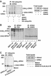
Mdm2 promotes GRK2 ubiquitination. (A) Wild-type GRK2, HA-ubiquitin and β2AR expression plasmids were transfected together with Mdm2 or an empty vector into HEK-293 cells. After 2 h of serum starving, cells were incubated with or without 10 μM isoproterenol for 15 min and lysed for ubiquitination assays as described in Materials and methods. The different GRK2 species present in the ubiquitin immunoprecipitates are indicated. Asterisk and arrowhead indicate mono- and bi-ubiquitinated GRK2 forms (see text for details). Blots depicting expression levels for Mdm2 and GRK2 in total lysates are shown. (B) Both basal and agonist-stimulated ubiquitination of endogenous GRK2 are blocked in either Mdm2- or β-arrestin-deficient cells. ∼50 × 106 wild-type MEFs or MEFs devoid of Mdm2 or β-arrestin proteins were serum-starved and challenged or not with 10 μM isoproterenol for 15 min. Cells were processed as indicated above and ubiquitinated GRK2 species were analyzed by immunoprecipitation of endogenous GRK2 with a specific monoclonal anti-GRK2 antibody (c5/1.1) and detection of endogenous ubiquitin conjugates with anti-ubiquitin antibody FK2. Membranes were stripped and immunoblotted (right panel) with a specific polyclonal anti-GRK2 antibody (C-15) to confirm equal GRK2 loading in each condition. (C) Mutation of critical lysine residues in the N-terminus of GRK2 impairs kinase ubiquitination in the presence of Mdm2. Wild-type GRK2 or the GRK2- K19,20,30,31R mutant was cotransfected with ubiquitin plasmids in the presence or absence of Mdm2 into HEK-293 cells and the kinase ubiquitination pattern was analyzed as above. Expression levels of GRK2 and Mdm2 were assessed in lysates by Western blot analysis. Gels in all panels are representative of 2–3 independent experiments.
Figure 4
Figure 4
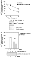
N-terminal lysine residues are critical for Mdm2-mediated GRK2 degradation. (A) Turnover of wild-type GRK2 and GRK2-K19,20,30,31R mutant proteins was assessed by pulse–chase experiments in HEK-293 cells as described in Materials and methods and Figure 2. (B) Similar experiments were performed in cells cotransfected with an Mdm2 construct. In both panels, data are mean±s.e.m. from three to four independent experiments (**P<0.01), and representative fluorographies are shown.
Figure 5
Figure 5
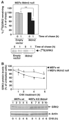
GRK2 turnover is blocked in Mdm2-deficient cells. (A) Mdm2-null MEFs were transfected with wild-type GRK2 in the presence or absence of Mdm2. Degradation of the heterologous GRK2 protein was assessed after 1 h of chase as in previous figures. (B) Stability of endogenous GRK2 is increased in the absence of Mdm2 expression. GRK2 protein levels were examined by immunoblot analysis in both wild-type and Mdm2-deficient MEFs upon treatment with cycloheximide (CHX) for the indicated times. The amount of GRK2 at 0 h was defined as 100%, and data normalized by actin protein levels. In both the panels, data are the mean±s.e.m. of four independent experiments (*P<0.05, **P<0.01). Representative gels are shown.
Figure 6
Figure 6
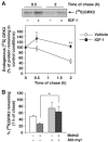
IGF receptor activation and Akt activity promote GRK2 stabilization. (A) Turnover of endogenous GRK2 was analyzed in MCF7 cells as detailed in Materials and methods in the presence of 50 ng/ml of IGF-1 or vehicle. Labeled proteins were immunoprecipitated with a specific anti-GRK2 antibody (C5/1.1). The density of the 35S-labeled GRK2 band after the pulse period was taken as 100%. Data are mean±s.e.m. of 3–5 independent experiments. *P<0.05; **P<0.01. A representative fluorography is shown. (B) HEK-293 cells were transfected with different combinations of GRK2, Mdm2 and a constitutively active Akt mutant (Akt-myr) as indicated. GRK2 degradation rate was determined by pulse–chase analysis. Results (mean±s.e.m.) from three independent experiments are shown. *P<0.05.
Figure 7
Figure 7
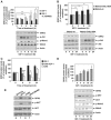
IGF-1 stimulation induces endogenous GRK2 protein accumulation in a PI3K-dependent manner. (A) MCF7 cells were serum-starved for 12 h and challenged with IGF-1 (50 ng/ml) for different times in the presence or absence of the PI3K inhibitor LY294002 (10 μM). Cells were subjected to immunoblot analysis with anti-GRK2, anti-Akt-pSer473, anti-Mdm2-pSer166 and anti-Mdm2 antibodies. GRK2 band densities were normalized by actin protein levels and expressed as percentage of GRK2 level in unstimulated conditions. Data are mean±s.e.m. of five independent experiments performed in duplicate. *P<0.05, **P<0.01. (B) IFG-1-dependent upregulation of GRK2 is prevented by expression of an Mdm2 mutant defective in Akt-mediated phosphorylation. MCF7 cells transiently overexpressing either wild-type Mdm2 or the ligase mutant Mdm2 S166, 186A were serum-starved as above and stimulated with IGF-1 (50 ng/ml) for the indicated times. Expression levels of GRK2, Akt-pSer473, Akt and actin were determined by immunoblotting. Data were analyzed and depicted as in panel A. Representative blots are shown. (C) IGF-1 increases GRK2 protein by decreasing its degradation rate without altering its protein synthesis. Endogenous GRK2 protein levels were determined by immunoblot analysis in MCF7 cells stimulated with IGF-1 as above in the presence or absence of cycloheximide and compared with cycloheximide treatment alone. Data are mean±s.e.m. of three independent experiments. *P<0.05; ***P<0.001. (D) GRK2 protein accumulation is induced by IGF-1 challenge, in the non-transformed mammary cell line MCF10A. Cells were treated as above and levels of GRK2, Akt-pSer473, Akt, Mdm2-pSer166, Mdm2 and actin immunodetected with specific antibodies. The patterns of Akt activation, Mdm2 phosphorylation and GRK2 protein increase are similar to those observed in MCF7 cells. (E) Transformed mammary cells display higher levels of Akt activity and GRK2 protein. Both malignant transformed (T, MDA-MB486 and MCF7) and non-transformed cells (N, MCF10A and 184B5) were lysed in RIPA buffer and expression levels of GRK2, Akt-pSer473, Akt and actin were determined as above. Representative blots from three independent experiments are displayed.
Figure 8
Figure 8
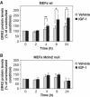
Knockdown of Mdm2 prevents endogenous GRK2 protein accumulation in response to IGF-1.Wild-type (A) and Mdm2-deficient MEFs (B) were treated with IGF-1 or vehicle for the indicated times as in Figure 7. GRK2 protein levels were determined by Western blot analysis and normalized by actin expression. Data are mean±s.e.m. of four experiments performed in triplicate, *P<0.05, **P<0.01.

Similar articles

Cited by

References

    1. Ashcroft M, Ludwig RL, Woods DB, Copeland TD, Weber HO, MacRae EJ, Vousden KH (2002) Phosphorylation of HDM2 by Akt. Oncogene 21: 1955–1962 - PubMed
    1. Bond GL, Hu W, Levine AJ (2005) MDM2 is a central node in the p53 pathway: 12 years and counting. Curr Cancer Drug Targets 5: 3–8 - PubMed
    1. Elorza A, Penela P, Sarnago S, Mayor F Jr (2003) MAPK-dependent degradation of G protein-coupled receptor kinase 2. J Biol Chem 278: 29164–29173 - PubMed
    1. Feng J, Tamaskovic R, Yang Z, Brazil DP, Merlo A, Hess D, Hemmings BA (2004) Stabilization of Mdm2 via decreased ubiquitination is mediated by protein kinase B/Akt-dependent phosphorylation. J Biol Chem 279: 35510–35517 - PubMed
    1. Ferguson SS (2001) Evolving concepts in G protein-coupled receptor endocytosis: the role in receptor desensitization and signaling. Pharmacol Rev 53: 1–24 - PubMed

Publication types

MeSH terms