. 2006;34(18):5217-31.
doi: 10.1093/nar/gkl677. Epub 2006 Sep 26.

Human RECQ5beta helicase promotes strand exchange on synthetic DNA structures resembling a stalled replication fork

Affiliations

Human RECQ5beta helicase promotes strand exchange on synthetic DNA structures resembling a stalled replication fork

Radhakrishnan Kanagaraj et al. Nucleic Acids Res. 2006.

Abstract

The role of the human RECQ5beta helicase in the maintenance of genomic stability remains elusive. Here we show that RECQ5beta promotes strand exchange between arms of synthetic forked DNA structures resembling a stalled replication fork in a reaction dependent on ATP hydrolysis. BLM and WRN can also promote strand exchange on these structures. However, in the presence of human replication protein A (hRPA), the action of these RecQ-type helicases is strongly biased towards unwinding of the parental duplex, an effect not seen with RECQ5beta. A domain within the non-conserved portion of RECQ5beta is identified as being important for its ability to unwind the lagging-strand arm and to promote strand exchange on hRPA-coated forked structures. We also show that RECQ5beta associates with DNA replication factories in S phase nuclei and persists at the sites of stalled replication forks after exposure of cells to UV irradiation. Moreover, RECQ5beta is found to physically interact with the polymerase processivity factor proliferating cell nuclear antigen in vitro and in vivo. Collectively, these findings suggest that RECQ5beta may promote regression of stalled replication forks to facilitate the bypass of replication-blocking lesions by template-switching. Loss of such activity could explain the elevated level of mitotic crossovers observed in RECQ5beta-deficient cells.


Figures
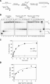
Figure 1
Figure 1
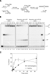
Strand-exchange by RECQ5β on synthetic forked DNA structure with homologous arms lacking the leading strand. (A) Scheme of the assay. The lengths of individual arms are indicated in nucleotides (nt) or base pairs (bp). The 3′ end of the lagging oligonucleotide is indicated by an arrow and the position of the 5′-32P label is marked by an asterisk. The homologous leading and lagging arms have a 5 nt heterology at the fork junction to prevent spontaneous strand exchange. (B) 1 nM 32P-labeled 30mer/60mer duplex (RK1/RK2) was incubated with 1 nM 60mer complementary oligonucleotide (RK3) in the presence of 40 nM RECQ5β to form the forked DNA structure. After 10 min, either ATP or ATPγS were added to a final concentration of 2 mM. Aliquots from different reaction time points, before and after addition of the nucleotide, were analyzed by non-denaturing PAGE followed by phosphorimaging. C1–C4, markers for the DNA substrate and the reaction products. (C) Quantification of the ATP and ATPγS containing reactions. Relative concentration of the 60mer duplex product is plotted versus reaction time. The data points represent the average values from three independent experiments. (D) Dependence of the strand-exchange activity of RECQ5β on Mg2+ ion concentration. Reaction mixtures contained 1 nM 3′-flap DNA substrate, 40 nM RECQ5β and 2 mM ATP with increasing concentrations of Mg(OAc)2. Relative concentration of the 60mer duplex product after 32 min was measured as in (B) and (C). The lines drawn are only to guide the eye.
Figure 2
Figure 2
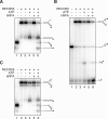
Strand-exchange by RECQ5β on synthetic forked DNA duplex with a leading-strand gap. (A) Scheme of the assay. The oligonucleotides used are the same as in Figure 1, except for an additional 20mer representing the leading strand. (B) 1 nM 32P-labeled 20mer/60mer duplex (RK1/RK2) was incubated with 1 nM 30mer/60mer duplex (RK3/RK4) in the presence of 40 nM RECQ5β to form the forked DNA structure. After 10 min, either ATP or ATPγS were added to a final concentration of 2 mM. Aliquots of the annealing and the nucleotide-driven reactions were analyzed by non-denaturing PAGE. C1–C3, markers for the DNA substrate and the reaction products. (C) Quantification of the ATP- driven reaction. The relative concentrations of the unwound 20mer oligonucleotide (leading strand; open circles) and subsequently formed 20mer/30mer partial duplex (leading/lagging duplex; closed circles) are plotted versus reaction time. The data points represent the average values from three independent experiments.
Figure 3
Figure 3
Effect of hRPA on RECQ5β-mediated strand exchange on forked DNA duplex lacking the leading strand (A) and forked DNA duplex with 10 nt leading-strand gap, radioactively labeled either on the leading-strand oligonucleotide (B) or on the lagging-strand template (C). The DNA substrates were prepared by RECQ5β mediated annealing as described in Materials and Methods and schematically indicated in Figures 1A and 2A. Reactions were carried out for 32 min as indicated. DNA, RECQ5β and hRPA were present at concentrations of 1, 40 and 20 nM, respectively. hRPA was added 2 min before the addition of ATP (2 mM).
Figure 4
Figure 4
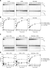
Strand exchange by BLM, WRN and RECQ5β in the absence and presence of hRPA. (A) Time course of reactions of 10 nM BLM, 5 nM WRN and 40 nM RECQ5β, respectively, with 1 nM forked DNA structure with homologous arms lacking the leading strand. (B) Quantification of reactions in (A). (C) Time course of reactions of 10 nM BLM, 5 nM WRN and 40 nM RECQ5β, respectively, with 1 nM DNA structure as in (A) in the presence of 20 nM hRPA. The DNA substrate was preincubated for 2 min with hRPA before addition of helicase. (D) Quantification of reactions in (C). The DNA substrate was the same as in Figure 1, except that it was prepared by spontaneous annealing of the component oligonucleotides as described in Materials and Methods. Reactions were analyzed as in Figure 1. In the graphs, relative concentrations of the products are plotted versus reaction time. The 60mer bubble duplex, filled circles; 3′-tailed 30mer/60mer duplex, open circles; free 60mer oligonucleotide, filled triangles. In all graphs, the data points represent the average values from three independent experiments.
Figure 5
Figure 5
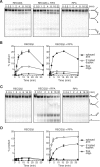
Unwinding of forked DNA structures with heterologous arms by RECQ5β. (A) Time course of RECQ5β-mediated unwinding of partial forked duplex lacking the leading strand (3′-flap duplex) in the absence and presence of 20 nM hRPA as indicated. Reaction containing RPA only is also shown as a control (B) Quantification of the RECQ5β-containing reactions in (A). The graph shows the relative concentrations of unwound products including the splayed arm (closed circles), the 3′-tailed duplex (open circles) and the free lagging-strand template (closed triangles). (C) Time course of RECQ5β-mediated unwinding of partial forked duplex lacking the lagging strand (5′-flap duplex) in the absence and presence of 20 nM hRPA. Reaction containing RPA only is also shown as a control (D) Quantification of the RECQ5β-containing reactions in (C). The graph shows the relative concentrations of unwound products including the splayed arm (closed circles) and the 3′-tailed duplex (open circles) and the free leading-strand template (closed triangles). All DNA substrates are derived from the same set of four oligonucleotides (RK1, RK2, RK5 and RK6) as described in Materials and Methods. The 3′ end of the leading and lagging strands are indicated by arrows. The position of the 5′-32P label in each substrate is indicated by an asterisk. For all reactions, DNA substrates were present at a concentration of 1 nM. RECQ5β was 100 nM in the reactions without hRPA and 80 nM in the reactions with hRPA. Reactions were carried out and analyzed as described in Materials and Methods. In all graphs, the data points represent the average values from three independent experiments.
Figure 6
Figure 6
Characterization of RECQ5β mutants. (A) A schematic representation of the human RECQ5β protein demonstrating the location of the DExH helicase (dark gray) and RecQ C-terminal (RQC) (light gray) regions conserved among RecQ helicases. The arrowheads indicate the positions of the C-terminal ends of the truncated RECQ5β polypeptides used in this study. The numbers refer to the amino acid sequence of RECQ5β. (B) Comparison of strand-exchange activities of RECQ5β and its deletion variants (40 nM each) on forked DNA structure lacking the leading strand (1 nM) pre-coated with hRPA (20 nM). The reactions were incubated for 32 min and analyzed as described in Materials and Methods. Relative concentration of the 60mer duplex product was calculated for each reaction, and the values obtained were corrected by subtracting the background value (reaction without protein). Strand exchange activity of the mutants is expressed as a fraction of the wild-type activity. The inset shows the scheme of the reaction. (C) Annealing of 50mer complementary oligonucleotides, each at a concentration of 1 nM, in the presence of 20 nM wild-type and mutant RECQ5β proteins as indicated. Reactions were incubated for 32 min and the relative concentration of the strand-annealing product was determined as described in Materials and Methods. Values determined for spontaneous (spont.) reaction are also plotted. The inset shows the scheme of the reaction. (D) Kinetics of unwinding of 1 nM forked structure lacking the leading strand by 100 nM RECQ5β and its deletion variants as indicated. Reactions were carried out and analyzed as described in Materials and Methods. The graph on the left shows relative concentrations of splayed arm product resulting from unwinding of the lagging arm. The graph on the right shows relative concentration of the 3′-tailed duplex generated by unwinding of the parental arm. All data points represent the average values from three independent experiments.
Figure 7
Figure 7
Co-localization of RECQ5β and PCNA in S phase nuclei of HeLa cells. Cells were synchronized at G1/S transition by treatment with hydroxyurea (HU) for 16 h and then released to S phase by adding fresh medium without HU. At indicated time points, cells were fixed with methanol, triply stained for RECQ5β (green), PCNA (red) and DNA (blue) as described in Materials and Methods, and analyzed by fluorescence microscopy. Representative images are shown. Yellow colour in the superimposed images (Merge) indicates co-localization of RECQ5β and PCNA staining. In parallel, cells were subjected to FACS analysis. The resultant cell cycle profiles for each time point are shown on the right. Arrowheads indicate cell population in G1 phase with a 2n DNA content and G2/M with 4n DNA content; S phase cells have DNA content between 2n and 4n.
Figure 8
Figure 8
Co-localization of RECQ5β and PCNA in HeLa cells following UV irradiation and CDDP treatment. Non-synchronized cells were UV-irradiated at 20 J/m2 and cultured for additional 4 h or treated with 20 μM CDDP for 6 h. After methanol fixation, cells were triply stained for RECQ5β (green), PCNA (red) and DNA (blue), and analyzed by fluorescence microscopy. Representative images are shown.
Figure 9
Figure 9
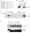
Interaction between RECQ5β and PCNA. (A) The putative PCNA-binding motif of human RECQ5β. The C-terminal amino acids 964–971 of RECQ5β are aligned with the PCNA-binding motifs identified in various PCNA-interacting proteins. The highly conserved residues are shown in boldface. (B) Direct interaction between RECQ5β and PCNA. Chitin beads coated with recombinant RECQ5β protein containing an intein-CBD tag were incubated with recombinant PCNA (1 μg) as described in Materials and Methods. In the control experiment, beads coated with the E.coli McrA endonuclease (mock beads) were used. Bound proteins were analyzed by SDS–PAGE and western blotting. Blots were probed with monoclonal anti-PCNA antibody (PC-10, Santa Cruz). (C) Mapping PCNA-interaction domain in RECQ5β. Glutathione beads coated with the GST-tagged RECQ5β deletion variants RECQ5542–991 and RECQ51–774, respectively, were incubated with increasing amounts of purified PCNA as indicated. Bound proteins were analyzed as in (B). (D) Co-immunoprecipitation of PCNA with RECQ5β from extracts of 293T cells: non-treated (NT) cells (lane 3), UV-irradiated (40 J/m2) cells incubated for 6 h (lane 4), cells treated with CDDP (20 μM) for 8 h (lane 5), cells arrested at G1/S by HU (2 mM, 16 h) and subsequently released to S phase for 3 h (lane 6). Extracts were immunoprecipitated using affinity-purified rabbit polyclonal anti-RECQ5β antibody as described in Materials and Methods. IgGs purified from corresponding pre-immune serum served as a control (lane 2). The immunoprecipitates were analyzed as in (A). The blots were probed with anti-PCNA and anti-RECQ5β antibodies.

Similar articles

Cited by

References

    1. Cox M.M. Recombinational DNA repair of damaged replication forks in Escherichia coli: questions. Annu. Rev. Genet. 2001;35:53–82. - PubMed
    1. Cordeiro-Stone M., Makhov A.M., Zaritskaya L.S., Griffith J.D. Analysis of DNA replication forks encountering a pyrimidine dimer in the template to the leading strand. J. Mol. Biol. 1999;289:1207–1218. - PubMed
    1. Lopes M., Foiani M., Sogo J.M. Multiple mechanisms control chromosome integrity after replication fork uncoupling and restart at irreparable UV lesions. Mol. Cell. 2006;21:15–27. - PubMed
    1. Hickson I.D. RecQ helicases: caretakers of the genome. Nat. Rev. Cancer. 2003;3:169–178. - PubMed
    1. Lambert S., Watson A., Sheedy D.M., Martin B., Carr A.M. Gross chromosomal rearrangements and elevated recombination at an inducible site-specific replication fork barrier. Cell. 2005;121:689–702. - PubMed

Publication types