. 2006 Oct 4;25(19):4490-502.
doi: 10.1038/sj.emboj.7601332. Epub 2006 Sep 21.

Smads orchestrate specific histone modifications and chromatin remodeling to activate transcription

Affiliations

Smads orchestrate specific histone modifications and chromatin remodeling to activate transcription

Sarah Ross et al. EMBO J. .

Abstract

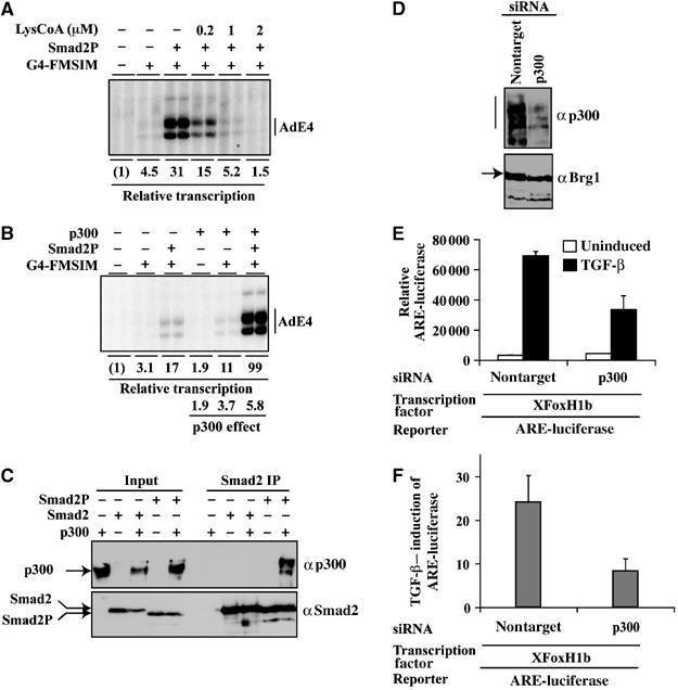
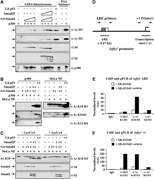
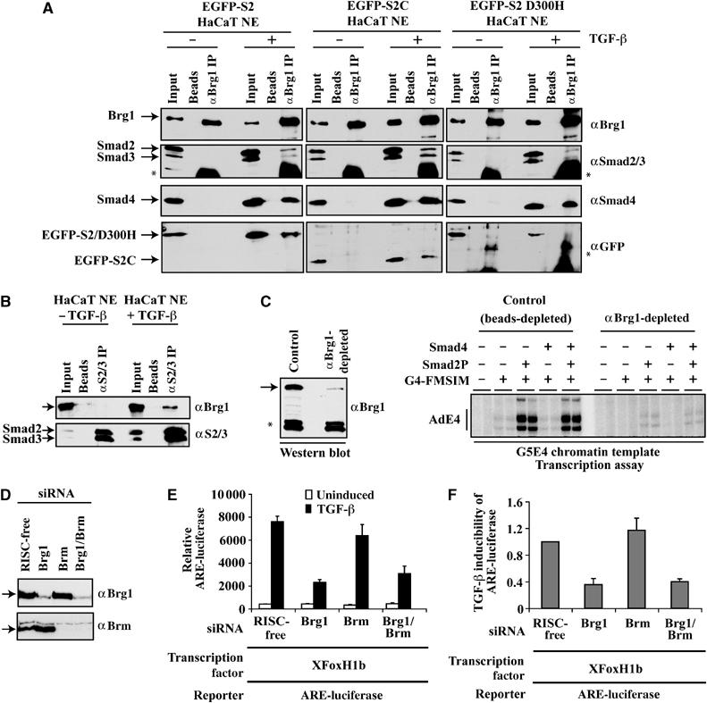
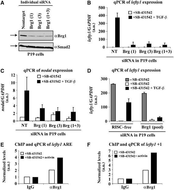
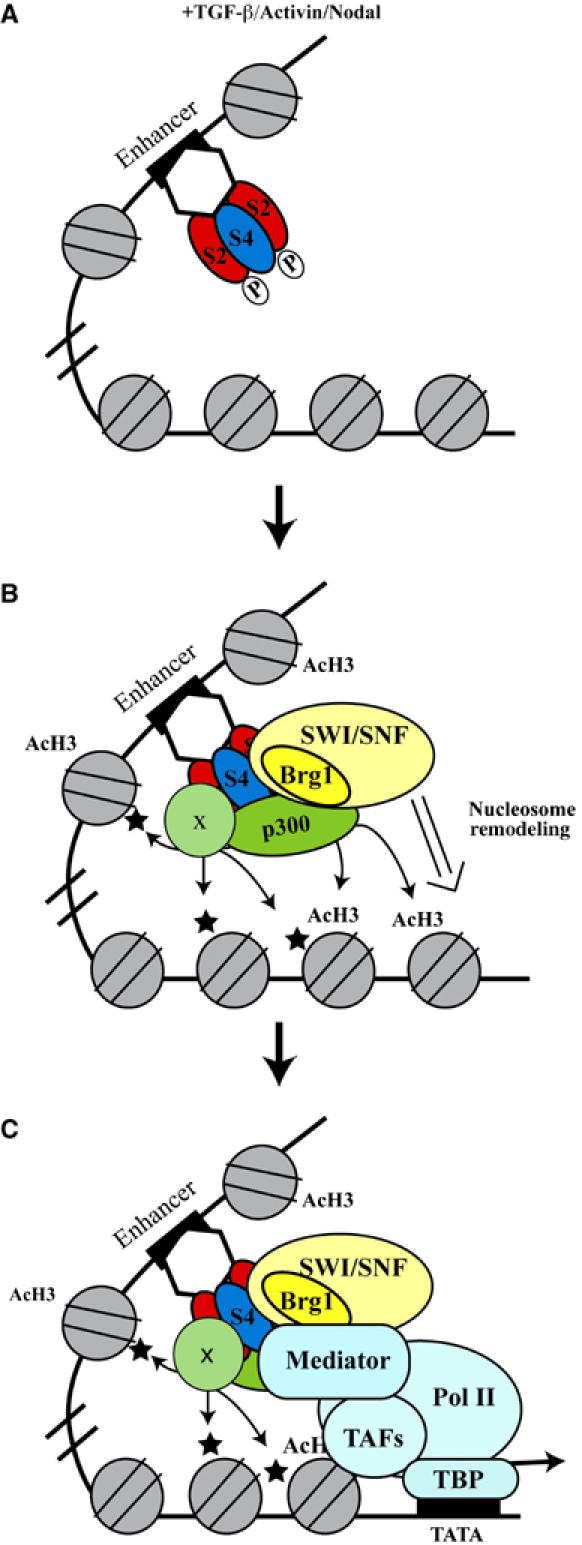
Smads are intracellular transducers for TGF-beta superfamily ligands, but little is known about the mechanism by which complexes of receptor-phosphorylated Smad2 and Smad4 regulate transcription. Using an in vitro transcription system, we have discovered that, unlike most transcription factors that are sufficient to recruit the basal transcription machinery and therefore activate transcription on both naked DNA and chromatin templates, the Smads only activate transcription from chromatin templates. We demonstrate that Smad2-mediated transcription requires the histone acetyltransferase, p300. Smad2-recruited p300 exhibits an altered substrate specificity, specifically acetylating nucleosomal histone H3 at lysines 9 and 18, and these modifications are also detected on an endogenous Smad2-dependent promoter in a ligand-induced manner. Furthermore, we show that endogenous Smad2 interacts with the SWI/SNF ATPase, Brg1, in a TGF-beta-dependent manner, and demonstrate that Brg1 is recruited to Smad2-dependent promoters and is specifically required for TGF-beta-induced expression of endogenous Smad2 target genes. Our data indicate that the Smads define a new class of transcription factors that absolutely require chromatin to assemble the basal transcription machinery and activate transcription.


Figures

Figure 1
Figure 1
An in vitro system for studying Smad2-dependent transcriptional activation. (A) A schematic of G4-FMSIM. (B) Luciferase reporter assay in NIH 3T3 cells transfected with (Gal4-OP)5-luciferase and plasmids expressing Gal4 (1–95) or G4-FMSIM. (C) Luciferase reporter assays in MDA-MB468 cells transfected with (Gal4-OP)5-luciferase and plasmids expressing G4-FMSIM alone or with HA-Smad4. (D, E) Recruitment of in vitro or endogenous Smad complexes by recombinant G4-FMSIM was assayed by bandshifts using a Gal4 binding site probe. Complexes were confirmed by supershifts with anti-Gal4 (G4), anti-Smad2 (S2) and anti-Smad4 (S4) antibodies. For in vitro Smad complexes, purified proteins were used; for endogenous complexes, nuclear extract from TGF-β-induced HaCaT cells (TGF-β ind.NE) was used. Asterisk indicates a nonspecific DNA-binding complex.
Figure 2
Figure 2
Phosphorylated Smad2-containing complexes activate transcription on chromatin templates, but not on naked DNA. (A) A schematic of the plasmid templates used in the in vitro transcription assays (top). Naked DNA templates were used with Gal4(1–95), G4-p53, G4-Smad2C and G4-FMSIM with recombinant Smad proteins (bottom). (BD) Transcription assays on chromatin templates with G4-FMSIM or Gal4 (1–95) and recombinant Smads as indicated. (E) G4-Smad4, G4-Smad4C and G4-Smad2C were analyzed for transcription activity on chromatin templates. (F) Smad2P was recruited to the chromatin template by G4-Smad4 or G4-Smad4C and transcriptional activity analyzed. Transcription assays were performed in duplicate and the level of activated transcription was quantitated relative to basal levels of transcription. Between experiments, a certain expected variability in transcriptional induction for G4-FMSIM alone that ranges from two to seven-fold is observed.
Figure 3
Figure 3
p300 is required for Smad2 complexes to activate transcription. (A) Transcription assays on chromatin templates using Smad2P recruited by G4-FMSIM in the presence of the p300 inhibitor, LysCoA. (B) p300 synergizes with Smad2P to induce transcription in vitro on chromatin templates. Transcription assays were performed in duplicate and the level of transcription quantitated relative to basal levels. (C) Interaction of recombinant p300 with recombinant Smad2 and Smad2P was assayed by immunoprecipitation with an anti-Smad2 antibody and Western blot analysis with anti-p300 and anti-Smad2 antibodies. The unphosphorylated Smad2 has a short N-terminal linker, which accounts for its slight decrease in mobility relative to Smad2P. (D) Proteins from NIH 3T3 cells transfected with siRNA pools against p300 or a nontarget control were analyzed by Western blotting using anti-p300 or anti-Brg1 antibodies. (E) Luciferase reporter assay in NIH 3T3 cells transfected with ARE-luciferase, a plasmid expressing XFoxH1b and siRNA pools against p300 or nontarget control. (F) Fold induction of ARE-luciferase in response to TGF-β from four independent siRNA experiments.
Figure 4
Figure 4
Smad2P-containing complexes recruit p300 to preferentially acetylate nucleosomal histone H3. (A) p300 histone acetylation assays performed on G5E4 dinucleosome templates or free core histones incubated with G4-p53 (250 ng) or G4-Smad4 alone or with Smad2P (50, 100, 250, 500 ng each). For (A) histones were prepared from HeLa cells. (B) p300 or HeLa nuclear extract-dependent histone acetylation assays on G5E4 dinucleosome templates incubated with 200 ng (+) or 500 ng (++) G4-p53, G4-Smad4 and Smad2P. (C) HeLa nuclear extract-dependent histone acetylation assays on G5E4 templates −/+ LysCoA (1 μM final concentration) with 500 ng (+) or 750 ng (++) G4-p53, G4-Smad4 and Smad2P. For (B) and (C) recombinant histone octamers were used. Western blotting was performed with anti-acetyl histone H3, anti-acetyl histone H4, anti-acetyl histone H3 K18, anti-acetyl histone H4 K8 and anti-acetyl histone H4 K12 antibodies as indicated. The levels of Smad2P, G4-Smad4 and p300 were confirmed as indicated. In (B) and (C), the asterisk indicates a non-specific band. (D) Schematic of the lefty1 promoter showing the location of the primers used in the ChIP assays. The ARE, which contains a FoxH1 (gray box) and Smad (white box) binding site is shown, and the start of transcription (+1). (E, F) qPCR of the lefty1 ARE region (E) or +1 transcription start site (F) from ChIP assays using IgG, anti-trimethyl histone H3 K4, anti-acetyl histone H3 K18 and anti-acetyl histone H4 K8 antibodies. ChIPs were performed on extracts from P19 cells treated with 10 μM SB-431542 overnight to abolish autocrine signaling and were either uninduced or induced with activin for 1 h. The data correspond to the average of triplicate PCRs normalized to IgG from a representative experiment. The IgG values were set to 1.
Figure 5
Figure 5
The SWI/SNF component, Brg1, interacts with Smad complexes and is required for TGF-β-activated transcription. (A, B) Proteins were immunoprecipitated with immobilized anti-Brg1 (A) or immobilized anti-Smad2/3 (αS2/3) (B) or protein G beads alone (A, B) from HaCaT nuclear extracts (HaCaT NE) that were either uninduced or induced for 1 h with TGF-β. In (A), the HaCaTs were stably expressing EGFP-tagged full-length Smad2 (EGFP-S2), Smad2 linker+MH2 (EGFP-S2C) or Smad2 (D300H) (EGFP-S2 D300H). Immunoprecipitated proteins, together with input protein samples, were analyzed by Western blotting using anti-Brg1, anti-Smad2/3, anti-Smad4 and anti-GFP antibodies as indicated. The asterisk indicates an IgG band. (C) Depletion of Brg1 from HeLa nuclear extract (left panel) inhibited Smad2P-dependent transcription in vitro (right panel). The asterisk indicates a background band. (D, E) NIH 3T3 cells were transfected with siRNA pools against Brg1, Brm or an RISC-free control and analyzed by Western blotting using anti-Brg1 or anti-Brm antibodies (D), or were further transfected with ARE-luciferase, a plasmid expressing XFoxH1b and analyzed for luciferase activity (E). (F) A graph representing the fold induction of ARE-luciferase in response to TGF-β from two independent siRNA experiments.
Figure 6
Figure 6
Brg1 is required for the TGF-β-induced expression of lefty1 and nodal in vivo and binds to the lefty1 promoter. (A) Proteins from P19 cells transfected with individual siRNA duplexes against Brg1 (1 or 3) or a nontargeting siRNA were analyzed by Western blotting with anti-Brg1 or anti-Smad2 antibodies. (B–D) Levels of lefty1 (B, D) or nodal (C) mRNA were measured by reverse transcription and qPCR of RNA isolated from P19 cells transfected with individual (Brg1 or Brg3) or a pool of siRNA duplexes targeting Brg1 and either nontargeting (NT) or RISC-free siRNA as controls. Following transfection, samples of cells were treated overnight with SB-431542 to abolish autocrine signaling, washed and then treated −/+ TGF-β for 2 h. The data for (B) and (C) represent the average of four PCR reactions from a representative experiment. The data for (D) correspond to duplicate PCR reactions from a representative experiment. All PCRs were performed in duplicate and quantitated relative to GAPDH. (E, F). qPCR of the lefty1 ARE region (E) or +1 transcription start site (F) from ChIPs with IgG or anti-Brg1 antibody. ChIPs were performed on extracts from P19 cells treated with SB-431542 overnight to abolish autocrine signaling and then treated −/+ activin for 1 h. The data correspond to the average of triplicate PCRs normalized to IgG from a representative experiment. The IgG values were set at 1.
Figure 7
Figure 7
Proposed model of Smad2P-mediated transcription on chromatin. (A) In response to TGF-β, Activin or Nodal, Smad2P-containing complexes are recruited to target promoters by transcription factors. (B) The Smad2P-containing complexes recruit p300, SWI/SNF and probably other modifiers (indicated by x) to modify (indicated by AcH3 and or a star for unknown modifications) and remodel (indicated by the white arrow) the chromatin. (C) The modified and remodeled chromatin together with Smad2P-containing complexes recruits the RNA Pol II transcription machinery to the promoter to activate transcription.

Similar articles

Cited by

References

    1. Alberts AS, Geneste O, Treisman R (1998) Activation of SRF-regulated chromosomal templates by Rho-family GTPases requires a signal that also induces H4 hyperacetylation. Cell 92: 475–487 - PubMed
    1. Cereghini S, Yaniv M (1984) Assembly of transfected DNA into chromatin: structural changes in the origin-promoter-enhancer region upon replication. EMBO J 3: 1243–1253 - PMC - PubMed
    1. Chacko BM, Qin BY, Tiwari A, Shi G, Lam S, Hayward LJ, De Caestecker M, Lin K (2004) Structural basis of heteromeric smad protein assembly in TGF-β signaling. Mol Cell 15: 813–823 - PubMed
    1. Cheung E, Schwabish MA, Kraus WL (2003) Chromatin exposes intrinsic differences in the transcriptional activities of estrogen receptors α and β. EMBO J 22: 600–611 - PMC - PubMed
    1. Conaway RC, Sato S, Tomomori-Sato C, Yao T, Conaway JW (2005) The mammalian Mediator complex and its role in transcriptional regulation. Trends Biochem Sci 30: 250–255 - PubMed

Publication types

MeSH terms