. 2006 Dec 1;577(Pt 2):497-511.
doi: 10.1113/jphysiol.2006.118141. Epub 2006 Sep 21.

Thyrotropin-releasing hormone increases GABA release in rat hippocampus

Affiliations

Thyrotropin-releasing hormone increases GABA release in rat hippocampus

Pan-Yue Deng et al. J Physiol. .

Abstract

Thyrotropin-releasing hormone (TRH) is a tripeptide that is widely distributed in the brain including the hippocampus where TRH receptors are also expressed. TRH has anti-epileptic effects and regulates arousal, sleep, cognition, locomotion and mood. However, the cellular mechanisms underlying such effects remain to be determined. We examined the effects of TRH on GABAergic transmission in the hippocampus and found that TRH increased the frequency of GABAA receptor-mediated spontaneous IPSCs in each region of the hippocampus but had no effects on miniature IPSCs or evoked IPSCs. TRH increased the action potential firing frequency recorded from GABAergic interneurons in CA1 stratum radiatum and induced membrane depolarization suggesting that TRH increases the excitability of interneurons to facilitate GABA release. TRH-induced inward current had a reversal potential close to the K+ reversal potential suggesting that TRH inhibits resting K+ channels. The involved K+ channels were sensitive to Ba2+ but resistant to other classical K+ channel blockers, suggesting that TRH inhibits the two-pore domain K+ channels. Because the effects of TRH were mediated via Galphaq/11, but were independent of its known downstream effectors, a direct coupling may exist between Galphaq/11 and K+ channels. Inhibition of the function of dynamin slowed the desensitization of TRH responses. TRH inhibited seizure activity induced by Mg2+ deprivation, but not that generated by picrotoxin, suggesting that TRH-mediated increase in GABA release contributes to its anti-epileptic effects. Our results demonstrate a novel mechanism to explain some of the hippocampal actions of TRH.


Figures
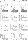
Figure 1
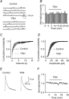
Figure 1. TRH increases sIPSC frequency in the hippocampus
A, TRH increased sIPSC frequency in CA1 region. Aa, upper trace, sIPSCs recorded from a CA1 pyramidal neuron before and during the application of TRH (1 μm) for 3–4 min. lower trace, time course of the sIPSC frequency averaged from 11 cells. Note that TRH increased sIPSC frequency. Ab, cumulative frequency distribution from a CA1 pyramidal neuron before and during the application of TRH for 3–4 min. Note that TRH reduced the intervals of the sIPSC (increased sIPSC frequency, P < 0.00001, Kolmogorov-Smirnov test). Ac, cumulative amplitude distribution from the same cell before and during the application of TRH for 3–4 min (P= 0.21, Kolmogorov-Smirnov test). Ad, summarized data from 11 CA1 pyramidal neurons. Note that TRH significantly increased sIPSC frequency without changing sIPSC amplitude. B, TRH increased sIPSC frequency in CA3 pyramidal neurons.. Note that TRH increased sIPSC frequency without altering sIPSC amplitude (n= 11). C, TRH increased sIPSC frequency in dentate gyrus granule cells without significantly changing sIPSC amplitude (n= 11). In B and C, the data were obtained in the same way as in A.
Figure 2
Figure 2. TRH increases sIPSC frequency via the activation of TRH receptors
A, concentration–response curve of TRH constructed from CA1 pyramidal neurons. Data between 0.03 and 1 μm were fitted by the Hill equation. Numbers in parentheses are the numbers of cells used. EC50 was 0.26 μm. B, TRH-induced increases in sIPSC frequency were blocked by the TRH receptor inhibitor, chlordiazepoxide (50 μm). Slices were pretreated with chlordiazepoxide and the extracellular solution contained the same concentration of chlordiazepoxide (n = 8).
Figure 3
Figure 3. TRH does not modulate mIPSCs recorded in the presence of TTX and the evoked IPSC amplitude recorded by placing a stimulation electrode in CA1 stratum radiatum
A, mIPSC current traces recorded from a CA1 pyramidal neuron before and during the application of TRH (0.5 μm). B, time course of mIPSC frequency summarized from five CA1 pyramidal neurons. C, cumulative frequency distribution of mIPSCs before and during the application of TRH (n = 5, p = 0.44, Kolmogorov-Smirnov test). D, cumulative amplitude distribution of mIPSCs before and during the application of TRH (n = 5, p = 0.27, Kolmogorov-Smirnov test). Note that TRH did not change the frequency or the amplitude of mIPSCs. E, evoked IPSC trace averaged from 10 IPSCs before and during the application of TRH. F, summarized time course of the evoked IPSC amplitude from 12 CA1 pyramidal neurons before, during and after the application of TRH (0.5 μm). Note that TRH did not change the amplitude of the evoked IPSCs.
Figure 4
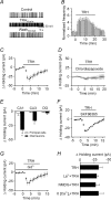
Figure 4. TRH increases the excitability of GABAergic interneurons in CA1 stratum radiatum
A, spontaneous action potentials recorded from an interneuron before, during and after the application of TRH (0.5 μm). B, time course of the action potential firing frequency averaged from 10 interneurons. Note that TRH increased the action potential firing frequency. C, application of TRH (0.5 μm) produced an inward current at −55 mV (n = 9, p = 0.002). Holding currents at −55 mV were initially recorded every 3 s in the presence of TTX (0.5 μm) and then averaged per minute. The averaged holding current for the last minute before the application of TRH was subtracted to calculate the change in holding current. D, before and during application of the TRH receptor inhibitor, chlordiazepoxide (50 μm), antagonized TRH-induced change in holding currents suggesting the involvement of TRH receptors (n = 7). E, TRH increased inward current at −55 mV in CA1 and CA3 stratum radiatum interneurons with no effects on interneurons in the hilus or any of the principal cells in the hippocampus. DG, dentate gyrus. F, before and during application of SKF96365 (100 μm), a receptor-operated cation channel inhibitor, failed to block TRH-induced change in holding currents (n = 6). G, before and during application of a non-selective cation channel blocker, Gd3+ (10 μm) did not block the effects of TRH (n = 6). H, TRH-induced depolarization was not affected by application of La3+ (10 μm, n = 10), replacement of the extracellular Na+ by NMDG (n = 6) or removal of extracellular Ca2+ (n = 5).
Figure 5
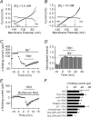
Figure 5. TRH inhibits K+ channels of the interneurons in CA1 stratum radiatum
A, voltage–current relationship recorded by a ramp protocol (from−140 to−40 mV, at a speed of 0.07 mV ms−1) before and during the application of TRH (0.5 μm) when the extracellular K+ concentration was 3.5 mm. Traces in the figure were averaged traces from eight cells. The TRH-induced net current has a reversal potential at ∼−84 mV, close to the calculated K+ reversal potential (∼−85 mV). B, voltage–current relationship recorded by the same protocol when the extracellular K+ concentration was 10 mm. Note that the reversal potential of the TRH-sensitive net current was ∼−64 mV, close to the calculated K+ reversal potential (∼−59 mV) suggesting that the net current was mediated by K+ ions. C, application of Ba2+ (2 mm) alone suppressed the holding current at −55 mV and attenuated TRH-induced change in current (n = 14). The averaged current 1 min prior to the application of TRH was subtracted from each time point (same for E). D, bath application of Ba2+ (2 mm) increased sIPSC frequency and significantly inhibited TRH-induced increase in sIPSC frequency (n = 8). E, bath application of ruthenium red (10 μm) failed to alter TRH-induced change in holding currents (n = 7). F, summarized results for the effects of K+ blockers on TRH-induced change in holding currents. Note that the TRH-induced changes in holding currents were insensitive to TEA, 4-aminopyridine, Cs+, tertiapin, Zn2+ and anandamide. Application of bupivacaine (Bup) at 500 μm significantly reduced TRH-induced increase in holding currents.
Figure 6
Figure 6. G-proteins are required for the effects of TRH on holding currents
A, inclusion of GDP-β-S (4 mm) in the recording pipettes significantly reduced the effects of TRH on holding currents (n = 12). Zero current level was defined as the current flowing prior to TRH application in each panel of this figure. B, TRH produced an irreversible change in current when GTP-γ-S (4 mm) was in the pipette (n = 7). C, intracellular infusion of antibody to Gαq/11 (20 μg ml−1) blocked TRH-induced change in holding currents (n = 11) whereas dialysis of antibody to Gβ (20 μg ml−1) had no effects (n = 8). D, intracellular application of the dynamin inhibitory peptide (QVPSRPNRAP, 50 μm) significantly slowed the desensitization of TRH-induced increase in holding currents (n = 9) compared with that recorded when a scrambled peptide (QPPASNPRVR, 50 μm) was included in the pipettes (n = 7).
Figure 7
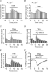
Figure 7. TRH-induced increases in GABA release are independent of intracellular signals and phosphorylation
A, application of TRH (0.1 μm) increased sIPSC frequency in eight slices from three wild-type (PLCβ1+/+) mice. B, application of TRH (0.1 μm) increased sIPSC frequency to the same level in 19 slices from three knock-out (PLCβ1−/−) mice. CE, TRH-induced increases in sIPSC frequency were not changed by the inhibitors of PKC (calphostin C, 0.5 μm, n = 10) (C), MAPK (PD 98059, 50 μm, n = 9) (D) or CAMK II (KN-62, 10 μm, n = 10) (E). F, omission of ATP from the intracellular solution had no effect on TRH-induced inward holding currents (n = 5).
Figure 8
Figure 8. TRH inhibits seizure activity induced by deprivation of extracellular Mg2+ with no effects on the seizure activity induced by picrotoxin
A, upper, seizure activity induced by bathing the hippocampal slices in extracellular solution containing 0Mg2+ before, during and after the application of TRH (0.5 μm). A single seizure event was shown in an enlarged scale below each trace. Lower, time course of the frequency of seizure activity averaged from 15 slices before, during and after the application of TRH. The numbers of the events were averaged for every minute and then normalized to the number of the events for the last minute before the application of TRH. B, upper, seizure activity evoked by application of the GABAA receptor blocker, picrotoxin (100 μm) in the hippocampal slices before, during and after the application of TRH (0.5 μm). A single seizure event was shown in an enlarged scale below each trace. Lower, summarized data from 12 slices. Note that TRH failed to alter the seizure activity induced by the picrotoxin seizure model.

Similar articles

Cited by

References

    1. Bannister RA, Melliti K, Adams BA. Differential modulation of CaV2.3 Ca2+ channels by Gαq/11-coupled muscarinic receptors. Mol Pharmacol. 2004;65:381–388. - PubMed
    1. Bayliss DA, Sirois JE, Talley EM. The TASK family: two-pore domain background K+ channels. Mol Interv. 2003;3:205–219. - PubMed
    1. Bayliss DA, Viana F, Berger AJ. Mechanisms underlying excitatory effects of thyrotropin-releasing hormone on rat hypoglossal motoneurons in vitro. J Neurophysiol. 1992;68:1733–1745. - PubMed
    1. Bayliss DA, Viana F, Berger AJ. Effects of thyrotropin-releasing hormone on rat motoneurons are mediated by G proteins. Brain Res. 1994;668:220–229. - PubMed
    1. Berg AP, Talley EM, Manger JP, Bayliss DA. Motoneurons express heteromeric TWIK-related acid-sensitive K+ (TASK) channels containing TASK-1 (KCNK3) and TASK-3 (KCNK9) subunits. J Neurosci. 2004;24:6693–6702. - PMC - PubMed

Publication types

MeSH terms

LinkOut - more resources