. 2006 Dec;80(23):11833-51.
doi: 10.1128/JVI.00857-06. Epub 2006 Sep 20.

Novel fiber-dependent entry mechanism for adenovirus serotype 5 in lacrimal acini

Affiliations

Novel fiber-dependent entry mechanism for adenovirus serotype 5 in lacrimal acini

Jiansong Xie et al. J Virol. 2006 Dec.

Abstract

The established mechanism for infection of most cells with adenovirus serotype 5 (Ad5) involves fiber capsid protein binding to coxsackievirus-adenovirus receptor (CAR) at the cell surface, followed by penton base capsid protein binding to alpha(v) integrins, which triggers clathrin-mediated endocytosis of the virus. Here we determined the identity of the capsid proteins responsible for mediating Ad5 entry into the acinar epithelial cells of the lacrimal gland. Ad5 transduction of primary rabbit lacrimal acinar cells was inhibited by excess Ad5 fiber or knob (terminal region of the fiber) but not excess penton base. Investigation of the interactions of recombinant Ad5 penton base, fiber, and knob with lacrimal acini revealed that the penton base capsid protein remained surface associated, while the knob domain of the fiber capsid protein was rapidly internalized. Introduction of rabbit CAR-specific small interfering RNA (siRNA) into lacrimal acini under conditions that reduced intracellular CAR mRNA significantly inhibited Ad5 transduction, in contrast to a control (nonspecific) siRNA. Preincubation of Ad5 with excess heparin or pretreatment of acini with a heparinase cocktail each inhibited Ad5 transduction by a separate and apparently additive mechanism. Functional and imaging studies revealed that Ad5, fiber, and knob, but not penton base, stimulated macropinocytosis in acini and that inhibition of macropinocytosis significantly reduced Ad5 transduction of acini. However, inhibition of macropinocytosis did not reduce Ad5 uptake. We propose that internalization of Ad5 into lacrimal acini is through a novel fiber-dependent mechanism that includes CAR and heparan sulfate glycosaminoglycans and that the subsequent intracellular trafficking of Ad5 is enhanced by fiber-induced macropinocytosis.


Figures
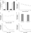
FIG. 1.
FIG. 1.
Ad5 transduction efficiency in LGAC and sensitivity to penton base, fiber, and knob protein. (A) The percentage of transduction efficiency of Ad-GFP in rabbit LGAC is approximately equivalent to that in HeLa cells, significantly higher than that in MDCK cells, and eight- to ninefold higher than that in CHO cells and Caco-2 cells. Rabbit LGAC were seeded onto Matrigel-coated 12-well plates at 2 × 10⁶ cells/well and used on day 2, while other cell lines were seeded onto noncoated 12-well plates at 1 × 10⁵ cells/well and used at 80% confluence. Ad-GFP was then added at an MOI of 2 PFU/cell, and the cells were incubated for 1 h at 4°C, washed, and then incubated for 10 to 12 h at 37°C. GFP positivity was determined by flow cytometry (n = 5). (B) Preincubation of LGAC with fiber substantially lowers Ad-GFP infectivity, while preincubation with penton base appears to have little effect (for penton base experiments, n = 7; for fiber experiments, n = 4). (C) Preincubation of HeLa cells with penton base results in reduced Ad-GFP infectivity (n = 5). (D) Preincubation of LGAC with knob substantially reduced Ad-GFP transduction efficiency, while preincubation with RGD and LDV had little effect (for RGD, n = 3; for LDV, n = 4; and for knob, n = 8). For the experiments for panels B to D, rabbit LGAC were seeded onto Matrigel-coated 12-well plates at 2 × 10⁶ cells/well and HeLa cells were seeded onto uncoated 12-well plates at 1 × 10⁵ cells/well. On day 2 of the culture (80% confluence for HeLa cells), the cells were preincubated with the indicated concentrations of recombinant knob, fiber, penton base, or the penton base-blocking peptides RGD or LDV for 2 h at 4°C. Ad-GFP was then added at an MOI of 2 PFU/cell, and the cells were incubated for 1 h at 4°C, washed, and incubated for 10 to 12 h at 37°C. GFP positivity was determined by flow cytometry. In all panels, *indicates significance at a P value of ≤0.05, based on results for the control. Error bars represent the standard error of the mean (SEM).
FIG. 2.
FIG. 2.
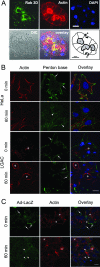
LGAC morphology and uptake of Ad5 and penton base protein. (A) The intracellular components of a reconstituted rabbit lacrimal gland acinus. Cells were triple stained with antibodies to Rab 3D (green), actin (red), or DAPI (4′,6′-diamidino-2-phenylindole) (blue) to show the localization of mature secretory vesicles, actin filaments, and nuclei, respectively. The diagram shows the location of lumina (L) bounded by the apical PM of epithelial cells (dark line). Basolateral membranes (B) of the epithelial cells are indicated by arrows. (B) Penton base uptake in HeLa and LGAC. HeLa cells or LGAC were seeded onto uncoated or Matrigel-coated 12-well plates at 1 × 10⁵ cells/well or 2 × 10⁶ cells/well, respectively. On day 2 (HeLa) or day 3 (LGAC) of the culture, control cells or cells exposed to penton base (20 μg/ml) for 0 min and 60 min at 37°C were fixed and processed to fluorescence label penton base (green), actin filaments (red), and nuclei (blue). Arrows, association of penton base with PM; arrowheads, penton base in perinuclear region. (C) Control rabbit LGAC or LGAC exposed to replication-deficient Ad-LacZ (MOI = 5) for 0 min and 60 min were fixed and processed to fluorescence label Ad5 proteins (green), actin filaments (red), and nuclei (blue). Arrows, association of Ad5 with basolateral membranes; arrowheads, accumulation of Ad5 in intracellular compartments; bar, 10 μm; *, apical/luminal region.
FIG. 3.
FIG. 3.
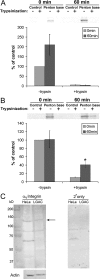
Penton base remains surface bound in LGAC. (A) Penton base is vulnerable to trypsin at 60 min after exposure in LGAC. Rabbit LGAC were seeded onto Matrigel-coated 12-well plates at 2 × 10⁶ cells/well. On day 2 of the culture, LGAC with or without exposure to penton base protein (20 μg/ml) for 0 min or 60 min at 37°C were treated with 0.2 mg/ml trypsin-EDTA for 1 h at 4°C or not treated and were lysed, using RIPA buffer. Cell lysates (150 μg/lane) were blotted with polyclonal antibody to Ad5 proteins and IRDye800-conjugated secondary antibody. Data were normalized in each preparation to that for penton base association at 0 min minus that for trypsin. Error bars represent SEM (n = 3). (B) Penton base acquires trypsin resistance after incubation in HeLa cells. HeLa cells were seeded on 12-well plates. When they reached 80% confluence, they were exposed to penton base protein (10 to 50 μg/ml) for 0 or 60 min at 37°C or not exposed and then were treated with trypsin-EDTA or not treated and were processed as described above. Data for each preparation were normalized to that for penton base association at 0 min minus that for trypsin. *, significant at P ≤ 0.05 based on results for cells at 0 min. Error bars represent SEM (n = 5). (C) LGAC express αv integrins. Western blots of αv integrin expression in rabbit LGAC and HeLa cells are shown. LGAC and HeLa cell lysates prepared using RIPA buffer (150 μg/lane) were blotted with a rabbit polyclonal antibody to αv integrin and IRDye 800-conjugated secondary antibody. The arrow depicts a band at 150 kDa, the reported molecular mass of the protein. The same lanes, reprobed for actin content, are shown below, while the signal associated with the secondary antibody (2o) alone is shown in the panel to the right.
FIG. 4.
FIG. 4.
Knob internalization in LGAC. (A) Knob protein internalized in LGAC as determined by immunofluorescence tagging. Rabbit LGAC were seeded onto Matrigel-coated 12-well plates at 2 × 10⁶ cells/well. On day 3 of the culture, control rabbit LGAC or LGAC exposed to knob protein (20 μg/ml) for 0 min and 120 min at 37°C were fixed and processed to fluorescence label knob (green) and actin filaments (red). Arrows, association of knob with basolateral PM; arrowheads, accumulation of knob in intracellular compartments; bar, 10 μm. (B) Knob protein acquired trypsin resistance after 60 min of incubation with LGAC. Rabbit LGAC were seeded in 150-mm petri dishes at a density of 2 × 10⁶ cells/ml. On day 2 of the culture, LGAC exposed to GFP-knob protein (20 μg/ml) for 0 min or 60 min were treated with 0.2 mg/ml trypsin-EDTA for 1 h at 4°C and lysed using RIPA buffer. Cell lysates (150 μg/lane) were blotted with monoclonal antibody to GFP and IRDye800-conjugated secondary antibody. The data for each preparation were normalized to that for GFP-knob association at 0 min minus that for trypsin (n = 3). *, significant at P ≤ 0.05 based on results for cells at 0 min. Error bars represent the SEM.
FIG. 5.
FIG. 5.
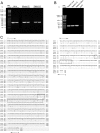
Rabbit CAR cDNA cloning. (A) CAR is expressed in rabbit LGAC as determined by RT-PCR. Total RNA was isolated from HeLa cells and mouse or rabbit LGAC, and cDNA was then synthesized as described in Materials and Methods. CAR gene expression was analyzed by PCR using primers designed according to a sequence conserved in human and mouse CAR. PCR products were separated on a 1.2% agarose gel. DNA bands of ∼400 bp were sliced, extracted, and then further amplified by PCR, using the CAR primer shown above. A 437-bp rabbit CAR fragment, from which a 357-bp reliable sequence was acquired (not shown), was isolated from the gel and sequenced. (B) Total RNA was isolated from rabbit liver, rabbit LG, rabbit LGAC, or CHO cells, and CAR expression was determined by RT-PCR as for Fig. 5A. (C) The entire coding sequence of rabbit lacrimal acinar CAR was determined and compared with known CAR sequences from human and mouse. Total RNA was isolated from rabbit LGAC as described in Materials and Methods. The 5′ and 3′ fragments of rabbit CAR were produced by RACE PCR, using the specially designed 5′-end RACE primer, modified oligo(T) (3′), and CAR-specific downstream and upstream primers whose design was based on the 357-bp reliable sequence. 5′- and 3′-RACE fragments of CAR were extracted and subjected to DNA sequencing analysis. The resulting 1,695-bp rabbit CAR sequence includes the entire coding sequence of 1,098 bp, which was aligned with the known nucleotide sequences of human and mouse CAR, using an NCBI BLAST tool (bl2seq). Dashed boxes show the ATG start codon or TAG stop codon, and the closed box highlights the nucleotide sequences predicted to encode transmembrane helices in protein analyzed with the TMHMM method. Rabbit LGAC CAR has 89% sequence identity with human CAR and 83% sequence identity with mouse CAR. The rabbit LGAC CAR protein sequence was predicted by analyzing the 1,098-bp nucleotide sequence using the ORF finder from NCBI, resulting in the generation of a 365-amino-acid sequence, which was aligned with the known protein sequences of human and mouse CAR using an NCBI BLAST tool (blastp) (right). The closed box highlights the predicted transmembrane helices. Rabbit LGAC CAR has 89% amino acid sequence identity with human CAR and 88% amino acid sequence identity with mouse CAR.
FIG. 6.
FIG. 6.
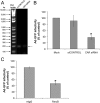
Knockdown of CAR with CAR-specific siRNA but not nontargeting control siRNA. (A) Treatment of rabbit LGAC with CAR-specific siRNA resulted in significantly reduced expression of CAR. Rabbit LGAC seeded at a density of 2 × 10⁶ cells/ml in 100-mm petri dishes were transfected with siRNA targeted to rabbit CAR or a siCONTROL nontargeting siRNA (100 nM) obtained from Dharmacon on day 1 of the culture, using the GeneSilencer siRNA transfection reagent. The CAR siRNA sense and antisense duplexes were designed according to the 1,098-bp CAR sequence shown in Fig. 5C with Dharmacon's online designer. After 48 h, the total RNA from the control and experimental cultures was isolated, treated, and used for cDNA synthesis as described in Materials and Methods. CAR gene expression in all samples was determined by PCRs within the linear range using primers designed according to the 1,098-bp rabbit lacrimal acinar CAR sequence. PCR products were separated on a 2% agarose gel. (B) Downregulation of CAR in rabbit LGAC reduced the transduction efficiency of Ad-GFP in those cells. Rabbit LGAC were seeded at a density of 1 × 10⁶ cells/well and transfected with siRNA as described above. After 48 h, Ad-GFP was added at an MOI of 2 PFU/cell, and the cells were incubated for 1 h at 4°C before being rinsed and recovered and incubated for 10 to 12 h at 37°C. GFP positivity was determined by flow cytometry (n = 4). (C) Reduction in the amount of surface CAR available on HeLa cells reduced the transfection efficiency of Ad-GFP to a comparable level. HeLa cells seeded on uncoated 12-well plates at 1 × 10⁵ cells/well and grown to 80% confluence were preincubated with anti-CAR RmcB monoclonal antibody or nonspecific mouse IgG (50 μg/ml) for 1 h at 4°C. Ad-GFP was added at an MOI of 2 PFU/cell, and the cells were incubated for 1 h at 4°C before being washed. The cells were then incubated for 10 to 12 h at 37°C. GFP positivity was determined by flow cytometry (n = 4). (*), significant at P ≤ 0.05, based on results from mock-treated acini. Error bars represent the SEM.
FIG. 7.
FIG. 7.
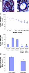
Involvement of HS-GAGs in Ad5 transduction. (A) GAGs are abundant at the basolateral membranes of acinar cells and within ductal regions of the lacrimal gland. Rabbit lacrimal glands were removed and immersed in optimal cutting temperature medium, rapidly frozen with liquid nitrogen, and cryosectioned at 5 μm with a Mikrom cryostat. Cryosections on glass coverslips were processed for Russell-Movat's pentachrome staining. In both panels, the blue to green staining shows GAGs beneath the basolateral membrane of some acinar clusters (arrow in left panel) and in ductal regions (arrowhead in right panel). The black-stained areas are nuclei. Bar, 20 μm. (B) Treatment of Ad-GFP with heparin reduces infection efficiency by approximately 40%, and the effect is additive to that of knob rather than competitive. Rabbit LGAC were seeded onto Matrigel-coated 12-well plates at 2 × 10⁶ cells/well. After 48 h, experimental cultures were incubated with recombinant knob protein (20 μg/ml) at an MOI of 2 PFU/cell and the controls were untreated. Ad-GFP that had been incubated with various concentrations of heparin for 1 h at 37°C was then added to control or experimental acinar cells for 1 h at 4°C. The cells were rinsed and incubated for 10 to 12 h at 37°C, and GFP positivity was determined by flow cytometry (n = 4). * in top panel, significant at P ≤ 0.05 based on results for the control; * in bottom panel, significant at P ≤ 0.05 based on results for knob-treated cells; error bars, SEM. (C) Treatment of rabbit LGAC with heparinases similarly reduced transduction efficiency of Ad-GFP. Rabbit LGAC were seeded onto Matrigel-coated 12-well plates at 2 × 10⁶ cells/well and allowed to adhere for 3 h. A heparinase III cocktail (10 U/ml; a mixture of heparinases I, II, and III) or control chondroitinase ABC (10 U/ml) was incubated with the LGAC for 2 h at 37°C. The cells were then rinsed well, Ad-GFP was added, and the procedures described for panel B were followed (n = 4). *, significant at P ≤ 0.05. Error bars represent the SEM.
FIG. 8.
FIG. 8.
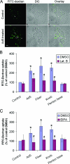
Effects of Ad5 and capsid proteins on FITC-dextran uptake. (A) LGAC labeled with FITC-dextran, as a measure of macropinocytosis, were stimulated by exposure to Ad5. Rabbit LGAC were seeded onto Matrigel-coated 12-well plates at 2 × 10⁶ cells/well. On day 3 of the culture, cells were incubated with or without Ad-LacZ (MOI = 100 PFU/cell) in binding buffer at 4°C for 1 h. The cells were washed, warmed to 37°C for 5 min, and pulsed with 1 mg/ml FITC-dextran at 37°C for 10 min. The cells were then washed extensively in ice-cold DPBS, fixed in 4% paraformaldehyde, and analyzed by confocal fluorescence microscopy. Arrowheads, accumulation of FITC-dextran in smaller intracellular vesicles (<1 μm); arrows, accumulation of FITC-dextran in larger intracellular vesicles (>1 μm); bar, 10 μm. (B) Significant reduction in Ad5-, fiber- or knob-stimulated FITC-dextran uptake with Lat B treatment suggests that Ad5-stimulated uptake is dependent on macropinocytosis. Rabbit LGAC were pretreated with DMSO or Lat B, incubated with Ad-LacZ or recombinant capsid proteins at cold temperatures, and analyzed for FITC-dextran uptake by flow cytometry as described in Materials and Methods. Penton base had no effect on uptake (n = 4). (C) Further confirmation that Ad5, fiber, and knob stimulate macropinocytosis was obtained by inhibiting virus-dependent uptake of FITC-dextran with the macropinocytosis inhibitor EIPA. Penton base had no effect on uptake. Rabbit LGAC were pretreated with DMSO or EIPA, incubated with Ad-LacZ or recombinant capsid proteins in cold temperatures, and analyzed for FITC-dextran uptake as described in Materials and Methods (n = 5). In panels B and C, * denotes significance at P values of ≤0.05 based on results for control acini and # denotes significance at P values of ≤0.05 based on results for the corresponding DMSO-treated acini.
FIG. 9.
FIG. 9.
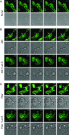
Time-lapse confocal fluorescence microscopy of membrane ruffling in LGAC treated with Ad5 and capsid proteins. Rabbit LGAC transduced to express GFP-actin as described in Materials and Methods were treated with DMSO or Lat B prior to being incubated with Ad-LacZ (MOI = 100 PFU/cell) or recombinant fiber (20 μg/ml) at 4°C for 1 h. The cells were warmed at 37°C for 5 min prior to the onset of the time-lapse sequence. Selected GFP-actin fluorescence and DIC images at regular intervals throughout the time-lapse sequence are shown. The arrows indicate basolateral actin remodeling. The boxed images were magnified and are shown in the insets. Bars, 10 μm.
FIG. 10.
FIG. 10.
Quantitation of membrane-ruffling events elicited by Ad5 and capsid proteins in LGAC. (A) A representative image of a transduced LGAC exposed to Ad-LacZ (MOI = 100 PFU/cell) from one frame within the 600-s analysis period is shown. The diameters of several membrane extensions in different stages of development are marked. (B) Treatment of LGAC with Ad5, fiber, and knob cause a significant increase in the number and size of membrane-ruffling events, while treatment with penton base does not. The histogram plot shows an average of the number of membrane-ruffling events per sequence (600-s analysis of an acinar cluster) under each experimental condition, with the ruffling events further grouped by maximum diameter. Data were collected from acinar clusters of comparable cell number and transduction efficiency within a 600-s period representing the period of highest membrane-ruffling activity in each sequence. Each membrane protrusion event occurring within that time frame was recorded at its largest diameter, using the LSM Image Browser overlay tools to measure diameter. These values were then classified by treatment group and divided into different maximal diameter ranges. The number of ruffling events in each range was grouped by treatment and divided by the number of sequences analyzed to obtain an average value per sequence for each treatment. Rabbit LGAC transduced to express GFP-actin as described in Materials and Methods were treated with recombinant proteins (20 μg/ml) or Ad-LacZ (MOI = 100 PFU/cell) at 4°C for 1 h and warmed at 37°C for 5 min prior to the onset of image acquisition (n = 16 for the control; n = 5 for penton base; n = 7 for knob; n = 5 for fiber; and n = 6 for Ad5). Panel C shows comparable values obtained from controls (n = 5), GST (n = 4), and Rab3DQL (n = 2). *, significant at P ≤ 0.05 based on results for the control. Error bars represent the SEM.
FIG. 11.
FIG. 11.
Effect of EIPA and Lat B on Ad5 infection efficiency and fiber entry. (A) Treatment with EIPA and Lat B significantly reduces Ad5 infection efficiency, suggesting that macropinocytosis may play a role in the infection process. Rabbit LGAC were seeded onto Matrigel-coated 12-well plates at 2 × 10⁶ cells/well. On day 2 of the culture, acinar cells were preincubated with DMSO, 500 μM EIPA, or 10 μM Lat B for 1 h at 37°C. Ad-GFP was then added at an MOI of 2 PFU/cell, and the cells were incubated for 1 h at 4°C before being washed and incubated for 10 to 12 h at 37°C. GFP positivity was determined by flow cytometry (n = 4 for EIPA; n = 5 for Lat B). *, significant at P ≤ 0.05 based on results for DMSO-treated acini. Error bars represent the SEM. (B) Inhibitors of macropinocytosis do not prevent fiber entry. Rabbit LGAC were seeded in 150-mm petri dishes at a density of 2 × 10⁶ cells/ml. On day 2 of the culture, vehicle-treated rabbit LGAC or LGAC pretreated with either Lat B or EIPA as described for panel A were incubated with Ad5 at an MOI of 15 PFU/cell for 0 or 60 min, followed by treatment with 0.2 mg/ml trypsin-EDTA for 1 h at 4°C and lysis using RIPA buffer. Cell lysates (150 μg/lane) were blotted with monoclonal antibody to fiber protein and IRDye800-conjugated secondary antibody (n = 3). Error bars represent the SEM. Values shown are fiber protein uptake at 60 min relative to that for the control (0 min, incubated only with Ad5).
FIG. 12.
FIG. 12.
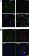
Colocalization of endocytotic markers with Ad5. (A) Localization of Ad5 after internalization reveals that its intracellular trafficking pathway diverges from the macropinocytosis pathway. Rabbit LGAC were seeded onto Matrigel-coated 12-well plates at 2 × 10⁶ cells/well. On day 3 of the culture, the cells were incubated with or without Ad-LacZ (MOI = 100 PFU/cell) in binding buffer at 4°C for 1 h. The cells were washed, warmed to 37°C for 5 min, and pulsed with 1 mg/ml FITC-dextran at 37°C for 10 min. The cells were then washed extensively in ice-cold DPBS, fixed in 4% paraformaldehyde, and processed to fluorescence label FITC-dextran (green), Ad5 proteins (red), and actin filaments (purple). A similar lack of colocalization was seen when lower doses of Ad5 were applied. *, apical/luminal region; bar, 10 μm. (B) Fiber-dependent Ad5 entry into LGAC appears to occur via the clathrin-dependent endocytotic pathway. Rabbit LGAC were seeded onto Matrigel-coated 12-well plates at 2 × 10⁶ cells/well. On day 3 of the culture, the cells were exposed to Ad-LacZ (MOI = 40 PFU/ml) for 60 min before being fixed and processed to fluorescence label Ad5 proteins (green), EEA1 (red), and nuclei (blue). The arrows point to a few of the many areas of colocalization of Ad5 and EEA1. Bar, 10 μm. A similar colocalization was seen when higher doses of Ad5 were applied (MOI up to 100 PFU/cell).

Similar articles

Cited by

References

    1. Ashbourne Excoffon, K. J. D., T. Moninger, and J. Zabner. 2003. The coxsackie B virus and adenovirus receptor resides in a distinct membrane microdomain. J. Virol. 77:2559-2567. - PMC - PubMed
    1. Awasthi, V., G. Meinken, K. Springer, S. C. Srivastava, and P. Freimuth. 2004. Biodistribution of radioiodinated adenovirus fiber protein knob domain after intravenous injection in mice. J. Virol. 78:6431-6438. - PMC - PubMed
    1. Baluska, F., J. Samaj, A. Hlavacka, J. Kendrick-Jones, and D. Volkmann. 2004. Actin-dependent fluid-phase endocytosis in inner cortex cells of maize root apices. J. Exp. Bot. 55:463-473. - PubMed
    1. Bergelson, J. M., J. A. Cunningham, G. Droguett, E. A. Kurt-Jones, A. Krithivas, J. S. Hong, M. S. Horwitz, R. L. Crowell, and R. W. Finbert. 1997. Isolation of a common receptor for coxsackie B viruses and adenoviruses 2 and 5. Science 275:1320-1323. - PubMed
    1. Borras, T. 2003. Recent developments in ocular gene therapy. Exp. Eye Res. 76:643-652. - PubMed

Publication types

Substances

Associated data

LinkOut - more resources