Endogenous retroviruses regulate periimplantation placental growth and differentiation
- PMID: 16980413
- PMCID: PMC1599973
- DOI: 10.1073/pnas.0603836103
Endogenous retroviruses regulate periimplantation placental growth and differentiation
Abstract
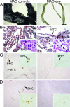
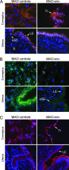
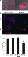
Endogenous retroviruses (ERVs) are fixed and abundant in the genomes of vertebrates. Circumstantial evidence suggests that ERVs play a role in mammalian reproduction, particularly placental morphogenesis, because intact ERV envelope genes were found to be expressed in the syncytiotrophoblasts of human and mouse placenta and to elicit fusion of cells in vitro. We report here in vivo and in vitro experiments finding that the envelope of a particular class of ERVs of sheep, endogenous Jaagsiekte sheep retroviruses (enJSRVs), regulates trophectoderm growth and differentiation in the periimplantation conceptus (embryo/fetus and associated extraembryonic membranes). The enJSRV envelope gene is expressed in the trophectoderm of the elongating ovine conceptus after day 12 of pregnancy. Loss-of-function experiments were conducted in utero by injecting morpholino antisense oligonucleotides on day 8 of pregnancy that blocked enJSRV envelope protein production in the conceptus trophectoderm. This approach retarded trophectoderm outgrowth during conceptus elongation and inhibited trophoblast giant binucleate cell differentiation as observed on day 16. Pregnancy loss was observed by day 20 in sheep receiving morpholino antisense oligonucleotides. In vitro inhibition of the enJSRV envelope reduced the proliferation of mononuclear trophectoderm cells isolated from day 15 conceptuses. Consequently, these results demonstrate that the enJSRV envelope regulates trophectoderm growth and differentiation in the periimplantation ovine conceptus. This work supports the hypothesis that ERVs play fundamental roles in placental morphogenesis and mammalian reproduction.
Conflict of interest statement
The authors declare no conflict of interest.
Figures


Similar articles
-
Application of next generation sequencing in mammalian embryogenomics: lessons learned from endogenous betaretroviruses of sheep.Anim Reprod Sci. 2012 Sep;134(1-2):95-103. doi: 10.1016/j.anireprosci.2012.08.016. Epub 2012 Aug 11. Anim Reprod Sci. 2012. PMID: 22951118 Free PMC article. Review.
-
Sheep endogenous betaretroviruses (enJSRVs) and the hyaluronidase 2 (HYAL2) receptor in the ovine uterus and conceptus.Biol Reprod. 2005 Aug;73(2):271-9. doi: 10.1095/biolreprod.105.039776. Epub 2005 Mar 23. Biol Reprod. 2005. PMID: 15788753
-
Endogenous retroviruses in trophoblast differentiation and placental development.Am J Reprod Immunol. 2010 Oct;64(4):255-64. doi: 10.1111/j.1600-0897.2010.00860.x. Epub 2010 May 26. Am J Reprod Immunol. 2010. PMID: 20528833 Free PMC article. Review.
-
Viral particles of endogenous betaretroviruses are released in the sheep uterus and infect the conceptus trophectoderm in a transspecies embryo transfer model.J Virol. 2010 Sep;84(18):9078-85. doi: 10.1128/JVI.00950-10. Epub 2010 Jul 7. J Virol. 2010. PMID: 20610723 Free PMC article.
-
Endogenous retroviruses of sheep: a model system for understanding physiological adaptation to an evolving ruminant genome.J Reprod Dev. 2012;58(1):33-7. doi: 10.1262/jrd.2011-026. J Reprod Dev. 2012. PMID: 22450282 Review.
Cited by
-
ROCK Inhibitor (Y-27632) Abolishes the Negative Impacts of miR-155 in the Endometrium-Derived Extracellular Vesicles and Supports Embryo Attachment.Cells. 2022 Oct 10;11(19):3178. doi: 10.3390/cells11193178. Cells. 2022. PMID: 36231141 Free PMC article.
-
Ancestral capture of syncytin-Car1, a fusogenic endogenous retroviral envelope gene involved in placentation and conserved in Carnivora.Proc Natl Acad Sci U S A. 2012 Feb 14;109(7):E432-41. doi: 10.1073/pnas.1115346109. Epub 2012 Jan 17. Proc Natl Acad Sci U S A. 2012. PMID: 22308384 Free PMC article.
-
A paradigm for virus-host coevolution: sequential counter-adaptations between endogenous and exogenous retroviruses.PLoS Pathog. 2007 Nov;3(11):e170. doi: 10.1371/journal.ppat.0030170. PLoS Pathog. 2007. PMID: 17997604 Free PMC article.
-
Capture of a Hyena-Specific Retroviral Envelope Gene with Placental Expression Associated in Evolution with the Unique Emergence among Carnivorans of Hemochorial Placentation in Hyaenidae.J Virol. 2019 Feb 5;93(4):e01811-18. doi: 10.1128/JVI.01811-18. Print 2019 Feb 15. J Virol. 2019. PMID: 30463979 Free PMC article.
-
Changes in population dynamics in mutualistic versus pathogenic viruses.Viruses. 2011 Jan;3(1):12-19. doi: 10.3390/v3010012. Epub 2011 Jan 17. Viruses. 2011. PMID: 21994724 Free PMC article.
References
Publication types
MeSH terms
Substances
Grants and funding
LinkOut - more resources
Full Text Sources

