. 2007 Jan;123(1):57-71.
doi: 10.1016/j.virusres.2006.08.003. Epub 2006 Sep 8.

The chemokine receptor homologue encoded by US27 of human cytomegalovirus is heavily glycosylated and is present in infected human foreskin fibroblasts and enveloped virus particles

Affiliations

The chemokine receptor homologue encoded by US27 of human cytomegalovirus is heavily glycosylated and is present in infected human foreskin fibroblasts and enveloped virus particles

Barry J Margulies et al. Virus Res. 2007 Jan.

Abstract

Human cytomegalovirus (HCMV), a member of the beta-herpesvirus family, encodes four homologues of cellular G protein-coupled receptors (GPCRs). One of these, the protein product of HCMV open reading frame (ORF) UL33, has been identified in HCMV-infected cells and virus particles and shown to be heat-aggregatable and N-glycosylated. Another, the product of ORF US28, has been functionally characterized as a beta-chemokine receptor. Here we report the use of RT-PCR, coupled in vitro transcription-translation, immunoprecipitation, and Western immunoassays to (i) show that RNA from the open reading frame US27 appears predominantly during the late phase of replication; (ii) identify the protein encoded by HCMV US27 in infected cells and enveloped virus particles; (iii) demonstrate that the US27-encoded protein is heterogeneously N-glycosylated and resolves as two species following treatment with peptide N-glycosidase F; and (iv) show that both the recombinant and deglycoylated infected cell US27 protein aggregate when heated in the presence of SDS prior to electrophoresis in polyacrylamide gels, a property which is abrogated with the addition of urea to sample buffer.


Figures
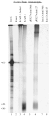
Fig. 1
Fig. 1
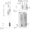
In vitro transcription–translation and immunoprecipitation of pUS27. The plasmid BJM2.1 (1 μg; BJM2.1; lanes 3 and 4) was transcribed and translated in vitro in a 25 μL TNT coupled reaction. Reactions containing no DNA (Blank; lane 2) or DNA encoding luciferase (Lucif.; lane 1) were also included. A small sample (1.5 μL) was removed from each reaction, solubilized, boiled (lanes 1–3) or not heated (lane 4), and subjected to SDS-PAGE and fluorography. Immunoprecipitations of in vitro-translated luciferase and pUS27 were done with Anti-27 (+Anti-27; lanes 6 and 8) or Anti-Neurothelin (+Anti-Nth; lanes 5 and 7) antiserum, and analyzed in parallel. Samples in lanes 4–8 were not heated prior to SDS-PAGE. An image of the resulting fluorograph was captured with Adobe Photoshop for Macintosh via a Umax PowerLook II scanner.
Fig. 2
Fig. 2
(A) HCMV-infected HFF cells express N-glycosylated GCR27. HFFs were infected with HCMV (infected; lanes 3 and 4) or not infected (mock; lanes 1 and 2). Total cell membranes were isolated 8 days after infection and incubated with or without PNGase F (±PNGase). Membrane protein (≈20 μg) was subjected to SDS-PAGE and analyzed by Western immunoassay with Anti-27. The brackets represent apparent molecular weight ranges of ≈ 80–130 and ≈ 150–180 kDa, respectively; molecular weights (in kDa) were estimated from molecular weight markers (See Blue Prestained Markers; Novex) included during SDS-PAGE that were electrotransfered to the Immobilon-P. Circles denote deglycosylated forms of GCR27. (B) Comparison of deglycosylated GCR27 and in vitro-synthesized pUS27. The same samples from lanes 1 to 4 were resolved in lanes 5–8, respectively, by 12% SDS-PAGE alongside TNT-derived pUS27 (lane 10) and a TNT reaction containing no DNA (lane 9). Western immunoassay was conducted with Anti-27. (C) Validating the specificity of Anti-27. The same samples from lanes 1 to 4 were resolved in duplicate, side-by-side, in lanes 11–14 and lanes 15–18, by 12% SDS-PAGE (“M” denotes mock-infected and “I” denotes HCMV-infected membrane samples). One-half of the gel was analyzed by a Western blot with Anti-27 (lanes 11–14); the other half was analyzed with Anti-33 (Margulies et al., 1996) (lanes 15–18). Molecular weights (in kDa) in parts B and C were estimated from pre-stained markers (Multi Mark; Invitrogen) included during SDS-PAGE that were electrotransfered to the Immobilon-P. Images of the resulting fluorographs and autoradiogram were captured with Adobe Photoshop for Macintosh via Umax PowerLook II and Epson 1200 U scanners.
Fig. 3
Fig. 3
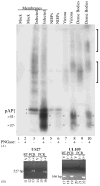
(A) N-Glycosylated GCR27 is present in HCMV extracellular enveloped particles. Cell membranes were prepared from non-infected (mock; lanes 1 and 2) or HCMV-infected (infected; lanes 3 and 4) HFF cells 8 days after infection and treated or not treated with PNGase F (±PNGase). HCMV enveloped extracellular virus particles, NIEPs (lanes 5 and 6), virions (lanes 7 and 8), and dense bodies (lanes 9 and 10), were purified from infected cells, treated or not treated with PNGase F (± PNGase), and subjected to SDS-PAGE, followed by Western immunoassay with a mixture of Anti-27 and Anti-C1, an antiserum specific for the CMV assembly protein precursor, pAP (Schenk et al., 1991; Welch et al., 1991b). Brackets denote two forms of glycosylated GCR27; circles denote deglycosylated forms of GCR27; brace and asterisks denote pAP (top) and pUL80.4, a slightly smaller protein also encoded by the HCMV UL80 nested genes (Casaday et al., 2004; Welch et al., 1991a). An image of the resulting fluorograph was captured with Adobe Photoshop for Macintosh via an Epson 1200 U scanner. (B) RT-PCR assay for UL27 and UL109 RNAs in virus particles. Clarified HCMV inoculum (100 μL) was treated with DNase-free RNase, then RNA was isolated from these samples (lanes 11, 14, 17, and 20) and subsequently treated with RNase-free DNase. Negative controls (lanes 12, 15, 18, and 21) contained no RNA; positive controls (lanes 13, 16, 19, and 22) contained approximately 50 ng of DNA from an HCMV (AD169) bacmid (Marschall et al., 2000), to show that RT-PCR and PCR did not fail because of poor enzyme cycling conditions. One-twentieth of the RNA isolated from virus inoculum was used for each experiment. Samples were amplified in the presence of an RT/Taq DNA polymerase mix (RT-PCR, lanes 11–13 and 17–19) or Platinum Taq DNA polymerase alone (PCR, lanes 14–16 and 20–22) for 60 cycles. PCR products were resolved on 2% agarose gels in SB buffer (Brody and Kern, 2004), stained with ethidium bromide, and photographed on a UV transilluminator as part of a Bio-Rad Molecular Imager Gel Doc XR system. Markers (1-kb markers or PCR markers, both from Promega) run in adjacent lanes allowed characterization of the 527-bp US27 PCR product and the 166-bp UL109 PCR product.
Fig. 4
Fig. 4
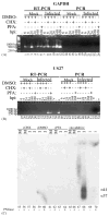
(A) Detection of RNA for the cellular housekeeping gene GAPDH. Total RNA was isolated from mock-infected (lanes 1–6 and 13–18) or HCMV-infected (lanes 7–12 and 19–22) human fibroblasts at 6 h (lanes 1, 2, 7, 8, 13, 14, 19, and 20), 24 h (lanes 3, 4, 9, 10, 15, 16, 21, and 22), and 168 h (lanes 5, 6, 11, 12, 17, 18, 23, and 24) after infection. These cells were treated 1 h prior to infection, then continuously until RNA was harvested, with either CHX in DMSO (lanes 2, 4, 8, 10, 14, 16, 20, and 22), DMSO alone (lanes 1, 3, 7, 9, 13, 15, 19, and 21), PFA (lanes 6, 12, 18, and 24), or no agent (lanes 5, 11, 17, and 23). A negative control (-) contained no RNA. Amplification was conducted in the presence of either an RT/Taq polymerase mix (RT-PCR; lanes 1–12) or only Platinum Taq DNA polymerase (PCR; lanes 13–24) for 30 cycles. One-twentieth of the RNA isolated from approximately 106 cells was used for each experiment. PCR products were resolved, stained, and imaged as in Fig. 3. Markers (1-kb markers, Promega) run in adjacent lanes allowed characterization of 1000-, 500-, and 250-base pair regions; sizes (bp) of marker DNAs are indicated beside each image. Primers were designed to produce an approximately 500-bp product from a target in the middle of the gene. (B) Time course of expression of the RNA produced from the viral US27 gene. One-Step RT-PCR and PCR were performed as in part A for 30 cycles. Primers were designed to produce a 527-bp product from the middle of US27. Virion RNA, isolated as described in Fig. 3B, was also included (V; lanes 37 and 52); negative controls (lanes 38 and 53) contained no RNA. A positive control (lanes 39 and 54) contained approximately 50 ng of DNA of an HCMV bacmid, as in Fig. 3. (C) Time course of expression of the GCR27 glycoprotein. Membranes were isolated from HCMV-infected (I; lanes 57, 58, 59, 60, 63, 64, 67, and 68) or mock-infected (M; lanes 55, 56, 61, 62, 65, and 66) cells treated with either CHX in DMSO (lanes 55–59), DMSO alone (DMSO; lanes 59 and 60), PFA (PFA; lanes 61–64), or no treatment (lanes 65–68) at either 24 h (for CHX and DMSO) or 168 h (for PFA and no additive) after infection. These membrane preparations were then either treated (lanes 56, 58, 60, 62, 64, 66, and 68) or not treated (lanes 55, 57, 59, 61, 63, 65, and 67) with PNGase F. A portion (50 μL) of the inoculum used for these experiments was similarly treated with PNGase F (i; lanes 69 and 70). Samples were resolved by SDS-PAGE, transferred to Immobilon-P, and analyzed by Western blot with the Anti-27 antiserum. Molecular weights (in kDa) were estimated from pre-stained markers (Multi Mark; Invitrogen) included during SDS-PAGE that were electrotransfered to the Immobilon-P. An image of the resulting fluorograph was captured with Adobe Photoshop for Macintosh via an Epson 1200 U scanner.
Fig. 5
Fig. 5
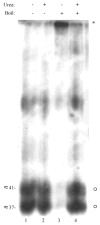
Urea abrogates the heat-induced aggregation of GCR27. A preparation of dense bodies was treated with PNGase F. Half was denatured in solubilizing buffer without urea, and either boiled (lane 3) or not boiled (lane 1). The other half of the preparation was denatured in solubilizing buffer containing 4 M urea, and either boiled (lane 4) or not boiled (lane 2). All samples were resolved by 12% SDS-PAGE and then analyzed by Western blot with Anti-27. Molecular weights (in kDa) were estimated from molecular weight markers (see Blue Prestained Markers; Novex) included during SDS-PAGE that were electrotransfered to the Immobilon-P. An image of the resulting fluorograph was captured with Adobe Photoshop for Macintosh via a Umax PowerLook II scanner.

Similar articles

Cited by

References

    1. AbuBakar S, Boldogh I, Albrecht T. Human cytomegalovirus stimulates arachidonic acid metabolism through pathways that are affected by inhibitors of phospholipase A2 and protein kinase C. Biochem Biophys Res Commun. 1990a;166(2):953–959. - PubMed
    1. AbuBakar S, Boldogh I, Albrecht T. Human cytomegalovirus: stimulation of [3H] release from [3H]-arachidonic acid prelabelled cells. Arch Virol. 1990b;113:255–266. - PubMed
    1. Albrecht T, Fons MP, Boldogh I, AbuBakar S, Deng CZ, Millinoff D. Metabolic and cellular effects of human cytomegalovirus infection. Transplant Proc. 1991;23(3):48S–55S. - PubMed
    1. Attwood TK, Findlay JBC. Design of a discriminating fingerprint for G-protein-coupled receptors. Protein Eng. 1994a;6(2):167–176. - PubMed
    1. Attwood TK, Findlay JBC. Fingerprinting G-protein-coupled receptors. Protein Eng. 1994b;7(2):195–203. - PubMed

Publication types

Substances