Review
. 2006 Sep;70(3):605-45.
doi: 10.1128/MMBR.00013-06.

The yeast actin cytoskeleton: from cellular function to biochemical mechanism

Affiliations
Review

The yeast actin cytoskeleton: from cellular function to biochemical mechanism

James B Moseley et al. Microbiol Mol Biol Rev. 2006 Sep.

Abstract

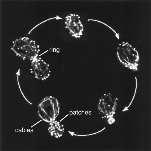
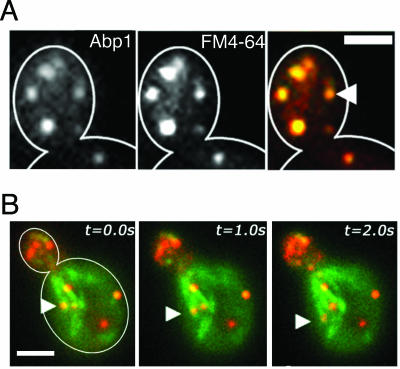
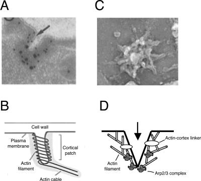
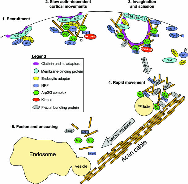
All cells undergo rapid remodeling of their actin networks to regulate such critical processes as endocytosis, cytokinesis, cell polarity, and cell morphogenesis. These events are driven by the coordinated activities of a set of 20 to 30 highly conserved actin-associated proteins, in addition to many cell-specific actin-associated proteins and numerous upstream signaling molecules. The combined activities of these factors control with exquisite precision the spatial and temporal assembly of actin structures and ensure dynamic turnover of actin structures such that cells can rapidly alter their cytoskeletons in response to internal and external cues. One of the most exciting principles to emerge from the last decade of research on actin is that the assembly of architecturally diverse actin structures is governed by highly conserved machinery and mechanisms. With this realization, it has become apparent that pioneering efforts in budding yeast have contributed substantially to defining the universal mechanisms regulating actin dynamics in eukaryotes. In this review, we first describe the filamentous actin structures found in Saccharomyces cerevisiae (patches, cables, and rings) and their physiological functions, and then we discuss in detail the specific roles of actin-associated proteins and their biochemical mechanisms of action.


Figures

FIG. 1.
FIG. 1.
Cell cycle-regulated organization of the S. cerevisiae actin cytoskeleton. Yeast cells at different stages in the cell cycle contain three visible F-actin structures: cortical actin patches, polarized actin cables, and a cytokinetic actin ring. While patches and cables are visible throughout the cell cycle, the ring is visible shortly before and during cytokinesis. Shown here are actively growing cells from an asynchronous culture that were chemically fixed and stained with rhodamine phalloidin to visualize filamentous actin structures. (Modified from reference with permission of the publisher.)
FIG. 2.
FIG. 2.
Cortical patches are sites of endocytosis and actin assembly. (A) Colocalization of Abp1-GFP (an actin patch marker) and FM4-64 (a membrane-binding dye/endosomal marker) at the yeast cell cortex. (B) Actin patches (Abp1-HcRed) undergo limited short-range movements while associated with the cell cortex and then transition to a phase of rapid movement upon internalization. The arrowhead tracks the rapid movement of a single actin patch moving over time. The patch moves in a retrograde manner (bud to mother cell) on the end of an actin cable (marked by Abp140-GFP). The rate of fast patch movement is approximately the same as the rate of cable movement (0.2 to 0.4 μm per s), suggesting that patches may be transported passively through attachment to cables. The molecular interactions linking patches to cables are unknown. Bars, 2 μm. (Modified from reference by copyright permission of The Rockefeller University Press.)
FIG. 3.
FIG. 3.
Electron micrographs of cortical actin patches. (A) Thin sectioning and electron microscopy reveal membrane invaginations at the yeast cell cortex that are enriched for actin (immunolabeled with 10-nm gold particles; arrow) and Abp1 (immunolabeled with 20-nm gold particles). (Reproduced from reference by copyright permission of the Rockefeller University Press.) (B) One model for actin filament organization at cortical patches based on the microscopy in panel A. (Reproduced from reference by copyright permission of the Rockefeller University Press.) (C) Rapid-freeze deep-etch electron microscopy reveals actin filaments at the cell cortex organized into branched, cone-shaped filamentous networks. Immunogold labeling (not shown) revealed that three known patch components (actin, Abp1, and Crn1) are distributed throughout these structures, whereas the Arp2/3 complex is restricted to the peaks or apexes of the mounds. (Reprinted from reference with permission of the publisher.) (D) Model for actin filament organization at patches based on the images in panel C. The arrow indicates the direction of membrane invagination during endocytosis.
FIG. 4.
FIG. 4.
Model for actin patch development. (Step 1) Receptors recruit early patch components, including clathrin, adaptors, and two NPFs (Pan1 and Las17), to the cell cortex to form a relatively immobile complex. (Step 2) Pan1 and Las17 recruit and activate the Arp2/3 complex to nucleate actin assembly, leading to slow patch movement at the cortex. During this stage, two additional NPFs are recruited, Abp1 and Myo3/Myo5. Abp1 in turn recruits the actin-regulating kinases Ark1 and Prk1, which phosphorylate Pan1 (and likely other patch proteins). Phosphorylation inhibits Pan1 activation of the Arp2/3 complex and triggers dissociation of the Pan1 complex (Pan1, End3, and Sla1). (Step 3) Pan1 phosphorylation and/or the activities of Rvs161, Rvs167, and the type I myosins Myo3 and Myo5 promote vesicle scission and internalization. The Arp2/3 complex, Abp1, Sac6, Cap1/Cap2, and likely other patch components maintain association with the internalized vesicle. Some early patch components are left at the cell cortex, including Las17, Pan1, Sla1, Sla2, Myo3, and Myo5. (Step 4) Endocytic vesicles associate with actin cables by an unknown mechanism and move passively with actin cable retrograde flow (bud to mother). (Step 5) Cable-associated vesicles fuse with an endosomal compartment concomitant with disassembly of the actin coat.
FIG. 5.
FIG. 5.
Formins are required for actin cable assembly at the bud tip and neck. (A) Localization of GFP-labeled yeast formins Bni1 and Bnr1 throughout the cell cycle. (Reprinted from reference with permission of the publisher.) (B) Formin function is required for the assembly and maintenance of actin cables. In a bnr1Δ background, point mutations in the FH2 domain of Bni1 render cells temperature sensitive for growth and actin cable formation. Shifting mutants from the permissive temperature (23°C, left panel) to the nonpermissive temperature (35°C, right panel) for 2 to 4 min results in complete loss of visible actin cables but does not affect actin patch distribution in budded (b) or unbudded (u) cells. (Reprinted from reference by permission from Macmillan Publishers Ltd.)
FIG. 6.
FIG. 6.
Models for regulation of actin cable assembly at the bud tip and neck. (A) Organization of formins (red), actin cables (gold), and septin structures (blue) in a budded yeast cell. Formin proteins (Bni1 at the bud tip and Bnr1 at the neck) directly nucleate actin cable formation. Septins serve as a diffusion barrier between bud and mother cell compartments and provide a scaffold for assembly of many bud neck components. (B) Interaction network at the bud neck: septins (blue); actin (tan); actin-binding proteins (red); all other factors (green). Solid lines represent known physical interactions; dashed lines denote septin-dependent localization. In G1/S, septins recruit other early components to the bud neck. Later in the cell cycle, just prior to cytokinesis, additional factors (late components) are recruited to the bud neck, including components of the bud tip “polarity cap” complex. (C) Model for regulation of actin cable assembly by polarity cap components at the bud tip and cable disassembly by cofilin and Aip1. The polarity cap may be linked to the plasma membrane via components that directly associate with phospholipids, including Cdc42, Cdc24, and Cla4. The actin filaments in cables are nucleated by the formin Bni1, using profilin- and Bud6-bound actin subunits as substrates. Bni1 activity may be regulated by interactions with Cdc42, Rho3, Rho4, Bud6, profilin, Spa2, and other factors (not pictured). Bni1 associates tightly with the fast-growing (barbed) end of the actin filament it nucleates, facilitating insertional growth while protecting ends from capping protein (Cap1/2). Cables are likely comprised of short staggered filaments connected by actin cross-linking proteins, such as Sac6 and Abp140. It is not yet clear what regulates the lengths of actin filaments within a cable. Cables are stabilized along their sides by tropomyosin, which competes with cofilin for binding F-actin. Genetic observations also suggest that Bni1 and Cap1/2 might cap the barbed ends of filaments in cables (see text). As rapidly as cables are formed, they must be disassembled, which recent evidence suggests is mediated by cofilin and Aip1. Barbed-end-directed type V myosins (Myo2 and Myo4) transport vesicles, organelles, and other cargos along cables to the bud tip.
FIG. 7.
FIG. 7.
Formation and contraction of an acto-myosin ring during yeast cytokinesis. (A) Prior to cytokinesis, actin filaments (rhodamine-phalloidin) and type II myosin (Myo1-GFP) assemble at the bud neck. (B) The actomyosin ring contracts, as demonstrated in consecutive frames of a Myo1-GFP time course at 1-minute intervals. Bar, 2 μm. (Reproduced from reference by copyright permission of the Rockefeller University Press.)
FIG. 8.
FIG. 8.
Assembly and turnover cycle of actin filaments. Actin filaments are polarized, with a fast-growing (barbed) end and a slow-growing (pointed) end. ATP-bound actin monomers (light gray) preferentially associate with the barbed end of the filament. ATP hydrolysis on an actin subunit is triggered by its addition to the end of a filament. The rate of this reaction varies for actins from different species. After hydrolysis, ADP and Pi remain bound to actin (medium gray), followed shortly by the release of Pi to yield ADP-bound actin (dark gray). Thus, steady-state F-actin exists as a mosaic of actin subunits in different nucleotide-bound states. At steady state, the pointed ends of filaments undergo a net loss of ADP-actin subunits, generating a pool of ADP-actin monomers (G-actin). Following their dissociation, free monomers rapidly exchange nucleotide (ATP for ADP) to replenish an assembly-competent pool of ATP-bound G-actin. The actin monomer-binding protein profilin is one factor that can accelerate the rate of nucleotide exchange on G-actin. Profilin binds to the barbed end of an actin monomer (see cocrystal in right panel), sterically blocking the addition of profilin-bound actin monomers to the pointed ends of actin filaments, but not the barbed ends. Based on these properties and the high abundance of profilin in cells, it is hypothesized that most in vivo actin assembly occurs specifically at filament barbed ends. Upon addition of a profilin-bound actin subunit to a filament barbed end, profilin disassociates rapidly, leaving the barbed end free for addition of the next subunit. The coordinates for the profilin-actin complex were obtained from reference .
FIG. 9.
FIG. 9.
The Arp2/3 complex mechanism of actin assembly. (A) Projection structures of free and ligand-bound yeast Arp2/3 complexes (each computed from 1,000 to 3,000 separate images). Electron microscopy and single-particle analyses were used to show that the Arp2/3 complex exists in an equilibrium among open, intermediate, and closed conformations (middle three panels), referring to the relative positions of the Arp2 and Arp3 subunits within the complex. Binding of an inhibitor (Crn1/coronin) promotes the open conformation (left panel), while binding of an NPF (Las17/WASp) promotes the closed conformation (316). The open conformation is thought to represent the inactive state, while the closed conformation is “primed” for nucleation. (B) Model for structural rearrangements during activation of the Arp2/3 complex. Arrows indicate conformational changes leading to the next structure in the activation sequence. In the transition from the open to the intermediate conformation, Arp2 and Arp3 subunits move toward the center of the gap separating the two subunits. Next, there is a further closure of the gap between Arp2 and Arp3 by a movement of Arp2 toward Arp3 and possibly a small movement of Arc18 toward the center of the gap, creating the closed conformation. WASp is shown binding to Arp2 and Arp3, and possibly also to p40, stabilizing the closed conformation. Coronin is shown binding to p35, stabilizing the open conformation. (C) Model for nucleation of branched actin filaments by the Arp2/3 complex. Solid black arrows indicate physical interactions; the dashed arrow depicts closure of the complex upon Las17 binding. The crystal structure of the bovine Arp2/3 complex (313) is docked into the three-dimensional EM reconstruction of the yeast Arp2/3 complex in its open inactive conformation (316). The complex associates with the side of an existing (mother) filament and nucleates formation of a new (daughter) filament at a 70° angle (large red arrow). Nucleation is triggered by association of the Arp2/3 complex with Las17 and the side of the mother filament. These interactions promote major structural rearrangements within the Arp2/3 complex, bringing Arp2 and Arp3 closer together to nucleate an actin filament. Nucleation also requires Las17 binding to an actin monomer, presumably to provide the first subunit of the daughter filament. (All reprinted from reference by permission from Macmillan Publishers Ltd.)
FIG. 10.
FIG. 10.
Yeast NPFs. (A) Schematics of each protein drawn to scale. Abbreviations: A, acidic; B, basic; CC, coiled-coil; EH, Eps15 homology; EVH1, Ena/VASP homology 1; IQ, IQ binding; LR, long repeat; PP or PPP, polyproline; SH3, Src homology 3; TH1/2, tail homology 1/2; WH1, WASp homology 1; WH2, WASp homology 2. The proposed actin-binding domain of each NPF is colored red; acidic domains are yellow. Las17 binds to G-actin, while Abp1, Pan1, and Myo3/5 all bind specifically to F-actin. Pan1 contains many Ark1/Prk1 consensus phosphorylation sites in LR1 and LR2 (indicated by the circled P). (B) Sequence alignment of acidic domains from each yeast NPF, with the consensus sequence below in bold.
FIG. 11.
FIG. 11.
Formin mechanism of actin assembly. (A) Domain organization of budding yeast formins Bni1 and Bnr1. SBD, Spa2-binding domain. (B) Crystal structure of the Bni1 FH2 dimer depicted by surface rendering, with a solid line separating the two functional halves (hemi-dimers). Residues are colored according to the degree of evolutionary conservation, with highly conserved residues in red and nonconserved residues in teal blue. Circled regions denote patches of conserved surface residues involved in actin binding, identified by mutational studies (348, 414) (Reprinted from reference with permission from Elsevier.) (C) Cocrystal structure of the Bni1 FH2 domain associated with tetramethylrhodamine-actin. Each hemi-dimer interacts with two actin subunits in the polymer. (Reprinted from reference by permission from Macmillan Publishers Ltd.) (D) Model for processive capping by the FH2 dimer, which maintains association with the rapidly growing barbed end of the actin filament. Alternating regions of the FH2 are displaced rapidly in response to subunit addition to allow insertion of the next subunit. Importantly, the precise biophysical mechanism of formin processive capping remains unclear. (E) While the formin caps the barbed end of the filament it has nucleated, the Arp2/3 complex caps the opposite (pointed) end of the filament, leaving the barbed end free. Thus, filaments nucleated specifically by formins are protected from early termination of elongation by capping protein. Directions of new filament growth are noted by red arrows.
FIG. 12.
FIG. 12.
Actin filament-bundling proteins. Schematics of each protein are drawn to scale. Abbreviations: CC, coiled-coil; CLR, calponin-like repeat; COOH, carboxyl-terminal domain. Known actin-binding domains are underlined. Yeast IQGAP (Iqg1) is included because mammalian IQGAP bundles actin filaments; however, this activity has not yet been demonstrated for yeast Iqg1. It is unknown how Abp140 bundles actin filaments, since its actin-binding domain(s) has not been defined.
FIG. 13.
FIG. 13.
Regulation of actin filament turnover by a network of interacting proteins. (A) Schematics of proteins discussed in the actin turnover section of the text. Abbreviations: β-propeller, forms β-propeller tertiary structure composed of WD repeats; CC, coiled-coil; α-helix, forms tertiary structure composed entirely of α-helices that resembles 14-3-3 domains (201, 238, 425); P, polyproline domain; WH2, WASp homology 2; β-sheet, forms tertiary structure composed entirely of β-sheets (78). (B) Model for the sequential events in actin turnover. Cofilin (orange) binds cooperatively to and severs older (ADP-bound) actin filaments. Aip1 (green) interacts with cofilin and F-actin to cap the barbed ends of severed filaments. Aip1-capped filaments disassemble rapidly from their pointed ends, generating a pool of cofilin-bound ADP-actin monomers that cannot exchange ATP for ADP. The dodecameric Srv2 complex (blue), tethered to F-actin via the SH3 domain of Abp1 (red), rapidly displaces cofilin and binds tightly to ADP-G-actin. This facilitates rapid nucleotide exchange (ATP for ADP) on actin. Srv2 has 100-fold-lower affinity for ATP-G-actin than ADP-G-actin and thus hands off ATP-G-actin to profilin. Profilin interacts directly with Srv2 and may accelerate nucleotide exchange on actin during this process. Profilin-bound ATP-actin monomers are available for rapid addition onto the barbed ends of filaments. (C) Relay of actin monomers from cofilin to Srv2 to profilin, facilitated by their specific affinities for ADP- versus ATP-actin. The proposed sequence of G-actin handoffs is boxed with arrows. Kd values are taken from references , , , , , and .

Similar articles

Cited by

References

    1. Abe, M., H. Qadota, A. Hirata, and Y. Ohya. 2003. Lack of GTP-bound Rho1p in secretory vesicles of Saccharomyces cerevisiae. J. Cell Biol. 162:85-97. - PMC - PubMed
    1. Adams, A., and J. Pringle. 1984. Relationship of actin and tubulin distribution to bud growth in wild-type and morphogenetic-mutant Saccharomyces cerevisiae. J. Cell Biol. 98:934-945. - PMC - PubMed
    1. Adams, A. E., D. Botstein, and D. G. Drubin. 1991. Requirement of yeast fimbrin for actin organization and morphogenesis in vivo. Nature 354:404-408. - PubMed
    1. Adams, A. E., D. Botstein, and D. G. Drubin. 1989. A yeast actin-binding protein is encoded by SAC6, a gene found by suppression of an actin mutation. Science 243:231-233. - PubMed
    1. Adams, A. E. M., and D. Botstein. 1989. Dominant suppressors of yeast actin mutations that are reciprocally suppressed. Genetics 121:675-683. - PMC - PubMed

Publication types

MeSH terms

LinkOut - more resources