

Kaposi's sarcoma-associated herpesvirus virions inhibit interferon responses induced by envelope glycoprotein gpK8.1
- PMID: 16956942
- PMCID: PMC1642153
- DOI: 10.1128/JVI.00846-06
Kaposi's sarcoma-associated herpesvirus virions inhibit interferon responses induced by envelope glycoprotein gpK8.1
Abstract
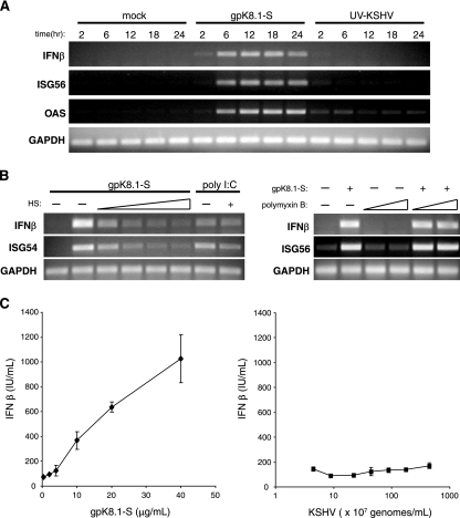
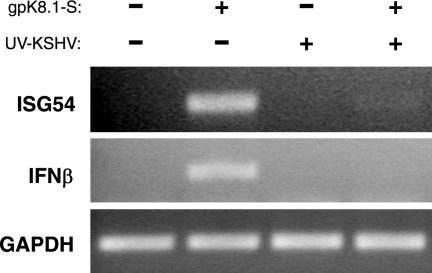
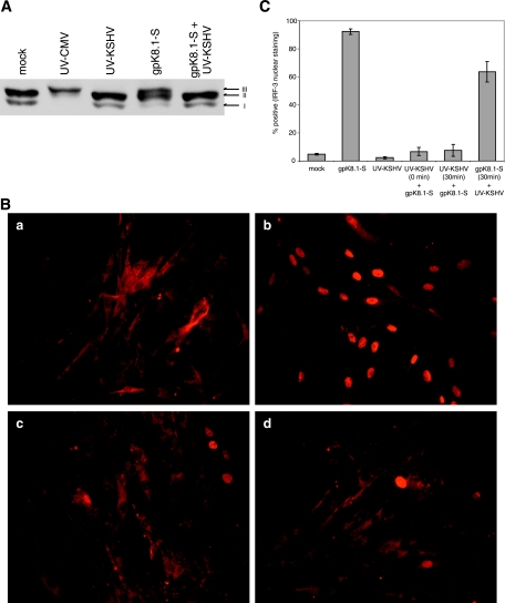
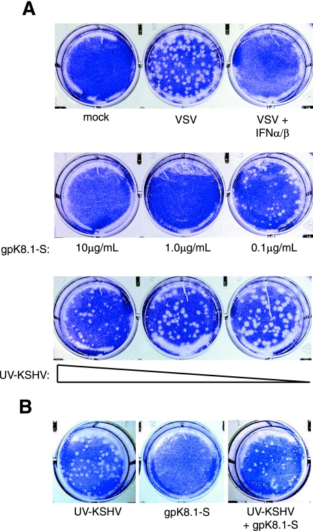
The Kaposi's sarcoma-associated herpesvirus (KSHV) envelope glycoprotein gpK8.1 contributes to cellular attachment through binding cell surface heparan sulfate proteoglycans. By using a soluble recombinant form of gpK8.1, we discovered that a consequence of gpK8.1 interaction with human fibroblasts is the induction of an antiviral response, as characterized by the activation of interferon regulatory factor 3 (IRF-3), production of interferon beta (IFN-beta), and expression of interferon-stimulated antiviral genes. In contrast, neither IFN-beta expression nor a functional antiviral response is observed in cells treated with KSHV virions. The interferon response induced by soluble gpK8.1 can be inhibited by simultaneous treatment with UV-inactivated virions, while the induction of an indicator inflammatory cytokine, interleukin-6, was readily evident in the response to both gpK8.1 and KSHV. In addition, KSHV virions abrogate gpK8.1-mediated activation of IRF-3, an early transcriptional regulator for cellular antiviral responses. Although innate immune responses are initiated during contact between gpK8.1 and cellular receptor(s), these results suggest that the virion contains one or more structural elements that selectively repress an effective antiviral response while allowing cellular responses favorable to the KSHV life cycle.
Figures


Similar articles
-
A Rhesus Rhadinovirus Viral Interferon (IFN) Regulatory Factor Is Virion Associated and Inhibits the Early IFN Antiviral Response.J Virol. 2015 Aug;89(15):7707-21. doi: 10.1128/JVI.01175-15. Epub 2015 May 13. J Virol. 2015. PMID: 25972548 Free PMC article.
-
Binding of Kaposi's sarcoma-associated herpesvirus K-bZIP to interferon-responsive factor 3 elements modulates antiviral gene expression.J Virol. 2007 Oct;81(20):10950-60. doi: 10.1128/JVI.00183-07. Epub 2007 Jul 25. J Virol. 2007. PMID: 17652396 Free PMC article.
-
Human herpesvirus 8 envelope glycoprotein K8.1A interaction with the target cells involves heparan sulfate.J Virol. 2001 Aug;75(16):7517-27. doi: 10.1128/JVI.75.16.7517-7527.2001. J Virol. 2001. PMID: 11462024 Free PMC article.
-
Molecular virology of Kaposi's sarcoma-associated herpesvirus.Philos Trans R Soc Lond B Biol Sci. 2001 Apr 29;356(1408):499-516. doi: 10.1098/rstb.2000.0777. Philos Trans R Soc Lond B Biol Sci. 2001. PMID: 11313008 Free PMC article. Review.
-
On the role of IRF in host defense.J Interferon Cytokine Res. 2002 Jan;22(1):59-71. doi: 10.1089/107999002753452665. J Interferon Cytokine Res. 2002. PMID: 11846976 Review.
Cited by
-
Hepatitis C virus infection induces the beta interferon signaling pathway in immortalized human hepatocytes.J Virol. 2007 Nov;81(22):12375-81. doi: 10.1128/JVI.01695-07. Epub 2007 Sep 5. J Virol. 2007. PMID: 17804510 Free PMC article.
-
Kaposi sarcoma-associated herpesvirus degrades cellular Toll-interleukin-1 receptor domain-containing adaptor-inducing beta-interferon (TRIF).J Biol Chem. 2011 Mar 11;286(10):7865-7872. doi: 10.1074/jbc.M110.191452. Epub 2011 Jan 6. J Biol Chem. 2011. PMID: 21212282 Free PMC article.
-
Kaposi sarcoma-associated herpesvirus latency-associated nuclear antigen inhibits interferon (IFN) beta expression by competing with IFN regulatory factor-3 for binding to IFNB promoter.J Biol Chem. 2010 Mar 5;285(10):7208-21. doi: 10.1074/jbc.M109.018838. Epub 2010 Jan 4. J Biol Chem. 2010. PMID: 20048166 Free PMC article.
-
Mechanisms of Kaposi's Sarcoma-Associated Herpesvirus Latency and Reactivation.Adv Virol. 2011;2011:193860. doi: 10.1155/2011/193860. Adv Virol. 2011. PMID: 21625290 Free PMC article.
-
Evasion and subversion of interferon-mediated antiviral immunity by Kaposi's sarcoma-associated herpesvirus: an overview.J Virol. 2011 Nov;85(21):10934-44. doi: 10.1128/JVI.00687-11. Epub 2011 Jul 20. J Virol. 2011. PMID: 21775463 Free PMC article. Review.
References
-
- Ankel, H., M. R. Capobianchi, C. Castilletti, and F. Dianzani. 1994. Interferon induction by HIV glycoprotein 120: role of the V3 loop. Virology 205:34-43. - PubMed
-
- Ankel, H., D. F. Westra, S. Welling-Wester, and P. Lebon. 1998. Induction of interferon-alpha by glycoprotein D of herpes simplex virus: a possible role of chemokine receptors. Virology 251:317-326. - PubMed
-
- Asou, H., J. W. Said, R. Yang, R. Munker, D. J. Park, N. Kamada, and H. P. Koeffler. 1998. Mechanisms of growth control of Kaposi's sarcoma-associated herpes virus-associated primary effusion lymphoma cells. Blood 91:2475-2481. - PubMed
Publication types
MeSH terms
Substances
Grants and funding
LinkOut - more resources
Full Text Sources
