. 2006 Sep;169(3):987-98.
doi: 10.2353/ajpath.2006.060180.

B and T lymphocytes are the primary sources of RANKL in the bone resorptive lesion of periodontal disease

Affiliations

B and T lymphocytes are the primary sources of RANKL in the bone resorptive lesion of periodontal disease

Toshihisa Kawai et al. Am J Pathol. 2006 Sep.

Abstract

Receptor activator of nuclear factor-kappaB (RANKL)-mediated osteoclastogenesis plays a pivotal role in inflammatory bone resorption. The aim of this study was to identify the cellular source of RANKL in the bone resorptive lesions of periodontal disease. The concentrations of soluble RANKL, but not its decoy receptor osteoprotegerin, measured in diseased tissue homogenates were significantly higher in diseased gingival tissues than in healthy tissues. Double-color confocal microscopic analyses demonstrated less than 20% of both B cells and T cells expressing RANKL in healthy gingival tissues. By contrast, in the abundant mononuclear cells composed of 45% T cells, 50% B cells, and 5% monocytes in diseased gingival tissues, more than 50 and 90% of T cells and B cells, respectively, expressed RANKL. RANKL production by nonlymphoid cells was not distinctly identified. Lymphocytes isolated from gingival tissues of patients induced differentiation of mature osteoclast cells in a RANKL-dependent manner in vitro. However, similarly isolated peripheral blood B and T cells did not induce osteoclast differentiation, unless they were activated in vitro to express RANKL; emphasizing the osteoclastogenic potential of activated RANKL-expressing lymphocytes in periodontal disease tissue. These results suggest that activated T and B cells can be the cellular source of RANKL for bone resorption in periodontal diseased gingival tissue.


Figures
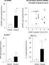
Figure 1
Figure 1
Protein concentrations of RANKL, OPG, and IL-1β in gingival tissues. Protein concentrations of RANKL and OPG in gingival tissues were measured by ELISA. Gingival tissue samples were homogenized in the presence of proteinase inhibitors (healthy, n = 3; disease, n = 11). The concentration of each protein, RANKL (A and B), OPG (C), and IL-1β (D), is expressed as pg per mg of gingival tissue (pg/mg). *Significantly higher than healthy control by Student’s t-test (P < 0.05). B: The concentrations of RANKL were positively correlated with the gingival pocket depth where the biopsy was sampled. **Positive correlation (n = 14, P < 0.01).
Figure 2
Figure 2
RANKL and OPG mRNA expression in gingival tissues. RT-PCR was performed to detect mRNA expression of RANKL and OPG in the total RNA extracted from whole gingival tissue samples. Expression of RANKL mRNA was detected in 67% of diseased periodontal tissues (solid bars, four of six), whereas no expression of RANKL mRNA was observed in the healthy gingival tissues tested (open bars, zero of four), whereas OPG mRNA message was observed from healthy patients (one of four) and from periodontal disease tissues (two of six). The relative intensity of each mRNA message in the acrylamide gel was scanned by densitometer using the AlphaImage analysis system and expressed as relative signal intensity (RSI) in the histograms (B, RANKL; C, OPG).
Figure 3
Figure 3
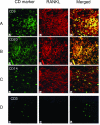
Confocal microscopic analyses of expression of RANKL in gingival tissues. Using double-color confocal microscopy, RANKL-expressing cells were identified in the gingival tissue of a periodontal disease patient (AC) and in healthy gingival tissue (D). CD3 (A and D), CD20 (B), or CD14 (C) expression was indicated by staining with various specific mouse mAbs followed by fluorescein isothiocyanate anti-mouse IgG (left column). RANKL expression was detected by OPG-biotin followed by streptavidin-Texas Red (middle column). Both specific CD marker-stained cells and RANKL+-stained cells were doubly exposed and expressed as yellow (merged; right column). CD20+ and CD14+ cells in healthy tissue (not shown) were as few as CD3+ cells in healthy gingival tissue (D). Scale bars = 20 μm.
Figure 4
Figure 4
Numbers of lymphocytes in the gingival tissues. The total number of CD3+, CD20+, and CD14+ cells in a confocal microscopic field of each tissue section was counted (×400, average cell number of at least three sections was enumerated as the number of cells per sample) and compared between healthy gingival tissues (open column) and disease gingival tissues (filled column). Mean cell number ± SD is shown. *Significantly higher than healthy gingival tissue by Student’s t-test (P < 0.01).
Figure 5
Figure 5
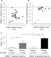
Expression of percentage of RANKL+ T cells or B cells in the gingival tissues. After computer capture of the confocal microscopic image of each gingival section (×400), the percentage of yellow cells (double positive for CD3/RANKL or CD20/RANKL) of the total number of green cells (CD3+ or CD20+) were calculated and are shown on scatter plots (A, CD3+ T cells; B, CD20+ B cells), according to the depth of gingival crevice from which the samples were collected (open triangles, healthy patients; filled triangles, periodontal disease patients). Both RANKL+ T cells and RANKL+ B cells showed positive correlation with the depth of gingival crevice (T cells: n = 34, r = 0.507, P < 0.01; B cells: n = 15, r = 0.702, P < 0.01). C: Percentage of RANKL+ T cells or B cells of the total T cells or B cells in the gingival tissues classified by the depth of the gingival crevice (GC depth) is shown. The data shown in A and B were converted to histograms, presented as mean ± SD of the percentage of RANKL+ T cells or B cells. *Statistically significant by Student’s t-test (P < 0.0001). Note: the percentage of RANKL+ CD14+ monocytes/total CD14+ monocytes (not shown) was 15.3 ± 16.8 and 59.5 ± 39.9 in healthy and disease gingival tissues, respectively, showing no statistical differences by t-test.
Figure 6
Figure 6
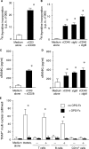
Activation of peripheral blood T cells or B cells induces sRANKL expression. Purified T cells isolated from a healthy subject PBMCs (2 × 10⁵/well) were activated for 7 days with immobilized anti-CD3 (×CD3) and anti-CD28 (×CD28) mAbs, and proliferation (A) and sRANKL production (C) were measured by [3H]thymidine incorporation assay and by ELISA, respectively. Purified B cells isolated from the PBMCs (2 × 10⁵/well) were also incubated in a 96-well plate for 7 days with immobilized anti-CD40 (×CD40), anti-IgM (×IgM), and anti-CD40 and anti-IgM antibodies, and proliferation (B) and sRANKL production (D) were measured by the same method as for T cells. E: Osteoclastogenesis induced by the activated peripheral blood T cells, B cells, and CD14+ monocytes is shown. The cultured T cells or B cells under the conditions shown in A or B were harvested and fixed on day 7. After activation of these CD14+ monocytes with fixed A. actinomycetemcomitans for 4 days, CD14+ monocytes produced IL-12 (143 + 23 pg/ml), but not RANKL or OPG. The fixed T cells, B cells, or CD14+ monocytes were co-cultured with MOCP-5 (104/well) in the presence of M-CSF (10 ng/ml) as described under Materials and Methods. The number of TRAP-positive multinuclear cells induced in each well was counted and expressed as mean ± SD of triplicate determinants. *Significantly higher than medium alone by Student’s t-test (P < 0.05). Other PBMC samples isolated from one periodontal diseased and two orally healthy patients showed similar results.
Figure 7
Figure 7
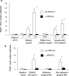
Osteoclast differentiation assay using the MOCP-5 osteoclast precursor cell line or human peripheral blood CD14+ monocytes. A: MOCP-5 osteoclast precursor cells were co-cultured with human recombinant RANKL, fresh nonstimulated PBMCs, or gingival MCs isolated from periodontal disease patients. PBMCs and gingival MCs were prefixed with formalin before co-culture with MOCP-5 (104/well). B: CD14+ monocytes isolated from PBMCs of a healthy subject (2 × 10⁴/well) were co-cultured with human recombinant RANKL or with fixed adherent or nonadherent gingival MCs isolated from periodontal disease patient. All culture medium contained recombinant human M-CSF (10 ng/ml) in the presence or absence of OPG-Fc (10 μg/ml) (both A and B). Half of the total culture medium was exchanged every 3 days. On day 8 (A) or on day 16 (B), the cultures were fixed with formalin, and TRAP staining was outperformed. The numbers of cells with more than three nuclei demonstrating TRAP+ staining were counted in each well under phase contrast microscopy. Similar results of TRAP+ cell induction were obtained using the MCs isolated from two different patients’ gingival tissues. *Significantly elevated compared to the control MOCP-5 (A); significantly elevated compared to human CD14+ monocytes cultured in medium alone without OPG-Fc (B) by Student’s t-test (P < 0.01).
Figure 8
Figure 8
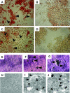
TRAP+ multinuclear cells induced from MOCP-5 osteoclast precursors by recombinant RANKL and patient’s gingival MCs. MOCP-5 osteoclast precursor cells were co-cultured with human recombinant RANKL or with gingival MCs isolated from the patient’s periodontal disease lesion. Patient’s gingival MCs fixed with formalin (104/well) were applied to the MOCP-5 culture. MOCP-5 cells were cultured on the oval-shape coverslip in a 96-well plate. Medium that contained M-CSF (10 ng/ml) was exchanged (50% of total culture volume) every 3 days. On day 8, the cultures were stopped, and the cells were fixed with formalin. The MOCP-5 cells adherent on the coverslip were reacted with TRAP reagent and mounted on glass slides. The figures demonstrate the appearance of TRAP staining of MOCP-5 co-cultured for 8 days with recombinant human RANKL (50 ng/ml) (A), recombinant human RANKL plus OPG-Fc (10 μg/ml) (B), fixed patient’s gingival MCs isolated from a diseased lesion (C), and fixed patient’s gingival MCs isolated from a diseased lesion plus OPG-Fc (10 μg/ml) (D). The bone-resorptive activities of TRAP+ multinuclear cells were examined using dentin disks (E–G) and the Osteologic system (H–J). The MOCP-5 was cultured in a medium supplemented with M-CSF (10 ng/ml) for 8 days in the following different conditions; medium alone (E, H), in the presence of recombinant RANKL (50 ng/ml) (F, I), in the presence of patient’s fixed gingival MCs (G, J). The arrows in A and C indicate the TRAP+ multinuclear cells. The arrows in E–J indicate the resorption pits formed by TRAP+ multinuclear cells. Of note, toluidine blue stains resportion pits dark brown (F, G), while the resorption pits in the Osteologic system are demonstrated as white clear areas (I, J).

Similar articles

Cited by

References

    1. McArthur WP, Clark WB. Specific antibodies and their potential role in periodontal diseases. J Periodontol. 1993;64:807–818. - PubMed
    1. Taubman MA, Ebersole JL, Smith DJ. Genco RJ, Mergenhangen SE, editors. Washington DC: American Society of Microbiology,; Association between systemic and local antibody and periodontal diseases. Host Parasite Interactions in Periodontal Diseases. 1982:pp 283–298.
    1. Ebersole JL, Taubman MA, Smith DJ, Frey DE, Haffajee AD, Socransky SS. Human serum antibody responses to oral microorganisms. IV. Correlation with homologous infection. Oral Microbiol Immunol. 1987;2:53–59. - PubMed
    1. Wu X, Pan G, McKenna MA, Zayzafoon M, Xiong WC, McDonald JM. RANKL regulates Fas expression and Fas-mediated apoptosis in osteoclasts. J Bone Miner Res. 2005;20:107–116. - PubMed
    1. Hofbauer LC, Khosla S, Dunstan CR, Lacey DL, Boyle WJ, Riggs BL. The roles of osteoprotegerin and osteoprotegerin ligand in the paracrine regulation of bone resorption. J Bone Miner Res. 2000;15:2–12. - PubMed

Publication types

MeSH terms

LinkOut - more resources