Nell-1-induced bone regeneration in calvarial defects
- PMID: 16936265
- PMCID: PMC1698834
- DOI: 10.2353/ajpath.2006.051210
Nell-1-induced bone regeneration in calvarial defects
Abstract
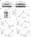
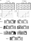
Many craniofacial birth defects contain skeletal components requiring bone grafting. We previously identified the novel secreted osteogenic molecule NELL-1, first noted to be overexpressed during premature bone formation in calvarial sutures of craniosynostosis patients. Nell-1 overexpression significantly increases differentiation and mineralization selectively in osteoblasts, while newborn Nell-1 transgenic mice significantly increase premature bone formation in calvarial sutures. In the current study, cultured calvarial explants isolated from Nell-1 transgenic newborn mice (with mild sagittal synostosis) demonstrated continuous bone growth and overlapping sagittal sutures. Further investigation into gene expression cascades revealed that fibroblast growth factor-2 and transforming growth factor-beta1 stimulated Nell-1 expression, whereas bone morphogenetic protein (BMP)-2 had no direct effect. Additionally, Nell-1-induced osteogenesis in MC3T3-E1 osteoblasts through reduction in the expression of early up-regulated osteogenic regulators (OSX and ALP) but induction of later markers (OPN and OCN). Grafting Nell-1 protein-coated PLGA scaffolds into rat calvarial defects revealed the osteogenic potential of Nell-1 to induce bone regeneration equivalent to BMP-2, whereas immunohistochemistry indicated that Nell-1 reduced osterix-producing cells and increased bone sialoprotein, osteocalcin, and BMP-7 expression. Insights into Nell-1-regulated osteogenesis coupled with its ability to stimulate bone regeneration revealed a potential therapeutic role and an alternative to the currently accepted techniques for bone regeneration.
Figures


Similar articles
-
Synergistic effects of Nell-1 and BMP-2 on the osteogenic differentiation of myoblasts.J Bone Miner Res. 2007 Jun;22(6):918-30. doi: 10.1359/jbmr.070312. J Bone Miner Res. 2007. PMID: 17352654 Free PMC article.
-
Nell-1, a key functional mediator of Runx2, partially rescues calvarial defects in Runx2(+/-) mice.J Bone Miner Res. 2011 Apr;26(4):777-91. doi: 10.1002/jbmr.267. J Bone Miner Res. 2011. PMID: 20939017 Free PMC article.
-
Craniosynostosis in transgenic mice overexpressing Nell-1.J Clin Invest. 2002 Sep;110(6):861-70. doi: 10.1172/JCI15375. J Clin Invest. 2002. PMID: 12235118 Free PMC article.
-
The role of NELL-1, a growth factor associated with craniosynostosis, in promoting bone regeneration.J Dent Res. 2010 Sep;89(9):865-78. doi: 10.1177/0022034510376401. Epub 2010 Jul 20. J Dent Res. 2010. PMID: 20647499 Free PMC article. Review.
-
Regulation of human cranial osteoblast phenotype by FGF-2, FGFR-2 and BMP-2 signaling.Histol Histopathol. 2002;17(3):877-85. doi: 10.14670/HH-17.877. Histol Histopathol. 2002. PMID: 12168799 Review.
Cited by
-
Pharmacokinetics and osteogenic potential of PEGylated NELL-1 in vivo after systemic administration.Biomaterials. 2015 Jul;57:73-83. doi: 10.1016/j.biomaterials.2015.03.063. Epub 2015 Apr 24. Biomaterials. 2015. PMID: 25913252 Free PMC article.
-
BMP2-induced inflammation can be suppressed by the osteoinductive growth factor NELL-1.Tissue Eng Part A. 2013 Nov;19(21-22):2390-401. doi: 10.1089/ten.TEA.2012.0519. Epub 2013 Jul 17. Tissue Eng Part A. 2013. PMID: 23758588 Free PMC article.
-
Efficacy of Intraperitoneal Administration of PEGylated NELL-1 for Bone Formation.Biores Open Access. 2016 Jun 1;5(1):159-70. doi: 10.1089/biores.2016.0018. eCollection 2016. Biores Open Access. 2016. PMID: 27354930 Free PMC article.
-
Delivery of lyophilized Nell-1 in a rat spinal fusion model.Tissue Eng Part A. 2010 Sep;16(9):2861-70. doi: 10.1089/ten.tea.2009.0550. Tissue Eng Part A. 2010. PMID: 20528102 Free PMC article.
-
Oligomerization-induced conformational change in the C-terminal region of Nel-like molecule 1 (NELL1) protein is necessary for the efficient mediation of murine MC3T3-E1 cell adhesion and spreading.J Biol Chem. 2014 Apr 4;289(14):9781-94. doi: 10.1074/jbc.M113.507020. Epub 2014 Feb 21. J Biol Chem. 2014. PMID: 24563467 Free PMC article.
References
-
- Canady JW, Zeitler DP, Thompson SA, Nicholas CD. Suitability of the iliac crest as a site for harvest of autogenous bone grafts. Cleft Palate Craniofac J. 1993;30:579–581. - PubMed
-
- Schlegel KA, Donath K, Rupprecht S, Falk S, Zimmermann R, Felszeghy E, Wiltfang J. De novo bone formation using bovine collagen and platelet-rich plasma. Biomaterials. 2004;25:5387–5393. - PubMed
-
- Kang Q, Sun MH, Cheng H, Peng Y, Montag AG, Deyrup AT, Jiang W, Luu HH, Luo J, Szatkowski JP, Vanichakarn P, Park JY, Li Y, Haydon RC, He TC. Characterization of the distinct orthotopic bone-forming activity of 14 BMPs using recombinant adenovirus-mediated gene delivery. Gene Ther. 2004;11:1312–1320. - PubMed
-
- Boden SD, Kang J, Sandhu H, Heller JG. Use of recombinant human bone morphogenetic protein-2 to achieve posterolateral lumbar spine fusion in humans: a prospective, randomized clinical pilot trial: 2002 Volvo Award in clinical studies. Spine. 2002;27:2662–2673. - PubMed
-
- Valentin-Opran A, Wozney J, Csimma C, Lilly L, Riedel GE. Clinical evaluation of recombinant human bone morphogenetic protein-2. Clin Orthop. 2002;1:110–120. - PubMed
Publication types
MeSH terms
Substances
Grants and funding
LinkOut - more resources
Full Text Sources
Other Literature Sources
Molecular Biology Databases
Research Materials

