Evidence for stroke-induced neurogenesis in the human brain
- PMID: 16924107
- PMCID: PMC1559776
- DOI: 10.1073/pnas.0603512103
Evidence for stroke-induced neurogenesis in the human brain
Abstract
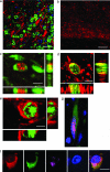
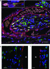
Experimental stroke in rodents stimulates neurogenesis and migration of newborn neurons from their sites of origin into ischemic brain regions. We report that in patients with stroke, cells that express markers associated with newborn neurons are present in the ischemic penumbra surrounding cerebral cortical infarcts, where these cells are preferentially localized in the vicinity of blood vessels. These findings suggest that stroke-induced compensatory neurogenesis may occur in the human brain, where it could contribute to postischemic recovery and represent a target for stroke therapy.
Conflict of interest statement
Conflict of interest statement: No conflicts declared.
Figures


Similar articles
-
Aging increases microglial proliferation, delays cell migration, and decreases cortical neurogenesis after focal cerebral ischemia.J Neuroinflammation. 2015 May 10;12:87. doi: 10.1186/s12974-015-0314-8. J Neuroinflammation. 2015. PMID: 25958332 Free PMC article.
-
Temporal profile of neurogenesis in the subventricular zone, dentate gyrus and cerebral cortex following transient focal cerebral ischemia.Neurol Res. 2009 Nov;31(9):969-76. doi: 10.1179/174313209X383312. Epub 2009 Jan 9. Neurol Res. 2009. PMID: 19138475
-
Immunodeficiency reduces neural stem/progenitor cell apoptosis and enhances neurogenesis in the cerebral cortex after stroke.J Neurosci Res. 2010 Aug 15;88(11):2385-97. doi: 10.1002/jnr.22410. J Neurosci Res. 2010. PMID: 20623538
-
Neurogenesis following Stroke Affecting the Adult Brain.Cold Spring Harb Perspect Biol. 2015 Nov 2;7(11):a019034. doi: 10.1101/cshperspect.a019034. Cold Spring Harb Perspect Biol. 2015. PMID: 26525150 Free PMC article. Review.
-
[Treatment of brain ischemic stroke by co-transplantation of neural stem cells and endothelial progenitor cells].Zhongguo Xiu Fu Chong Jian Wai Ke Za Zhi. 2007 Feb;21(2):204-8. Zhongguo Xiu Fu Chong Jian Wai Ke Za Zhi. 2007. PMID: 17357472 Review. Chinese.
Cited by
-
Exosomes in stroke pathogenesis and therapy.J Clin Invest. 2016 Apr 1;126(4):1190-7. doi: 10.1172/JCI81133. Epub 2016 Apr 1. J Clin Invest. 2016. PMID: 27035810 Free PMC article. Review.
-
Function of neural stem cells in ischemic brain repair processes.J Cereb Blood Flow Metab. 2016 Dec;36(12):2034-2043. doi: 10.1177/0271678X16674487. Epub 2016 Oct 14. J Cereb Blood Flow Metab. 2016. PMID: 27742890 Free PMC article. Review.
-
Single-Cell Transcriptome Reveals Cell Type-Specific Molecular Pathology in a 2VO Cerebral Ischemic Mouse Model.Mol Neurobiol. 2024 Aug;61(8):5248-5264. doi: 10.1007/s12035-023-03755-4. Epub 2024 Jan 5. Mol Neurobiol. 2024. PMID: 38180614 Free PMC article.
-
Post-stroke remodeling processes in animal models and humans.J Cereb Blood Flow Metab. 2020 Jan;40(1):3-22. doi: 10.1177/0271678X19882788. Epub 2019 Oct 23. J Cereb Blood Flow Metab. 2020. PMID: 31645178 Free PMC article. Review.
-
Transcriptional regulation of neurogenesis: potential mechanisms in cerebral ischemia.J Mol Med (Berl). 2007 Jun;85(6):577-88. doi: 10.1007/s00109-007-0196-z. Epub 2007 Apr 11. J Mol Med (Berl). 2007. PMID: 17429598 Review.
References
-
- Eriksson P. S., Perfilieva E., Bjork-Eriksson T., Alborn A. M., Nordborg C., Peterson D. A., Gage F. H. Nat. Med. 1998;4:1313–1317. - PubMed
-
- Gu W., Brannstrom T., Wester P. J. Cereb. Blood Flow Metab. 2000;20:1166–1173. - PubMed
-
- Zhang R. L., Zhang Z. G., Zhang L., Chopp M. Neuroscience. 2001;105:33–41. - PubMed
Publication types
MeSH terms
Grants and funding
LinkOut - more resources
Full Text Sources
Other Literature Sources
Medical
Miscellaneous

