. 2006 Oct;26(20):7791-805.
doi: 10.1128/MCB.00022-06. Epub 2006 Aug 21.

Different routes of bone morphogenic protein (BMP) receptor endocytosis influence BMP signaling

Affiliations

Different routes of bone morphogenic protein (BMP) receptor endocytosis influence BMP signaling

Anke Hartung et al. Mol Cell Biol. 2006 Oct.

Abstract

Endocytosis is important for a variety of functions in eukaryotic cells, including the regulation of signaling cascades via transmembrane receptors. The internalization of bone morphogenetic protein (BMP) receptor type I (BRI) and type II (BRII) and its relation to signaling were largely unexplored. Here, we demonstrate that both receptor types undergo constitutive endocytosis via clathrin-coated pits (CCPs) but that only BRII undergoes also caveola-like internalization. Using several complementary approaches, we could show that (i) BMP-2-mediated Smad1/5 phosphorylation occurs at the plasma membrane in nonraft regions, (ii) continuation of Smad signaling resulting in a transcriptional response requires endocytosis via the clathrin-mediated route, and (iii) BMP signaling leading to alkaline phosphatase induction initiates from receptors that fractionate into cholesterol-enriched, detergent-resistant membranes. Furthermore, we show that BRII interacts with Eps15R, a constitutive component of CCPs, and with caveolin-1, the marker protein of caveolae. Taken together, the localization of BMP receptors in distinct membrane domains is prerequisite to their taking different endocytosis routes with specific impacts on Smad-dependent and Smad-independent signaling cascades.


Figures
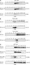
FIG. 1.
FIG. 1.
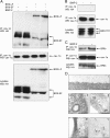
BMP receptors float with DRM domains. BRIa, BRIb, and BRII distribution was studied in 293T (A and D) and C2C12 (B and C) cells. CHAPS lysates were subjected to Optiprep gradient fractionation and analyzed by Western blotting with anti-cav-1 or anti-HA antibodies. (A) BRIa, analyzed in 293T cells transfected with HA-BRIa and HA-cav-1α, is detected in DRM fractions. The distribution of BRIa was not altered when cav-1α was not transfected (bottom panel). (B) In C2C12 cells stably expressing HA-BRIb, the receptor cofractionates with endogenous cav-1α in DRM fractions. (C) C2C12 cells stably expressing HA-BRII-LF or (D) 293T cells transiently transfected with HA-BRII-LF and HA-cav-1α show a wide receptor distribution. Western blotting with IL-2Rβ-specific antibody verifies fraction 6 as the main DRM fraction. The distribution of BRII was not altered when no cav-1α was transfected (bottom panel). WB, Western blotting.
FIG. 2.
FIG. 2.
BMP receptors interact and colocalize with endogenous cav-1α. cav-1α was immunoprecipitated from C2C12 cells stably expressing HA-tagged BRII-LF, BRII-SF, BRII-TC1, or BRIb (A to C). (A) BRII-LF and BRII-SF interact with cav-1α. BMP-2 stimulation shows no effect. (B) BRII-TC1 is not associated with cav-1α. (C) BRIb interacts with cav-1α, which is unaffected by BMP-2 stimulation. (D) Immunogold electron microscopy analysis of BRII in C2C12 cells stably expressing HA-BRII-LF. BRII-LF was labeled as described in Materials and Methods, using secondary antibodies conjugated with 12-nm colloidal gold. For images I and II, cells were treated with antibodies before fixation and embedding. BRII-LF is observed at the cell surface (I) or in vesicles, which were identified as CCV (II). For images III to V, cell pellets were fixed and embedded before antibody application. BRII-LF is located in clusters of 50 to 75 nm in diameter. In images IV and V, sections were additionally incubated with an antibody which recognizes endogenous cav-1α, followed by secondary antibody conjugated with 6-nm colloidal gold. BRII-LF (arrows) is located in clusters together with cav-1α (arrowheads), indicating the localization of BRII in caveolae/caveosomes. Bar, 100 nm. IP, immunoprecipitation; WB, Western blotting.
FIG. 3.
FIG. 3.
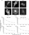
Quantitative measurement of BMP receptor endocytosis. COS7 cells (A to D) or C2C12 cells (E) were transiently transfected with HA-BRIb (A to C) or myc-BRII-LF (D and E). After 48 h, live cells were labeled at 4°C with anti-HA or anti-myc IgG followed by Alexa 546 goat anti-mouse Fab′. After being washed, cells were incubated either at 4°C (time zero) or for the indicated periods at 37°C, shifted back to 4°C, and fixed. (A) Typical images of HA-BRIb in COS7 cells during endocytosis. As a control (A, lower rightmost panel), cells incubated at 37°C for 30 min were acid stripped to remove noninternalized fluorescent Fab′. Bar, 20 μm. (B to E) Quantitative measurements of BRIb and BRII-LF internalization. Measurements were done using the point confocal method (see Materials and Methods). (C) Control experiment where the cells were fixed with 4% paraformaldehyde in phosphate-buffered saline and quenched by three incubations with 50 mM glycine in phosphate-buffered saline prior to the labeling with antibodies, thus eliminating endocytosis and enabling the evaluation of the dissociation of the fluorescent antibodies from the surface during the incubation at 37°C (no significant dissociation occurred). The laser beam was focused on defined spots in the plasma membrane focal plane, away from vesicular staining (typical spots where the beam was focused are shown as white circles in the panel marked 20 min in panel A). The results shown at each time point are means ± standard errors of the means (SEM) of measurements for 80 to 150 cells. Addition of ligand (50 nM BMP-2; 1 h of preincubation at 4°C, keeping the ligand in during all subsequent incubations with antibodies and warming to 37°C) had no significant effects on the endocytosis rates of either receptor (data not shown).
FIG. 4.
FIG. 4.
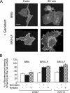
Influence of inhibitors of caveola-like endocytosis on the internalization of BMP receptors. COS7 or C2C12 cells were transiently transfected with HA-BRIb or myc-BRII-LF as described for Fig. 3. After 48 h, they were incubated with nystatin (25 μg/ml, 30 min) or genistein (200 μM, 60 min) at 37°C. The surface receptors were then labeled at 4°C with fluorescent antibodies as for Fig. 3, followed by a 20-min incubation at 37°C or 4°C (control) in media containing inhibitors. Cell surface receptor levels were quantified by the point confocal method. (A) Typical images (COS7 cells) showing inhibition by genistein of the shift in HA-BRIb and myc-BRII-LF to vesicular endocytic patterns. Analogous results (not shown) were obtained with C2C12 cells or by using nystatin in place of genistein. Bar, 20 μm. (B) Quantification of the internalization studies. The reduction in cell surface labeling due to internalization was quantified by the point confocal method as described for Fig. 3. The fluorescence intensity in identically treated cells at time zero was taken as 100% for each treatment individually to eliminate the contribution of potential alterations in the steady-state surface levels of the receptors in the presence of the inhibitors. The results are means ± SEM of measurements of 80 to 140 cells in each case. In a comparison of the results from pairs of treated versus untreated cells for each group, neither genistein nor nystatin had significant effects on HA-BRIb internalization (P > 0.05, Student's t test); similar results (not shown) were obtained for HA-BRIb in C2C12 cells. However, both treatments had significant inhibitory effects on myc-BRII-LF internalization either in COS7 cells (P < 10−12 for genistein and P < 10−15 for nystatin) or in C2C12 cells (P < 10−12 and P < 10−17 for genistein and nystatin, respectively).
FIG. 5.
FIG. 5.
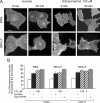
Treatments that disrupt CCP-mediated endocytosis inhibit the internalization of BMP receptors. COS7 or C2C12 cells were transfected with HA-BRIb or myc-BRII-LF as described for Fig. 3. In some experiments, the cells were cotransfected with a sixfold excess of dyn2-K44A along with the BMP receptor construct. Hypertonic treatment, cytosol acidification, and treatment with CP were employed as described in the text. (A) Typical images of the effects of hypertonic treatment (sucrose) or CP (100 μM, 15 min) on the endocytosis of HA-BRIb and myc-BRII-LF in COS7 cells. Similar levels of inhibition were observed following cytosol acidification (data not shown), as well as in cells cotransfected with dyn2-K44A and in C2C12 cells (see panel B). Bar, 20 μm. (B) Quantification by the point confocal method. The fluorescence intensity measured at time zero on similarly treated cells was taken as 100% for each specific treatment. Bars represent means ± SEM of measurements conducted on 80 to 140 cells in each case. A comparison of pairs on treated versus untreated cells demonstrates highly significant effects of all the treatments on both COS7 and C2C12 cells (P < 10−12 and in some cases P < 10−25). Analogous results (not shown) were obtained for BRIb in C2C12 cells.
FIG. 6.
FIG. 6.
Influence of cholesterol-enriched plasma membrane regions on BMP signaling pathways. C2C12 cells were treated with 50 μM lovastatin and 50 μM mevalonate and stimulated with BMP-2 as described in the text. (A) Western blot (WB) analyses using an anti-phospho- Smad1/5 (P-Smad1/5) antibody show that Smad1/5 phosphorylation is not altered by LM. For a loading control, the membrane was incubated with anti-β-actin antibody. (B) C2C12 cells were transfected with p(BRE)4-luc and pRLTK before metabolic inhibition using LM was accomplished. BMP signaling is only slightly reduced by LM. Error bars represent standard deviations from three different assays. (C) RT-PCR of ALP expression levels shown as the level of induction (n-fold) upon BMP-2 stimulation; cholesterol depletion by LM results in a reduction of the ALP transcript by 80%. Error bars represent standard deviations (SD) from duplicates. The experiment was done two times. (D) ALP activity measurements after cholesterol depletion by LM (this experiment was performed in parallel with the experiment whose results are shown in panel C) show reduction by 75%. Error bars represent SD from two independent experiments, each with triplicates. w/o, without.
FIG. 7.
FIG. 7.
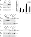
Effect of cav-1α, cav-1-siRNA, or wt dyn2 and -K44A expression on BMP signaling. (A) C2C12 cells were transfected with HA-tagged cav-1α and stimulated with 20 nM BMP-2 for 30 min. Western blot analyses using anti-P-Smad1/5-specific antibodies reveal no significant change in Smad1/5 phosphorylation. For a loading control, the same membrane was incubated with anti-β-actin antibody. (B) Cells were transiently transfected with cav-1α, BRII-LF, and BRIa as indicated, together with reporter constructs. cav-1α expression significantly reduced both ligand-independent and -dependent BMP signaling. Error bars represent standard deviations from three different assays. (C) cav-1-specific siRNA was transfected in C2C12 cells to knock down endogenous cav-1 expression. A P-Smad1/5 Western blot shows no influence of cav-1 downregulation on the phosphorylation level. (D) Expression of HA-tagged wt dyn2 and the dyn-K44A mutant shows no significant alteration in the level of phosphorylated Smad1/5. Controls confirm wt dyn2, dyn2-K44A, and β-actin expression. WB, Western blotting; w/o, without.
FIG. 8.
FIG. 8.
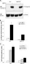
Influence of CP on BMP signaling. C2C12 cells were treated with 5 μM CP and stimulated with BMP-2 as described in the text. (A) Western blot analyses with anti-P-Smad1/5 antibody show that the level of phosphorylated Smad1/5 was not altered by CP treatment. The loading control is shown by anti-β-actin antibody (lower panel). (B) C2C12 cells were transfected with p(BRE)4-luc and pRLTK and treated with CP. This results in a 65%-reduced BMP response. Error bars represent standard deviations (SD) from three different assays. (C) ALP activity was measured after CCP-mediated endocytosis was blocked by CP. This treatment reduces ALP activity by around 80%. Error bars represent SD from three different assays. WB, Western blotting; w/o, without.
FIG. 9.
FIG. 9.
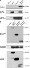
Association of BRII with Eps15R. 293T cells were transfected with HA-tagged BRII-LF, BRII-SF, or BRII-TC1 and EGFP-tagged Eps15R. Immunoprecipitation (IP) and Western blotting (WB) were accomplished as indicated in the text. (A) Eps15R and BRII-LF form a complex in 293T cells. (B) All receptor variants are verified in the Eps15R precipitate with HA antibody. Eps15R interacts also with BRII-TC1.
FIG. 10.
FIG. 10.
Smad-dependent signaling and Smad-independent BMP signaling originate from distinct endocytic routes of the BMP receptors. Smad-dependent signaling is initiated upon BMP-2 binding to PFCs, which exist in an inactive state at the cell surface prior to ligand binding (45). Activation of this signaling cascade by BMP-2 binding results in the phosphorylation of Smad1/5 by the type I receptor. This step in Smad signaling occurs while the receptors reside at the plasma membrane; inhibition of endocytosis (treatment with chlorpromazine or expression of dominant negative dynamin) does not affect BMP-2-mediated Smad phosphorylation. Subsequent endocytosis of the receptors through CCPs, however, is needed to transmit the Smad signal into the nucleus to control the transcriptional activity of target genes. Treatment with lovastatin to perturb cholesterol-dependent plasma membrane regions does not affect Smad signaling but reduces BMP-2-mediated ALP induction through a Smad-independent cascade. This signaling pathway is initiated by the binding of BMP-2 to high-affinity BRI, and a subsequent recruitment of BRII into the signaling receptor complex BISC occurs (45). From the data presented here, we conclude that Smad-independent signaling resulting in ALP induction starts from receptors (BISCs) residing in cholesterol-enriched plasma membrane regions.

Similar articles

Cited by

References

    1. Anderson, R. G. 1998. The caveolae membrane system. Annu. Rev. Biochem. 67:199-225. - PubMed
    1. Anderson, R. G., and K. Jacobson. 2002. A role for lipid shells in targeting proteins to caveolae, rafts, and other lipid domains. Science 296:1821-1825. - PubMed
    1. Anderson, R. G., B. A. Kamen, K. G. Rothberg, and S. W. Lacey. 1992. Potocytosis: sequestration and transport of small molecules by caveolae. Science 255:410-411. - PubMed
    1. Aoki, T., R. Nomura, and T. Fujimoto. 1999. Tyrosine phosphorylation of caveolin-1 in the endothelium. Exp. Cell Res. 253:629-636. - PubMed
    1. Brown, D. A., and E. London. 2000. Structure and function of sphingolipid- and cholesterol-rich membrane rafts. J. Biol. Chem. 275:17221-17224. - PubMed

Publication types

MeSH terms

LinkOut - more resources