Myeloid lineage progenitors give rise to vascular endothelium
- PMID: 16920790
- PMCID: PMC1559769
- DOI: 10.1073/pnas.0604203103
Myeloid lineage progenitors give rise to vascular endothelium
Abstract
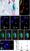
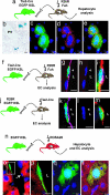
Despite an important role in vascular development and repair, the origin of endothelial progenitors remains unknown. Accumulating evidence indicates that cells derived from the hematopoietic system participate in angiogenesis. However, the identity and functional role of these cells remain controversial. Here we show that vascular endothelial cells can differentiate from common myeloid progenitors and granulocyte/macrophage progenitors. Endothelial cells derived from transplanted bone marrow-derived myeloid lineage progenitors expressed CD31, von Willebrand factor, and Tie2 but did not express the hematopoietic markers CD45 and F4/80 or the pericyte markers desmin and smooth muscle actin. Lineage tracing analysis in combination with a Tie2-driven Cre/lox reporter system revealed that, in contrast to bone marrow-derived hepatocytes, bone marrow-derived endothelial cells are not the products of cell fusion. The establishment of both hematopoietic and endothelial cell chimerism after parabiosis demonstrates that circulating cells can give rise to vascular endothelium in the absence of acute radiation injury. Our findings indicate that endothelial cells are an intrinsic component of myeloid lineage differentiation and underscore the close functional relationship between the hematopoietic and vascular systems.
Conflict of interest statement
Conflict of interest statement: No conflicts declared.
Figures


Comment in
-
My O'Myeloid, a tale of two lineages.Proc Natl Acad Sci U S A. 2006 Aug 29;103(35):12959-60. doi: 10.1073/pnas.0606018103. Epub 2006 Aug 21. Proc Natl Acad Sci U S A. 2006. PMID: 16924095 Free PMC article. No abstract available.
Similar articles
-
Bone marrow monocyte lineage cells adhere on injured endothelium in a monocyte chemoattractant protein-1-dependent manner and accelerate reendothelialization as endothelial progenitor cells.Circ Res. 2003 Nov 14;93(10):980-9. doi: 10.1161/01.RES.0000099245.08637.CE. Epub 2003 Oct 2. Circ Res. 2003. PMID: 14525810
-
Hematopoietic stem cells give rise to perivascular endothelial-like cells during brain tumor angiogenesis.Stem Cells Dev. 2005 Oct;14(5):478-86. doi: 10.1089/scd.2005.14.478. Stem Cells Dev. 2005. PMID: 16305333
-
Gene signature of stromal cells which support dendritic cell development.Stem Cells Dev. 2008 Oct;17(5):917-27. doi: 10.1089/scd.2007.0170. Stem Cells Dev. 2008. PMID: 18564035
-
Contribution of circulating vascular progenitors in lesion formation and vascular healing: lessons from animal models.Curr Opin Lipidol. 2008 Oct;19(5):498-504. doi: 10.1097/MOL.0b013e32830dd566. Curr Opin Lipidol. 2008. PMID: 18769231 Review.
-
Endothelial progenitor cells: characterization and role in vascular biology.Circ Res. 2004 Aug 20;95(4):343-53. doi: 10.1161/01.RES.0000137877.89448.78. Circ Res. 2004. PMID: 15321944 Review.
Cited by
-
Versatile cell ablation tools and their applications to study loss of cell functions.Cell Mol Life Sci. 2019 Dec;76(23):4725-4743. doi: 10.1007/s00018-019-03243-w. Epub 2019 Jul 29. Cell Mol Life Sci. 2019. PMID: 31359086 Free PMC article. Review.
-
Myeloid cells contribute to tumor lymphangiogenesis.PLoS One. 2009 Sep 17;4(9):e7067. doi: 10.1371/journal.pone.0007067. PLoS One. 2009. PMID: 19759906 Free PMC article.
-
TNFalpha accelerates monocyte to endothelial transdifferentiation in tumors by the induction of integrin alpha5 expression and adhesion to fibronectin.Mol Cancer Res. 2011 Jun;9(6):702-11. doi: 10.1158/1541-7786.MCR-10-0484. Epub 2011 May 2. Mol Cancer Res. 2011. PMID: 21536688 Free PMC article.
-
TGF-β1 signaling and Krüppel-like factor 10 regulate bone marrow-derived proangiogenic cell differentiation, function, and neovascularization.Blood. 2011 Dec 8;118(24):6450-60. doi: 10.1182/blood-2011-06-363713. Epub 2011 Aug 9. Blood. 2011. PMID: 21828131 Free PMC article.
-
Identification of three molecular and functional subtypes in canine hemangiosarcoma through gene expression profiling and progenitor cell characterization.Am J Pathol. 2014 Apr;184(4):985-995. doi: 10.1016/j.ajpath.2013.12.025. Epub 2014 Feb 11. Am J Pathol. 2014. PMID: 24525151 Free PMC article.
References
-
- Rafii S., Lyden D. Nat. Med. 2003;9:702–712. - PubMed
-
- Jain R. K., Duda D. G. Cancer Cell. 2003;3:515–516. - PubMed
-
- Coultas L., Chawengsaksophak K., Rossant J. Nature. 2005;438:937–945. - PubMed
-
- Gariano R. F., Gardner T. W. Nature. 2005;438:960–966. - PubMed
-
- Ferrara N., Kerbel R. S. Nature. 2005;438:967–974. - PubMed
Publication types
MeSH terms
Grants and funding
LinkOut - more resources
Full Text Sources
Other Literature Sources
Medical
Research Materials
Miscellaneous

