

Focal adhesion kinase targeting using in vivo short interfering RNA delivery in neutral liposomes for ovarian carcinoma therapy
- PMID: 16914580
- PMCID: PMC3144499
- DOI: 10.1158/1078-0432.CCR-06-0021
Focal adhesion kinase targeting using in vivo short interfering RNA delivery in neutral liposomes for ovarian carcinoma therapy
Erratum in
-
Correction: Focal Adhesion Kinase Targeting Using In vivo Short Interfering RNA Delivery in Neutral Liposomes for Ovarian Carcinoma Therapy.Clin Cancer Res. 2019 May 15;25(10):3194. doi: 10.1158/1078-0432.CCR-19-1132. Clin Cancer Res. 2019. PMID: 31092618 No abstract available.
Abstract
Purpose: Focal adhesion kinase (FAK) plays a critical role in ovarian cancer cell survival and in various steps in the metastatic cascade. Based on encouraging in vitro results with FAK silencing, we examined the in vivo therapeutic potential of this approach using short interfering RNA (siRNA) in the neutral liposome 1,2-dioleoyl-sn-glycero-3-phosphatidylcholine (DOPC).
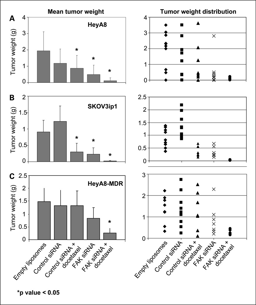
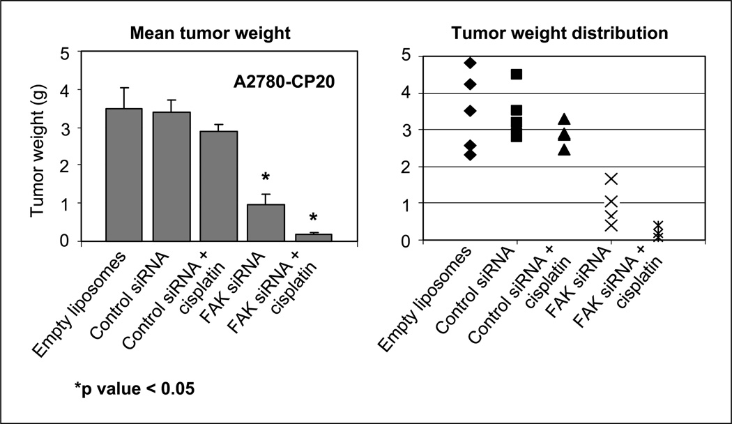
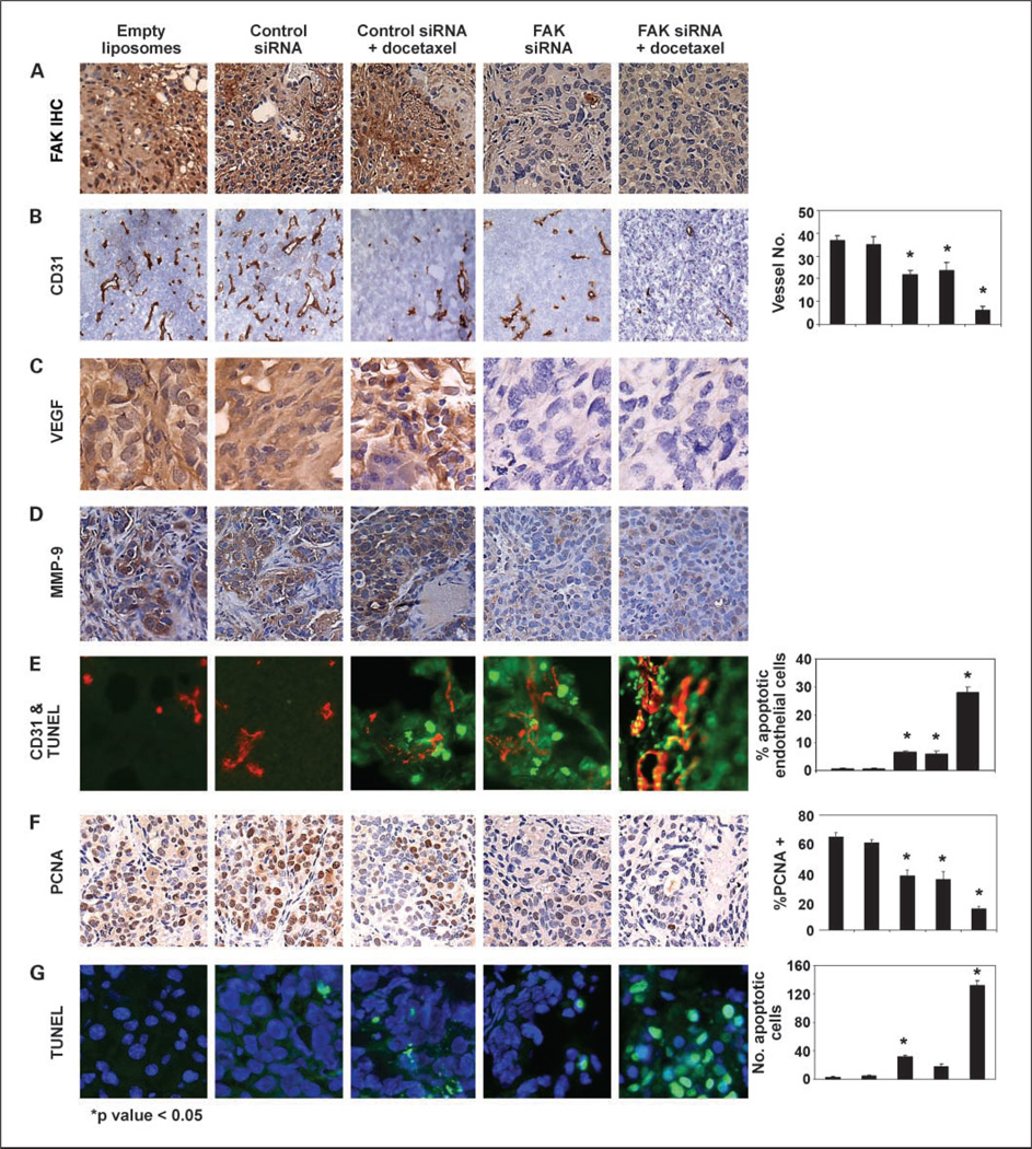
Experimental design: Therapy experiments of FAK siRNA with or without docetaxel were done using human ovarian cancer cell lines SKOV3ip1, HeyA8, and HeyA8MDR in nude mice. Additional experiments with a cisplatin-resistant cell line (A2780-CP20) were also done. Assessments of angiogenesis (CD31), cell proliferation (proliferating cell nuclear antigen), and apoptosis (terminal deoxynucleotidyl transferase-mediated dUTP nick end labeling) were done using immunohistochemical analysis.
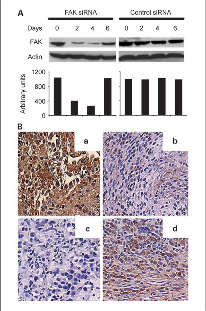
Results: A single dose of FAK siRNA-DOPC was highly effective in reducing in vivo FAK expression for up to 4 days as assayed by Western blot and immunohistochemical analysis. Therapy experiments were started 1 week after injection of the ovarian cancer cells. Treatment with FAK siRNA-DOPC (150 mug/kg twice weekly) reduced mean tumor weight by 44% to 72% in the three cell lines compared with the control group (Ps < 0.05 for HeyA8, A2780-CP20, and SKOV3ip1). When FAK siRNA-DOPC was combined with docetaxel, there was even greater reduction in mean tumor weight in all models (all Ps < 0.05). Similar results were observed in combination with cisplatin. Treatment with FAK siRNA-DOPC plus docetaxel resulted in decreased microvessel density, decreased expression of vascular endothelial growth factor and matrix metalloproteinase-9, and increased apoptosis of tumor-associated endothelial cells and tumor cells.
Conclusions: Taken together, these findings suggest that FAK siRNA-DOPC plus docetaxel or platinum might be a novel therapeutic approach against ovarian cancer.
Figures
Similar articles
-
Therapeutic EphA2 gene targeting in vivo using neutral liposomal small interfering RNA delivery.Cancer Res. 2005 Aug 1;65(15):6910-8. doi: 10.1158/0008-5472.CAN-05-0530. Cancer Res. 2005. PMID: 16061675
-
Dual targeting of EphA2 and FAK in ovarian carcinoma.Cancer Biol Ther. 2009 Jun;8(11):1027-34. doi: 10.4161/cbt.8.11.8523. Epub 2009 Jun 24. Cancer Biol Ther. 2009. PMID: 19395869 Free PMC article.
-
Effect of interleukin-8 gene silencing with liposome-encapsulated small interfering RNA on ovarian cancer cell growth.J Natl Cancer Inst. 2008 Mar 5;100(5):359-72. doi: 10.1093/jnci/djn024. Epub 2008 Feb 26. J Natl Cancer Inst. 2008. PMID: 18314475 Free PMC article.
-
Focal Adhesion Kinase in Ovarian Cancer: A Potential Therapeutic Target for Platinum and Taxane-Resistant Tumors.Curr Cancer Drug Targets. 2019;19(3):179-188. doi: 10.2174/1568009618666180706165222. Curr Cancer Drug Targets. 2019. PMID: 29984656 Review.
-
Nanomedicine based approaches for the delivery of siRNA in cancer.J Intern Med. 2010 Jan;267(1):44-53. doi: 10.1111/j.1365-2796.2009.02191.x. J Intern Med. 2010. PMID: 20059643 Review.
Cited by
-
FAK deletion promotes p53-mediated induction of p21, DNA-damage responses and radio-resistance in advanced squamous cancer cells.PLoS One. 2011;6(12):e27806. doi: 10.1371/journal.pone.0027806. Epub 2011 Dec 14. PLoS One. 2011. PMID: 22194793 Free PMC article.
-
Inhibition of focal adhesion kinase (FAK) activity prevents anchorage-independent ovarian carcinoma cell growth and tumor progression.Clin Exp Metastasis. 2013 Jun;30(5):579-94. doi: 10.1007/s10585-012-9562-5. Epub 2012 Dec 30. Clin Exp Metastasis. 2013. PMID: 23275034 Free PMC article.
-
Targeting V600EB-Raf and Akt3 using nanoliposomal-small interfering RNA inhibits cutaneous melanocytic lesion development.Cancer Res. 2008 Sep 15;68(18):7638-49. doi: 10.1158/0008-5472.CAN-07-6614. Cancer Res. 2008. PMID: 18794153 Free PMC article.
-
Phase I Study of the Focal Adhesion Kinase Inhibitor BI 853520 in Japanese and Taiwanese Patients with Advanced or Metastatic Solid Tumors.Target Oncol. 2019 Feb;14(1):57-65. doi: 10.1007/s11523-019-00620-0. Target Oncol. 2019. PMID: 30725402 Free PMC article. Clinical Trial.
-
New aspects of gene-silencing for the treatment of cardiovascular diseases.Pharmaceuticals (Basel). 2013 Jul 19;6(7):881-914. doi: 10.3390/ph6070881. Pharmaceuticals (Basel). 2013. PMID: 24276320 Free PMC article.
References
-
- Jemal A, Murray T, Ward E, et al. Cancer statistics, 2005. CA Cancer J Clin. 2005;55:10–30. - PubMed
-
- McGuire WP, Hoskins WJ, Brady MF, et al. Cyclophosphamide and cisplatin compared with paclitaxel and cisplatin in patients with stage III and stage IV ovarian cancer.[comment] N Engl J Med. 1996;334:1–6. - PubMed
-
- du Bois A, Luck HJ, Bauknecht T, et al. First-line chemotherapy with epirubicin, paclitaxel, and carboplatin for advanced ovarian cancer: a phase I/II study of the Arbeitsgemeinschaft Gynakologische Onkologie Ovarian Cancer Study Group. J Clin Oncol. 1999;17:46–51. - PubMed
-
- Kohno M, Hasegawa H, Miyake M, et al. CD151 enhances cell motility and metastasis of cancer cells in the presence of focal adhesion kinase. Int J Cancer. 2002;97:336–343. - PubMed
-
- Schaller MD, Parsons JT. Focal adhesion kinase: an integrin-linked protein tyrosine kinase. Trends Cell Biol. 1993;3:258–262. - PubMed
Publication types
MeSH terms
Substances
Grants and funding
LinkOut - more resources
Full Text Sources
Other Literature Sources
Medical
Miscellaneous
